Cohesive Strength of Sound Dentin and a Resin-based Composite
*e-mail: [email protected]
Microtensile Bond Strength of Current Adhesive
Systems when Compared to Cohesive Strength of
Sound Dentin and a Resin-based Composite
Paulo Eduardo Capel Cardosoa*, Fernanda Tranchesi Sadekb, Eliane Placidob, José Fortunato Ferreira Santosc
aProfessor, Department of Dental Materials, Faculty of Dentistry, University of São Paulo,
Av. Lineu Prestes, 2227, 05508-900 São Paulo, Brazil
bGraduate student, Department of Dental Materials, Faculty of Dentistry,
University of São Paulo, São Paulo, Brazil
cProfessor, Department of Dental Materials, Faculty of Dentistry,
University of São Paulo, São Paulo, Brazil Visiting Professor, Department of Operative Dentistry,
UNC Chapel Hill, North Carolina, USA.
Received: March 28, 2004; Revised: September 8, 2004
Purpose:To evaluate the microtensile bond strength (µTBS) to dentin of 4 adhesive systems, the micromorphology of the adhesive/dentin interface and to compare the results to the µTS (co-hesive strength) of sound dentin and resin composite. Occlusal surfaces of 24 extracted caries-free human molars were cut flat to expose the dentin surface. They were randomly assigned to 4 groups (n = 6): Adper Scotchbond Multi Purpose Plus (MP), Adper Single Bond (SB), Clearfil Protect Bond (CP) and Adper Prompt (AP). Adhesive systems were applied and “crowns” were built using Z100. Other 5 human molars were sectioned to obtain square-shaped dentin blocks and 5 resin blocks were built using a composite resin, Z100. After storage in distilled water at 37°C for 24 h, stick-shaped specimens were obtained for all groups (n = 5) with 0.8 mm2 and subjected
to µTBS or µTS test. Results were analyzed using One-Way ANOVA and Tukey’s test at p < 0.05. The remaining bonded teeth were cut in two halves perpendicularly to the bonded interface and prepared for SEM analyses. No significant differences were found among CP (45.6 + 7.4 MPa), SB (43.3 + 6.5 MPa) and MP (35.1 + 5.9 MPa) (p > 0.05). The lowest result was found for AP (27.4 + 4.7 MPa), although not statistically different from MP. Most specimens (89.4%) showed predominant adhesive failure. None of the systems tested reached the µTS values of dentin (108.5 + 9.4 MPa) and Z100 (86.5 + 3.6 MPa). Bonded interfaces showed lower µTBS than those µTS of dentin and resin composite blocks. The all-in-one self-etching adhesive had the lowest µTBS.
Keywords: bond strength, cohesive strength, resin, dentin
1. Introduction
Successful adhesion to dentin is one of the requirements when choosing tooth-colored materials, specifically
resin-based composites1,2. A great demand for more simplified
application techniques, with reduced clinical steps3 and
lower technique sensitivity4 is also noticed.
Several studies regarding the efficacy of new adhesive systems can be found in the literature showing promising
results2,5,6. However, due to the complexity of dentin, such
as the high percentage of organic components7, the
vari-ability in surface moisture8, varied regional tubule
orienta-tion9,10, differences in permeability11,12 and presence of
scle-rotic dentin13-15, a wide range of results are seen among
dif-ferent studies.
structure6. The procedure basically consists in the use of a
30-40% acid phosforic that removes the smear layer plugs over intertubular dentin, as well as peritubular dentin, opens dentinal tubules and demineralizes the intertubular dentin. Primer resins, like as 2-Hydroxyethyl methacrylate (HEMA), bisphenyl-dimethacrylate (BPDM), Glycerol-propano-dimethacrylate (GPDM), Polymethyl methacrylate (PAMM), among others, are then applied, followed by meth-acrylate-based bonding resins. The resin monomer penetra-tion into the demineralized dentin forming a resin-infiltrated
zone (hybrid layer)16 seems to be responsible for the greater
longevity and stability of resin restorations, and for the
higher bond strength values observed in several studies17-19.
Nowadays, bonding systems have been directed towards the use of more simplified bonding procedures. In addition to one-bottle adhesives, self-etching primers and all-in-one self-etching systems have been proposed. In self-etching primer systems, an acidic primer is applied and the bonding agent is subsequently applied as a second coat (with no need for the rinsing and drying steps). In the all-in-one self-etch-ing adhesive systems, etchself-etch-ing, primself-etch-ing and bondself-etch-ing
proce-dures are combined in a single application step20. Their
for-mulations are based on the need for diminishing technique sensitivity and incorporating or modifying the smear layer instead of removing it. The acidic monomers penetrate, modify or incorporate this layer, and interact with struc-tures from deep areas of dentin, finally being polymerized
in situ6,21,22.
Improvement in bond strength values has been achieved with these new systems, but the number of cohesive
fail-ures in dentin has also increased23. This does not mean that
the bonding interface is stronger than the intrinsic strength of the substrate, but that stresses were applied non-uniformly
during the tests1,24. This explains why shear and tensile
strength tests have been thoroughly criticized by some
au-thors18,25. Moreover, the absence of a standardized
method-ology to be followed by all research centers makes it
diffi-cult to compare results obtained by different authors26,27.
Aiming at a better stress distribution in the same speci-men and achieving tensile strength values that might repre-sent what really occurs along the tooth/restoration interface, some authors have proposed the microtensile bond strength test18,24,28 with specimens with a cross sectional area of
ap-proximately 1 mm2. Such a test has been used by several
authors to study the effect of variations along the dentin
substrate on the bond strength10,29,30. A better distribution of
forces is achieved with this test, as well as a lower
coeffi-cient of variation25,and fewer occurrence of cohesive
frac-tures along dentin31,32.
Therefore, the objective of the present in vitro study was
to analyze differences in microtensile bond strength of speci-mens made with four adhesive systems [a multi-step, a one-bottle and two self-etching systems (a one- and a two-step
product)], and compare them with two ‘homogeneous’ substrates, one made from a bulk of resin composite and the other from sound dentin.
2. Methods and Materials
Twenty four sound extracted human molars stored in
distilled water at 37°C for a maximum period of three
months were cleaned of debris and used to evaluate microtensile bond strength and the micro-morphological resin-adhesive-dentin interface. They were partially embed-ded in PVC tubes, using chemically-activated acrylic resin, leaving their crown exposed. This was done to facilitate posterior procedures for specimen fixation and sectioning. Occlusal surfaces were cut by means of a low-speed dia-mond disc (Extec Co., USA) under water cooling, so that superficial dentin (approximately 2-3 mm below the den-tin-enamel junction) was exposed. Surrounding enamel was removed using nº 3100 diamond bur (KG Sorensen, Bra-zil). Dentin surfaces were consecutively polished using sand-papers grits 220, 320 and 400 for 10 s each, and finally 600-grit sandpaper for 60 s, providing a standardized smear layer formation.
The teeth were then randomly divided into four groups (n = 6), according to the adhesive systems presented in Ta-ble 1, and applied as described below:
Group 1 – Adper Scotchbond Multi Purpose Plus [MP] (3M ESPE, St. Paul, MN, USA). Dentin surfaces were acid etched by 35% phosphoric acid (Scotchbond Ecthing Gel, 3M ESPE, St. Paul, MN, USA) for 15 s, thoroughly washed and gently air-dried for 2 s, thus remaining visibly moist. One drop of primer was applied to the whole surface and air-dried. Finally, a coat of bonding agent was applied and light-cured for 10 s using an Optilux 500 light-curing unit (Demetron/Kerr Corp., Orange, CA, USA), whose light
in-tensity was in the range of 600 mW/cm2.
Group 2 – Adper Single Bond [SB] (3M ESPE, St. Paul, MN, USA). Dentin surfaces were etched following the same procedures as the previous Group 1. On the visible moist dentin, two consecutive coats were then applied, gently air-dried and light-cured for 20 s.
Group 3 – Clearfil Protect Bond [CP] (Kuraray Medical Inc., Osaka, Japan). After the removal of excess moisture from the dentin surfaces, the acidic primer was applied, left undisturbed for 20 s and then dried with an air-blow. The adhesive was applied, air-dried and light-cured for 10 s.
Group 4 – Adper Prompt [AP] (3M ESPE, St. Paul, MN, USA). One drop of solution A and one drop of solution B were mixed in a dappen dish. The adhesive was scrubbed with microbrush on the dentin surface for 15 s, gently air-dried, and light-cured for 10 s.
Cohesive Strength of Sound Dentin and a Resin-based Composite
surface of dentin. Each increment was light-cured for 40 s using the same light-curing unit as mentioned above. At the end of this procedure, a resin composite block approximately 5 mm high was obtained. Teeth were then stored in distilled
water at 37°C for 24 h.
To evaluate the cohesive strength of dentin, which we denominated as dentin microtensile strength [SD], five sound
extracted human molars, stored in distilled water at 37°C
for a maximum period of three months, were sectioned to obtain square-shaped blocks with 5 mm of thickness. To evaluate the cohesive strength of Z100 resin composite, also denominated as Z100 microtensile strength [RC], five square-shaped blocks were built with the aid of a plastic matrix, in five increments of 1 mm, each one being light-cured for 40 s. Both dentin and composite resin blocks were
stored in distilled water at 37°C for 24 h.
Stick-shaped specimens with a rectangular
cross-sec-tional area of 0.8 mm2 were obtained for all groups (n = 5)
(bonded restorations, dentin and resin composite blocks) by means of sequential crossed perpendicular cuts using a double-faced diamond disc in a cutting machine (Labcut 1010 – Extec Co., USA). The number of sticks obtained for each group after sectioning is shown in Table 2. Specimens were fixed to a metal claw using cyanoacrylate glue. The claw/specimen set was positioned in a universal testing machine (Instron model 5565, Canton, MA, USA), so that microtensile forces were applied parallel to the long axis of each specimen at a crosshead speed of 0.5 mm/min. The mode of failure for the adhesive interfaces was analyzed under a stereomicroscope with 25 X magnification, to verify
the occurrence of adhesive failures. Only the specimens that exhibited an adhesive failure mode were included for sta-tistical analysis.
The distribution of µTBS and µTS data was first checked for normality with the Kolmogorov-Smirnov test. Since the sample showed a normal distribution, the results were sub-jected to One-Way ANOVA and Tukey’s test to determine possible statistical differences among the groups tested in this study (p < 0.05).
For SEM analyses, the remaining bonded tooth of each
Table 1. Composition and corresponding batch number for each adhesive system tested in this study.
Adhesive Composition Batch # Manufacturer
Adper Scotchbond Plus PRIMER – HEMA/polyalkenoic acid/water 3008 3M ESPE
Multi Purpose BOND – HEMA/Bis-GMA/amines 7543 (Minneapolis, MN, USA)
Adper Single Bond Bis-GMA / HEMA/ethanol/water/dimethacrylates/ 1FB 3M ESPE
polyalkenoic acid/amines/Methacrylate functional (Minneapolis, MN, USA)
copolymer of polyacrylic and polyitaconic acid
Clearfil Protect Bond PRIMER – MDPB/MDP/HEMA/hydrophilic 000914 Kuraray Medical Inc.
dimethacrylate/solvent (water)/initiators/ (Osaka, Japan)
N,N-Diethanol-p-toluidine
BOND – MDP/HEMA/Bis-GMA/hydrophobic 001018
dimethacrylate/silanated colloidal silica/initiators/ N,N-Diethanol-p-toluidine
Adper Prompt self-etching LIQUID 1 – Methacrylated phosphoric esters/ EXM-618 3M ESPE
Bis-GMA/Initiators/stabilizers (Minneapolis, MN, USA)
LIQUID 2 – Water/HEMA/stabilizers/ polyalkenoic acid
HEMA – 2-Hydroxyethyl methacrylate
Bis-GMA – Bis-phenol A diglycidylmethacrylate MDPB – 12-methacryloyloxydodecylpyridinium bromide MDP – 10-methacryloyloxdecyl dihydrogen phosphate
Table 2. Microtensile strength values obtained for the adhesives systems, dentin and resin composite. Values are in MPa (number of sticks).
UTS (MPa) SD
Sound Dentin 108.5a 9.4
(32)
Resin Composite 86.5b 3.6
(29)
Clearfil Protect Bond 45.6c 7.4
(61)
Adper Single Bond 43.3c 6.5
(65)
Adper Scotchbond MP Plus 35.1c,d 5.9
(54)
Adper Prompt 27.4d 4.7
(46)
UTS - Ultimate tensile strength = force at failure/cross-sectional area. SD - Standard deviation.
adhesive group was cut in two halves perpendicularly to the bonded interface using a low-speed diamond saw. Each half was polished with sandpaper 600, 1200 and 2000-grit under refrigeration. The final polish was obtained with in-creasingly fine diamond pastes (1 and 0.25 mm; Buehler, Lake Bluff, IL USA). Debris were ultrasonically removed for 10 min between each polishing step. The interface
sec-tions were demineralized in 50% H3PO4 for 5 s,deproteinized
in 1% NaOCl for 10 min, and finally dehydrated with silica. Each sample was mounted on aluminum stubs, sputter-coated and observed in a scanning electron microscope (Philips XL30; Philips, Eindhoven, The Netherlands). Photomicrographs were taken in order to analyze the mor-phological characteristics of the hybrid layer and resin pen-etration into the dentin substrate.
3. Results
Mean microtensile bond strength values obtained after statistical analysis are presented in Table 2. Among the ad-hesive systems tested in this study, the highest mean bond strength value was obtained for Clearfil Protect Bond (45.6 + 7.4 MPa), although it was not statistically different from Adper Single Bond (43.3 + 6.5 MPa) and Adper Scotchbond MP Plus (35.1 + 5.9 MPa) (p < 0.05). Adper Prompt re-sulted in the lowest mean value (27.4 + 4.7 MPa), and was statistically similar to Scotchbond MP Plus (p > 0.05). None of the systems tested was able to reach the mean microtensile cohesive strength values obtained for sound dentin (108.5 + 9.4 MPa) or for Z100 resin composite (86.5 + 3.6 MPa). Sound dentin also showed statistically higher values than Z100 (p < 0.0001).
Of the bonded specimens, the number of sticks
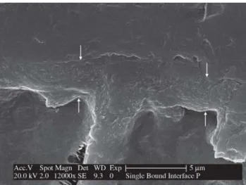
consid-Figure 1. SEM micrograph of resin-dentin interface created by Adper Scotchbond Multi Purpose Plus. The hybrid layer (arrows) and the resin tag formation can be observed.
Figure 2. SEM micrograph of resin-dentin interface created by Adper Single Bond. The hybrid layer (arrows) and the resin tag formation can be observed.
ered for statistical analysis is shown within parenthesis in Table 2. Approximately 20% of specimens, which were not included in the statistical analysis, not uniformly distrib-uted among the groups, were lost during preparation, re-sulting in an uneven number of specimens for each group (Table 2),
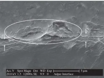
The resin-dentin interfaces of the four adhesive systems are illustrated in the scanning electron micrographs shown in Figs. 1 to 4. The hybrid layer could be observed in all specimens, although the thickness varied depending on the bonding system used. Resin penetrations into dentinal tu-bules and intimate adaptation to the underlying dentin were also clearly observed in all specimens, with exception of Adper Prompt that showed a non-uniform hybrid layer, a poorly formed resin tag and a discontinued area between adhesive layer and the resin (Fig. 4).
Self-etching systems (Clearfil Protect Bond and Adper Prompt) produced a thinner hybrid layer (Figs. 3 and 4) when compared to the total-etch systems (Figs. 1 and 2).
4. Discussion
Among the systems that require previous etching of the substrate, Adper Scotchbond Multi-Purpose Plus is a repre-sentative of conventional systems and Adper Single Bond can be considered as its simplified version, as they belong to the same manufacturer. In this study, both materials ob-tained similar microtensile bond strength values, a result
that is in agreement with previous studies4,25. A possible
Cohesive Strength of Sound Dentin and a Resin-based Composite
to the operator performance33. Adper Single Bond also
con-tains ethanol which acts as a water chaser. Because of this, it is less sensitive to different moisture conditions, and thus
facilitates adequate wettability16,34. Its reduced viscosity also
adds better spreading properties to the material. This can explain its tendency towards better results and better pen-etration into the demineralized dentin, when compared to MP: 43.3 MPa for SB and 35.1 MPa for MP (Figs. 1 and 2), Although statistically similar results were found among the above cited materials and Clearfil Protect Bond, the latter showed the highest mean bond strength values: 45.6 MPa. It has been reported that the use of phosphoric acid for dentin etching can result in a porous zone beneath the hybrid layer, possibly due to over etching and collapse of the collagen
net-work4,35, which could not be found in the photomicrographs
of this present study. Self-etching primers are an excellent alternative to acid etching systems, by reducing the technique sensitivity, as the intermediate steps of washing and drying
of the substrate are eliminated36. Their mechanism of action
is based on the dissolution of the smear layer and its incorpo-ration in the formed hybrid layer, which is thinner than that obtained with other systems, as can be observed in Figs. 3 and 4. However, the thickness of the hybrid layer does not seem to be relevant for the resultant bond strength, as com-parable bond strength values have been observed for
one-bottle and conventional systems5,34,37 (Table 2).
The major simplification of the adhesive technique was achieved with all-in-one self-etching adhesives, in which the acidic primer and bonding agent are combined in a real single step application. Etch&Prime 3.0 and Prompt L Pop can be cited as two of the first introduced materials in the market. However, they did not show satisfactory results in
many in vivo and in vitro studies reported in the
litera-ture2,25,38,39. The new version of Prompt L Pop, the Adper
Prompt, is considered by the manufacturer as an improved formulation of the first. Nonetheless, in this study, it also showed low bond strength values compared to the other systems, although statistically similar to SBMP. Further-more, the SEM photomicrograph (Fig. 4) shows areas of discontinuity between the adhesive line and the composite resin being, probably, the cause of bond strength low val-ues. This material contains methacrylated phosphoric es-ters as part of the acidic monomers. These compounds are necessary in self-etching systems to dissolve the smear layer and expose the collagen fibrils, but they can be unstable due to hydrolysis by aqueous solutions with an acid pH
value19. This may explain the results obtained using this
system. In the case of Clearfil Protect Bond the manufac-turer introduced 10-methacryloyloxdecyl dihydrogen phos-phate (MDP) as acidic monomer, which retains its hydro-lytic stability in conjunction with highly acidic pH values, and thus more reliable bond strength values can be
ob-tained40.
According to Urabe & others41, to assure an adequate
and durable adhesive restoration, it is important to verify the strength of tooth structures, which can be referred as the true measure for restored teeth. Also according to these authors, the tooth and restoration should be considered as a ‘monolithic structure’, if a sufficient adhesion to tooth struc-tures is achieved.
It was demonstrated in this study that bond strength val-ues for current adhesive systems cannot be compared to the microtensile strength of a bulk structure of dentin or resin composite. When one considers a bonded restoration, the
Figure 4. SEM micrograph of resin-dentin interface created by Adper Prompt. A non-uniform hybrid layer (arrows) and the poorly formed resin tag can be observed. Areas of discontinuity can also be noted (circle).
weakest spot will always be the interface, as we are bond-ing a very heterogeneous structure, as dentin, to a more ‘ho-mogeneous’ material, as the resin composite. Clinically, it is important to consider that the adhesive interface contin-ues to be the weakest point in aesthetic resin composite res-torations, and the clinician should be careful when working on it. Further studies seem of great importance to evaluate the bonding stability, for bond strength values should be maintained throughout a certain period of time.
In this study values obtained for dentin (108.5 MPa) were similar to those reported in the literature with the same
method42. An interesting fact to be pointed out is the lower
standard deviation found for Z100 when compared to den-tin and the adhesive systems tested. This reinforces the lack of uniformity in the dentin/adhesive interface, and the dentinal tissue as a whole structure. Intrinsic defects such as microcracks can be found in both cases, as well as, the influence of other characteristics that are inherent to this
substrate. Different tubule direction9,43 and diameter,
depend-ing on the area to be tested (deep or superficial dentin), presence of sclerotic tissue, and the high percentage of
or-ganic components7, can influence the results and lead to
greatly variable values with high coefficient of variation, which also depends on the handling by the operator.
5. Conclusions
Interfaces bonded with the four adhesive systems showed lower microtensile strength than those of dentin and resin composite blocks. The self-etching adhesive had the lowest bond strength value (27.4 MPa) compared to the other tested systems, and still require further testing to be considered effective for clinical use.
Agradecimentos
The authors are grateful to Núcleo de Apoio à Pesquisa em Materiais Dentários (NAPEM - FOUSP).
References
1. Eick, J.D.; Gwinnett, A.J.; Pashley, D.H. ; Robinson, S.J.
Crit Rev Oral Biol Med, v. 8, n. 3, p. 306-335, 1997. 2. Frankenberger, R.; Perdigao, J.; Rosa, B.T.; Lopes, A.
Dental Materials, v. 17, n. 5, p. 373-380, 2001.
3. Ferrari, M.; Goracci, G.; Garcia-Godoy, F. Am J Dent,
v. 10, n. 5, p. 224-230, 1997.
4. Toledano, M.; Osorio, R.; de Leonardi, G.; Rosales-Leal,
J.I.; Ceballos, L.; Cabrerizo-Vilchez, M.A. Am J Dent,
v. 14, n. 4, p. 205-210, 2001.
5. Hannig, M.; Reinhardt, K.J.; Bott, B. Oper Dent, v. 24,
n. 3, p. 172-180, 1999.
6. Van Meerbeek, B.; Perdigao, J.; Lambrechts, P.; Vanherle, G. J Dent, v. 26, n. 1, p. 1-20, 1998.
7. Marshall Jr., G.W., Quintessence Int, v. 24, n. 9, p.
606-617, 1993.
8. Pashley, D.H.; Carvalho, R.M. J Dent, v. 25, n. 5, p.
355-372, 1997.
9. Giannini, M.; Carvalho, R.M.; Martins, L.R.; Dias, C.T.;
Pashley, D.H. J Adhes Dent, v. 3, n. 4, p. 315-324, 2001.
10. Yoshiyama, M.; Carvalho, R.M.; Sano, H.; Horner, J.A.;
Brewer, P.D.; Pashley, D.H. J Dent, v. 24, n. 6, p.
435-442, 1996.
11. Pashley, E.L.; Tao, L.; Matthews, W.G.; Pashley, D.H.
Dent Mater, v. 9, n. 1, p. 19-22, 1993.
12. Yoshiyama, M.; Carvalho, R.; Sano, H.; Horner, J.;
Brewer, P.D.; Pashley, D.H. Am J Dent, v. 8, n. 6, p.
297-302, 1995.
13. Duke, E.S.; Lindemuth, J. Am J Dent, v. 3, n. 6, p.
264-270, 1990.
14. Prati, C.; Chersoni, S.; Mongiorgi, R.; Montanari, G.;
Pashley, D.H. Oper Dent, v. 24, n. 2, p. 66-72, 1999.
15. Tay, F.R.; Kwong, S.M.; Itthagarun, A.; King, N.M.;
Yip, H.K.; Moulding, K.M.; Pashley, D.H. J Adhes
Dent, v. 2, n. 1, p. 9-28, 2000.
16. Gwinnett, A.J. Am J Dent, v. 7, n. 3, p. 144-148, 1994.
17. Ferrari, M.; Mason, P.N.; Vichi, A.; Davidson, C.L. Am
J Dent, v. 13, n. 6, p. 329-336, 2000.
18. Pashley, D.H.; Carvalho, R.M.; Sano, H.; Nakajima, M.; Yoshiyama, M.; Shono, Y.; Fernandes, C.A.; Tay, F. J Adhes Dent, v. 1, n. 4, p. 299-309, 1999.
19. Vargas, M.A.; Cobb, D.S.; Denehy, G.E. Dent Mater,
v. 13, n. 5, p. 316-324, 1997.
20. Lopes, G.C.; Baratieri, L.N.; de Andrada, M.A.; Vieira,
L.C. Quintessence Int, v. 33, n. 3, p. 213-224, 2002.
21. Kwong, S.M.; Tay, F.R.; Yip, H.K.; Kei, L.H.; Pashley,
D.H. J Dent, v. 28, n. 7, p. 515-528, 2000.
22. Watanabe, I.; Nakabayashi, N.; Pashley, D.H. J Dent
Res, v. 73, n. 6, p. 1212-1220, 1994.
23. Pashley, D.H.; Sano, H.; Ciucchi, B.; Yoshiyama, M.;
Carvalho, R.M. Dent Mater, v. 11, n. 2, p. 117-125,
1995.
24. Phrukkanon, S.; Burrow, M.F.; Tyas, M.J. Dental
Ma-terials, v. 14, n. 3, p. 212-221, 1998.
25. Cardoso, P.E.; Braga, R.R.; Carrilho, M.R. Dent Mater,
v. 14, n. 6, p. 394-398, 1998.
26. Sudsangiam, S.; van Noort, R. J Adhes Dent, v. 1, n. 1,
p. 57-67, 1999.
27. Watanabe, I.; Nakabayashi, N. J Dent, v. 22, n. 2, p.
67-72, 1994.
28. Sano, H.; Shono, T.; Sonoda, H.; Takatsu, T.; Ciucchi,
B.; Carvalho, R.; Pashley, D.H. Dent Mater, v. 10, n. 4,
p. 236-240, 1994.
29. Shono, Y.; Ogawa, T.; Terashita, M.; Carvalho, R.M.;
Pashley, E.L.; Pashley, D.H. J Dent Res, v. 78, n. 2,
p. 699-705, 1999.
30. Shono, Y.; Terashita, M.; Pashley, E.L.; Brewer, P.D.;
Cohesive Strength of Sound Dentin and a Resin-based Composite
31. Chen, K.K.; Shono, Y.; Ogawa, T.; Kozono, Y.; Terashita, M. Dent Mater J, v. 20, n. 4, p. 315-324, 2001. 32. Schreiner, R.F.; Chappell, R.P.; Glaros, A.G.; Eick, J.D.
Dent Mater, v. 14, n. 3, p. 194-201, 1998.
33. Finger, W.J.; Inoue, M.; Asmussen, E. Am J Dent, v. 7,
n. 1, p. 35-38, 1994.
34. Zheng, L.; Pereira, P.N.R.; Nakajima, M.; Sano, H.;
Tagami, J. Operative Dentistry, v. 26, n. 1, p. 97-104,
2001.
35. Phrukkanon, S.; Burrow, M.F.; Tyas, M.J. J Dent, v. 27,
n. 4, p. 265-274, 1999.
36. Perdigao, J.; Lopes, M. J Adhes Dent, v. 1, n. 3, p.
191-209, 1999.
37. Nakajima, M.; Ogata, M.; Okuda, M.; Tagami, J.; Sano,
H.; Pashley, D.H. Am J Dent, v. 12, n. 6, p. 309-314,
1999.
38. Brackett, W.W.; Covey, D.A.; St Germain, H.A., Jr. Oper
Dent, v. 27, n. 3, p. 218-222, 2002.
39. Cardoso, P.E.; Placido, E.; Moura, S.K. Am J Dent, v. 15,
n. 3, p. 164-168, 2002.
40. Hayakawa, T.; Kikutake, K.; Nemoto, K. Dent Mater,
v. 14, n. 2, p. 99-105, 1998.
41. Urabe, I.; Nakajima, S.; Sano, H.; Tagami, J. Am J Dent,
v. 13, n. 3, p. 129-135, 2000.
42. Sano, H.; Takatsu, T.; Ciucchi, B.; Russell, C.M.; Pashley,
D.H. J Dent Res, v. 74, n. 4, p. 1093-1102, 1995.
43. Schüpbach, P.; Krejci, I.; Felix, L. European Journal