Arq Bras Cardiol volume 74, (nº 2), 2000
Clinicopathologic Session
1 6 3 Session editor: Alfredo José Mansur
Associate editors: Desiderio Favarato - Vera Demarchi Aiello
Mailing address: Alfredo José Mansur – Incor – Av. Dr. Enéas C. Aguiar, 44 – 05403-000 – São Paulo, SP, Brazil
Case 1 / 0 0 – The patient is a 6 5 -year-old w oman w ith heart failure and proteinuria (Instituto do Cora çã o do Hospita l da s Clínica s – FM USP)
Clinicopathologic Session
Clinicopathologic Session
A 65-year-old woman sought medical assistance due to dyspnea on mild exertion and edema.
She complained that 6 months earlier, dyspnea on mo-derate exertion appeared, rapidly evolving to dyspnea on mild exertion and even at rest. Simultaneously, dizziness and edema of the lower limbs appeared and she lost 10 kg. She denied precordial pain, palpitation, and syncope during this time, as well as hypertension and diabetes mellitus. Twenty-five years earlier, she underwent a partial thyroidec-tomy because of multinodular goiter.
On physical examination, her heart rate was 74 bpm, her blood pressure 110/70 mmHg, and the jugular venous pressure was increased (++/4+). Examination of the lungs revealed crepitant rales at both bases. Heart examination showed reduction in the intensity of heart sounds. The liver was tender on palpation and could be felt 4cm from the right costal margin and 6cm from the xiphoid process. Mar-ked edema of the lower limbs occurred.
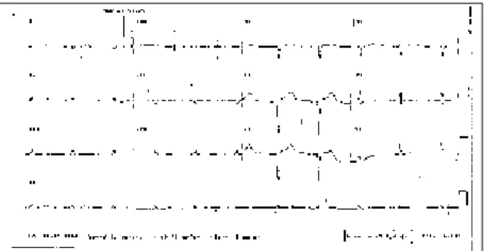
Electrocardiogram showed atrial rhythm with a heart rate of 63 bpm, QRS duration of 0.08s; QRS axis of +120º back-wards, a low voltage in the frontal plane, a small progression of the R wave from V1 to V3, a probably inactive lateral area, and diffuse alterations of ventricular repolarization (fig. 1).
Chest X-ray showed a moderate increase in the car-diac silhouette (++/4+) and bilateral pleural effusion.
Laboratory tests were as follows: hemoglobin – 14g/ dL; hematocrit - 42%; glycemia – 105mg/dL; creatinine – 0.8 mg/dL; total cholesterol – 155 mg/dL (HDL - 27mg/dL; LDL – 105mg/dL; VLDL – 23mg/dL); triglycerides – 117mg/dL. Urine analysis revealed density of 1028, pH of 5.0, proteinu-ria of 3.2g/L, and a normal sediment except for 660 cylin-ders/mL.
The diagnosis of heart failure due to cardiomyopathy with ventricular dilation was established and the investiga-tion for anatomical and etiologic characterizainvestiga-tion of the cli-nical findings was started. The patient was prescribed 80mg of furosemide, 50mg of hydrochlorothiazide in association with 5mg of amiloride and 20mg of enalapril.
One week later, she sought medical assistance due to severe dyspnea even at rest and orthopnea. On physical
exa-mination, the heart rate was 64bpm and the blood pressure was 80/50mmHg. Lung examination revealed crepitant rales in both pulmonary bases. Heart examination showed reduction in intensity of the cardiac sounds, no abnormal murmurs or cardiac sounds. The liver could be palpated 6cm from the costal margin. Edema was present in the lower limbs (+++/4+). Dopamine at the dosage of 30µg/kg/min and furosemide, 40mg twice a day at 6-hour intervals, were administered.
Electrocardiogram showed an atrial fibrillation rhythm with a heart rate of 80bpm, QRS duration of 0.12 s, QRS axis of +120º backwards, small progression of the R wave from V1 to V3, and an electrically negative area in the lateral wall (fig. 2). A few hours after admittance to the hospital, the pa-tient developed tachycardia with widened QRS, probably of ventricular origin, arterial hypotension followed by brady-cardia of 30 bpm, cardiogenic shock with inaudible blood pressure, and irreversible cardiopulmonary arrest.
Discussion
Clinical features – According to data from the
anamne-sis and physical examination, the case fits a congestive heart failure syndrome involving right and left cardiac chambers.
The contrast between the mild increase in the cardiac silhouette on chest X-ray and the severity of the clinical fin-dings is notable. The clinical finfin-dings included jugular stasis (++/4+), tender hepatomegaly, and marked edema of the lower limbs, suggesting the anatomical diagnosis of restrictive cardiomyopathy. A second hypothesis was constrictive pericarditis, whose differential diagnosis is dif-ficult because of the similarity of the hemodynamic findings. Other causes, such as ischemic heart disease, hypothyroi-dism, chronic hypocalcemia, and heart tumors are conside-red less probable hypotheses.
Restrictive cardiomyopathy is characterized by the following findings: reduction in the diastolic filling and ventricular distensibility; an ejection fraction that may be preserved; normal or reduced diastolic volumes; normal or increased ventricular thickness; and atrial dilation that may be almost always found 1,2. Signs and symptoms are those
of systemic and pulmonary venous congestion indicating restrictive physiopathological findings, and the disease is clinically suspected in those cases with normal heart si-lhouette.
endo-1 6 4
Clinicopathologic Session Arq Bras Cardiol
volume 74, (nº 2), 2000
myocardial causes. Among the infiltrative myocardial cau-ses we can cite: amyloidosis, sarcoidosis, Gaucher’s disea-se, Hurler’s diseadisea-se, Fabry’s diseadisea-se, and hemochroma-tosis. Among the noninfiltrative myocardial causes we can cite: idiopathic cardiomyopathy, familial scleroderma, and familial cardiomyopathy. Among the endomyocardial cau-ses we can cite: endomyocardial fibrosis, Loeffler’s endo-carditis, carcinoid syndrome, toxicity due to antracycline, and radiation.
Among all the cited causes, amyloidosis is the most common form found out of tropics. On the other hand, in some regions of India, Africa, South and Central America, endomyocardial fibrosis is the most commonly found form of the disease 2.
Endomyocardial fibrosis is characterized by fibrous thickening of the endocardium and involvement of the un-derlying myocardium, apex of the heart and ventricular inlet. Fibrosis of the papillary muscles is frequent causing dys-functions of the atrioventricular valves. All these features limit ventricular filling therefore leading to the clinical mani-festations and hemodynamic disorders of the disease. Gros-sly, the heart appearance is varied, usually with ventricles of normal size and very enlarged atria.
In regard to the microscopic changes, 50% of the pati-ents show fibrosis on the right ventricular endomyocardial biopsy 3.
Clinical findings depend on the type of involvement (right ventricle or left ventricle or both). The etiology of
en-domyocardial fibrosis is unknown and among the factors triggering the process we can cite the following: viral, bac-terial, and parasitic infections, and malnutrition 4.
In regard to amyloidosis, it comprises a heterogeneous group of hereditary, inflammatory, and neoplastic disorders that lead to accumulation of amyloid fibrils in various organs, such as the heart, kidneys, and nervous system. The amyloid fibrils are considered protein subunits of low molecular weight derived from normal or abnormal serum proteins.
The classification of amyloidosis is based on the chemical structure of the amyloid fibrils. Primary amyloidosis results from deposition of light chain immunoglobulins pro-duced by plasma cells, such as in multiple myeloma. Secon-dary amyloidosis results from a reactive deposition of pro-teins, which are not immunoglobulins, and are called amyloid. It may be associated with chronic infectious and inflamma-tory diseases. Familial and senile amyloidosis are distinct en-tities and should not be mistaken for secondary amyloidosis. In the familial type, amyloid fibrils are variations of the plasma protein transthyretin and more than 40 different mutations as-sociated with the deposit have been described. Most of them are of dominant autosomal transmission with ascending pe-ripheral neuropathy and cardiac amyloidosis 2. Usually
pati-ents with this form of the disease are older.
With age progression, the incidence of senile amyloidosis increases. This type of amyloidosis can be classified into the 3 following forms: isolated atrial amyloidosis, senile systemic amyloidosis, and isoleucine transthyretin 122 amyloidosis. Patients with the isolated atrial form are more prone to develop heart arrhythmias, mainly atrial fibrillation. Approximately 50% of the individuals older than 90 years have the systemic senile type of cardiac amyloidosis, once other sites may also show these deposits. In this type of presentation, the mean survival time is longer than that in the primary cases. The third type of amyloidosis is caused by mutation of the transthyretin gene that results in replacement of isoleucine for valine and it attacks mostly African American individuals 5.
Cardiac involvement in amyloidosis occurs indepen-dently of the cause of the amyloid infiltration and is usually associated with involvement of other organs. Amyloid deposition in the heart may occur in the contractile ele-ments, conducting system, valves, and coronary arteries. In other cases, the extracellular deposit of amyloid causes atrophy of the adjacent myocytes. Cardiac involvement oc-curs in up to 1/3 of the cases of amyloidosis and in most of the cases of the primary type 6.
Clinical forms of presentation are varied and comprise the following: congestive heart failure, "myocardial hyper-trophy", atrial fibrillation, disorders of the conducting system, ventricular tachycardia, heart valvular dysfunc-tions, syncope, pulmonary and systemic embolism, and even sudden death due to ventricular fibrillation 5.
A study7 of 236 patients has shown that most of them
were in the group of primary amyloidosis and that 2/3 of the causes of secondary amyloidosis were associated with rheu-matoid arthritis and osteomyelitis. In addition, in 1/3 of the patients, the amyloid material was located in one single or-gan, and in 50% of the cases in the urinary bladder, lungs,
Fig. 1 – Low voltage complexes in the frontal plane, reduction of the septal forces, and diffuse changes of ventricular repolarization.
Arq Bras Cardiol volume 74, (nº 2), 2000
Clinicopathologic Session
1 6 5
skin, or larynx. In the primary and secondary amyloidosis, the major symptoms were weight loss, fatigue, edema, dyspnea, headache or syncope and paresthesia. Extracardiac ma-nifestations, such as macroglossia, carpal tunnel syndrome, nephrotic syndrome, intestinal bleeding, orthostatic hy-potension, and mononeuropathy multiplex should always be investigated because they may be diagnostic clues 7. On
physical examination hepatomegaly occurred in more than 50% of the cases, and approximately 50% of the patients had renal failure at the time of the diagnosis with proteinuria in 90%. Hepatic, renal, and carpal tunnel biopsies were positive in more than 90% of the cases and, curiously, the rectal biopsy showed the amyloid protein in 84% of the cases 7.
Our patient had weight loss, signs and symptoms of biventricular congestive heart failure, and the laboratory tests showed high levels of proteinuria.
Usually, amyloidosis shows on electrocardiogram low voltage complexes, small progression of the R wave in the precordial leads, presence of Q waves, as in the case descri-bed, in addition to disorders of heart conduction.
Postmortem examination of 11 patients with restrictive cardiomyopathy showed the existence of several renal ab-normalities, from thickening of the capillary walls to dense subendothelial deposits with a wide clinical spectrum, de-pending on the cause 8. Systemic primary amyloidosis
should be considered in conditions where congestive heart failure is associated with proteinuria and pleural effusion 9.
The association of proteinuria and heart failure was studied in 1983 10. In situations where proteinuria is higher
than 1 gram in 24 hours or does not reverse in 2 weeks of successful treatment for congestive heart failure, intrinsic renal disease should be suspected.
All these data stress the hypothesis of amyloidosis as the main etiologic diagnosis in our case.
Diagnostic hypothesis: restrictive cardiomyopathy
due to amyloidosis.
(Dr. Adriana Regina Perez)
Autopsy
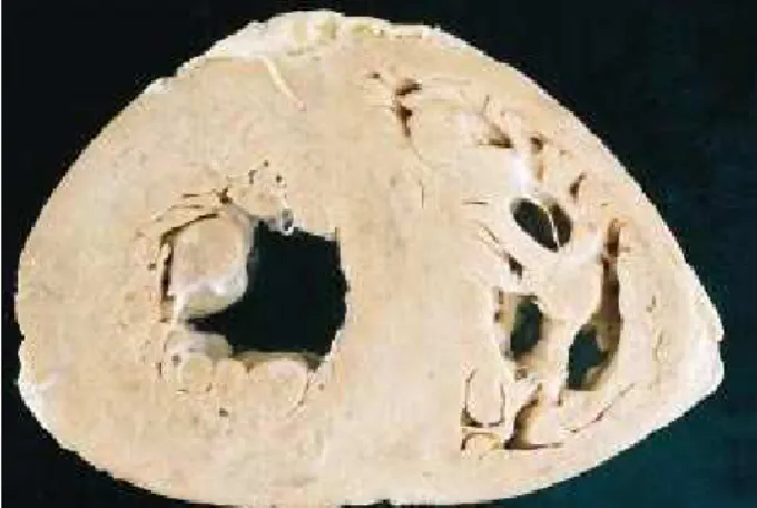
The heart weighed 560g, being slightly enlarged due to diffuse thickening of the walls (Fig. 3). The ventricles had vo-lume within the normal range and the atria were slightly dilated. The heart had a rubbery consistency and the color was brown with a grayish tone. No infectious disease or neoplasia was found. The liver, lungs, spleen, and kidneys were slightly enlarged, the first 2 with passive congestion findings.
Microscopically, the heart showed amorphous extra-cellular deposits slightly eosinophilic with the hematoxylin and eosin stain and weakly bluish with the Masson’s tri-chrome stain. These deposits widened the interstitial space therefore separating the myocardial fibers (Fig. 4). The Con-go red stain was performed in an attempt to highlight amy-loid deposits. This stain, except for one or another focal and irregular spots of positivity, was negative. This may happen in approximately 30% of the cases with this type of accumu-lation. The diagnosis was confirmed, however, by electron
microscopy, which revealed in the myocardial interstitium the presence of fibrils, characterizing the amyloid substan-ce (fig. 5). Restrictive heart failure is one of the major forms of heart amyloidosis presentation 11.
Other deposits with the same microscopic characteristics were found in the pulmonary, hepatic, and splenic vessels, in the glomerular loops, and in the tongue. The thyroid, which
Fig. 3 – Cross section of the heart at the level of the ventricles. Myocardium is diffusely thickened and the cavities have normal volume.
Fig. 4 - Histologic section of the myocardium stained with the Masson’s trichrome stain. Myocardial fibers, stained in red, are separated due to the presence, in the interstitium, of a deposit weakly stained in blue. (original magnification: 4x)
1 6 6
Clinicopathologic Session Arq Bras Cardiol
volume 74, (nº 2), 2000
1. Macedo AJ, Henrickson J, Kaku S, Cabral A, Pinto E, Lima M. Miocardiopatia restritiva na criança. Rev Port Cardiol 1995; 14: 401-8.
2. Kushwaha S, Fallon JT, Fuster V. Restrictive cardiomyopathy. N Engl J Med 1997; 2336: 267-76.
3. Barreto ACP, Dauar D. Cardiomiopatia restritiva. Rev Bras Med 1986; 6: 29-31. 4. Vianna CB, Barreto ACP, Belloti G. Correlações entre eosinofilia e
endomocar-diofibrose. Situação atual. Arq. Bras. Cardiol. 1990; 54: 247-50.
5. McCarthy RE, Kasper EK. A Review of amiloidosis that infiltrate the heart. Clin Cardiol 1998; 21: 547-52.
6. Giles TD, Kemnnt EK. Restritive and obliterative cardiomyopathy. In: Parmley WW, Chartterjje K. Cardiology. Baltimore: Lippincott-Raven 1996: 1-19.
7. Kyle RA, Bayrd ED. Amyloidosis. Review of 236 cases. Medicine 1975; 54: 271-99.
References
8. Date A, Parameswaran A, Bhaktaviziam A. Renal lesions in the obliterative cardiomyopathies: endomyocardial fibrosis and Loffler’s. J Pathol 1993; 140: 113-22.
9. Spyridopoulos I., Helber U, Voelker W. Primary systemic amyloidosis leading to advanced renal and cardiac involvement in a 30-year old man. Clin Investig 1994; 72: 462-5.
10. Albright R, Brensilver J, Cortell S. Proteinuria in congestive heart failure. Am J Nephrol 1983; 3: 272-5.
11. Barretto ACP, Precoma D, Serro-Azul JB, et al. Amiloidose cardíaca. Uma doença de muitas faces e diferentes prognósticos. Arq Bras Cardiol 1997; 69: 89-93.
12. Sinha RN, Plehn JF, Kinlaw WB. Amyloid goiter due to primary systemic amyloi-dosis: a diagnostic challenge. Thyroid 1998; 8: 1051-4.
had been previously partially resected, exhibited adenoma-tous goiter, but no amyloid, which was absent in the remaining organs. Goiter may be found in patients with amyloidosis 12, but
apparently this was not what happened in our case.
Anatomicopathological diagnoses – Systemic
amyloi-dosis, mainly cardiovascular, with restrictive heart failure.