PHENOTYPIC AND GENOTYPIC DIVERSITY OF PSEUDOMONAS AERUGINOSA STRAINS ISOLATED FROM HOSPITALS IN SIEDLCE (POLAND)
Katarzyna Wolska1*; Barbara Kot 1; Antoni Jakubczak2
1
University of Natural Sciences and Humanities in Siedlce, Department of Microbiology, Poland; .2State College of Computer
Science and Business Administration in Łom a, Food Technology Institute, Poland.
Submitted: December 10, 2010; Returned to authors for corrections: March 22, 2011; Approved: June 06, 2011.
ABSTRACT
A total of 62 Pseudomonas aeruginosa strains isolated from two hospitals in Siedlce (Poland) were studied
by repetitive element based PCR (rep-PCR) using BOX primer. BOX-PCR results revealed the presence of 7
numerous genotypes and 31 unique patterns among isolates. Generally, the strains of P. aeruginosa were
characterized by resistance to many antibiotics tested and by differences in serogroups and types of growth
on cetrimide agar medium. However, the P. aeruginosa strains isolated from faeces showed much lower
phenotypic and genotypic variations in comparison with strains obtained from other clinical specimens. It
was observed that genetic techniques supported by phenotypic tests have enabled to conduct a detailed
characterization of P. aeruginosa strains isolated from a particular environment at a particular time.
Key words: Pseudomonas aeruginosa, BOX-PCR, antibiotic resistance, serotyping, cetrimide agar.
INTRODUCTION
Pseudomonas aeruginosa is a ubiquitous pathogen
prevalent in hospital environments. It can cause severe
nosocomial infections, particularly among
immunocompromised patients. People with respiratory,
gastrointestinal, urinary tract, and wound infections as well as
burn victims, individuals with cancer, and patients hospitalized
in intensive care units are affected by P. aeruginosa mostly due
to nosocomial spread and cross contaminations (9, 10, 14). P.
aeruginosa accounts for 10% of all hospital acquired
infections, a site specific prevalence which may vary from one
unit to another and from study to study (11). Various possible
sources of P. aeruginosa infection in hospitals have been
identified, i.e., tap water, disinfectants, food, sinks, mops,
medical equipment, hospital personnel and others (7, 14, 19).
P. aeruginosa can be internally divided into subgroups by
classical methods such as: biotyping, serotyping, pyocin
typing, phage typing and antibiotic sensitivity of tested strains.
However, the discriminatory power is much lower than that
obtained by molecular typing methods. DNA typing methods
have been frequently used to investigate the diversity of
collections of P. aeruginosa (20). These methods include
pulsed-field gel electrophoresis (PFGE) (8, 21, 22), ribotyping
(6, 8), restriction fragment length polymorphic DNA analysis
(RFLP) (6), random amplified polymorphic DNA assay
(RAPD) (8, 13, 21), arbitrary primed PCR (AP-PCR) (4),
amplified fragment length polymorphism (AFLP) (21), and
repetitive element based PCR (rep-PCR) (6, 22). Rep-PCR is a
method for fingerprinting bacterial genomes, which
examines strain-specific patterns obtained from PCR
amplification of repetitive DNA elements present within
bacterial genomes. Three main sets of repetitive elements are
used for typing purposes: the repetitive extragenic palindromic
(REP) sequence, the enterobacterial repetitive intergenic
consensus sequence (ERIC) and the BOX elements (16).
The aim of this work was to estimate intra-species
differentiation of P. aeruginosa strains isolated from two
hospitals in Siedlce (Poland) using phenotypic methods
(serotyping, susceptibility to chemotherapeutic agents, and type
of growth on cetrimide agar medium) and the genotypic
method (BOX-PCR).
MATERIALS AND METHODS
Bacterial strains
A total of 62 strains of P. aeruginosa, were originally
isolated from a variety of clinical specimens: faeces (26), urine
(12), blood (1), bronchial washings (8), sputum (1), wound
swab (9), throat swab (2), ulceration swab (1), swab from skin
round tracheotomy (1) and from ear (1). The bacteria were
obtained from 62 patients from different wards of the
municipal hospital, main hospital and outpatients department
in Siedlce (Poland), between December 2005 and March 2006.
The strains were identified as P. aeruginosa on the basis of
typical morphology by gram-negative staining, a positive
oxidase reaction, growth at 42oC and conventional biochemical
tests using the Api 20NE system (Bio-Mérieux, France). We
also identified P. aeruginosa by PCR amplification of 16 S
ribosomal RNA (12). All isolates resulted in a positive
reaction. The control strain of P. aeruginosa NCTC 6749 was
also examined. Stock cultures were stored in tripticase soy
broth (TSB, Difco, USA) containing 20% glycerol at -80oC.
Genetic analysis
Isolates were grown in TSB at 37oC for 24 h and DNA was
extracted by using the Genomic DNA Pre Plus (A&A
Biotechnology, Poland). Rep-PCR fingerprinting was carried
out using one BOX primer of sequence 5’ - CTA CGG CAA
GGC GAC GCT GAC G - 3’. Amplification was carried out
with a 10x PCR buffer (100 mM Tris-HCl, 1 mM DTT, 0.1
mM EDTA, 100 mM KCl, 0.5% Nonidet P40, 0.5% Tween 20)
in a total reaction of 50 µL containing 2.5 mM dNTP, 20 mM
MgCl2, 100 pmol of primer, 2 µL of genomic template DNA,
and 1 unit of Taq DNA polymerase (DNA Gdansk, Poland).
Rep-PCR typing was carried out according to Dawson et al. (6)
using a PTC-100 Programmable Thermo Controller (MJ
Research, USA) according to the following procedure. Initial
denaturation at 94oC for 5 min followed by 35 cycles of PCR
consisting of denaturation at 94oC for 1 min, annealing at 48oC
for 2 min, and extension at 72oC for 2 min; in the last cycle, the
extension time was 5 min. The PCR product (10 µl) was
analysed using a 2% agarose gel in the TBE buffer [5.4 g l-1
Tris, 2.75 g l-1 Boric acid, 0.37 g l-1 EDTA (pH 8.0)] and
photographed under the UV light. The size of the products was
analyzed in comparison to a M100-1000 bp ladder M.W. size
marker (DNA Gdansk, Poland).
Phenotypic study
Pyocin production was tested on selective Cetrimide Agar
(Merc, Germany). Serotyping was determined by the slide
agglutination test with 16 monovalent antisera numbered O1 to
O16 and 4 polyvalent antisera [PMA (O1 + O3 + O4 + O6),
PME (O2 + O5 + O15 + O16), PMF (O7 + O8 + O11 + O12),
PMC (O9 + O10 + O13 + O14)] (Sanofi Diagnostics Pasteur,
France) as recommended by the manufacturer. Susceptibility to
antibacterial drugs was studied by the disk diffusion method
according to CLSI (Clinical and Laboratory Standards
Institute) (3) for 12 following antimicrobial agents
(Bio-Mérieux, France): carbenicillin (CB, 100 µg), mezlocillin (MZ,
75 µg), piperacillin (PIP, 100 µg), piperacillin-tazobactam
(TZP, 100 µg+10 µg), aztreonam (ATM, 30 µg), ceftazidime
(CAZ, 30 µg), imipenem (IMP, 10 µg), meropenem (MEM, 30
µg), gentamicin (CN, 10 µg), netilmicin (NET, 30 µg),
amikacin (AN, 30 µg) and ciprofloxacin (CIP, 5 µg).
RESULTS
BOX-PCR fingerprinting revealed 38 genetic patterns,
among them 7 main genotypes, containing 3 to 8 isolates and
31 other unique patterns. The clusters were shown in 2 to 11
8 bands per pattern. The most characteristic products of PCR
for P. aeruginosa were the following: 200, 420, 650, 1200 and
1400 bp (Fig. 1). Two of the genotypes (8 and 21) consisted of
7 (11.3%) and 8 (12.9%) isolates, respectively. The next two
numerous genotypes (4 and 13) contained 4 (6.45%) isolates.
All these isolates were obtained from faeces of patients
hospitalized in the Paediatric Ward of the Main and Infectious
Ward of the Municipal hospitals. The remaining three
numerous genotypes (11, 23 and 5) consisted of isolates from
wound (3 isolates) of patients of Orthopaedic and
Orthopaedic-Traumatical Ward; bronchial washings (3) of patients of
Neurological Ward, and from urine (2), and wound (1) of
patients being treated in Orthopaedic, Urologic and
Rehabilitation wards of the Main Hospital. Other unique types
were collected from the following clinical specimens: urine
(83.3%), wound (55.5%), bronchial washings (62.5%), faeces
(11.5%) and from sputum (1), throat swab (2), ulceration swab
(1), swab from skin round tracheotomy (1), blood (1), and from
ear (1). This data demonstrated that isolates from urine, wound
and bronchial washings were highly heterogeneous; among 12,
9 and 8 isolates, 11, 7 and 6 respectively different clusters
appeared. While the group of isolates from faeces showed
slightly genetic variation; in the group of 26 isolates we
Figure 1. BOX-PCR fingerprinting of Pseudomonas aeruginosa strains. Lane M: Molecular weight marker (MW100-1000 bp, DNA-GDANSK). A - Lines 1 to 9 - P. aeruginosa strains isolated from urine (1, 2, 6, 7), wound (3) and faeces (4, 5, 8). B – Lines 10 to 18 - P.
aeruginosa strains isolated from faeces (10, 11, 12, 13, 14), NCTC 6749 (15) and wound (16, 17, 18). C – Lines 19 to 41 - P. aeruginosa
strains isolated from bronchial washings (19, 28, 34, 35), faeces (20, 21, 22, 25, 26, 31, 36, 37, 38, 41), throat swab (23), skin (24),
wound (27, 29) and urine (30, 32, 39). D – Lines 42 to 63 - P. aeruginosa strains isolated from throat swab (42), faeces (43, 45, 47, 50,
52, 53, 59), urine (44, 49, 57, 62), wound (46, 55), bronchial washings (48, 51, 56, 61), ulceration (54), blood (60) and ear (63).
Detailed data on comparison of genotypic and phenotypic
strain features are presented in Table 1.
All tested strains were agglutinable. Forty three (69.3%) of
62 strains gave agglutination with the monovalent O6 serum.
They were isolated from faeces (100%), urine (66.7%),
bronchial washings (37.5%), wound (44.4%) and single strains
from throat swab and swab from skin round tracheotomy. Six
(9.7%) strains obtained from wound (33.3%), urine (16.7%)
and sputum (1) reacted with serum O1. Eight (12.9%) strains
isolated from bronchial washings (50.0%), wound (22.2%),
throat swab (1) and ulceration (1) were typed only by
polyvalent sera: PMA (5), PMF (2) and PMC (1). Individual
isolates from urine, bronchial washings and blood were
assigned to following sera: O9, O10, O15 and O16. A variety
of serotypes were demonstrated among 12 isolates from urine
(O6, O1, O9, O10, PMA), 9 isolates from wound (O6, O1,
PMF) and 8 isolates from bronchial washings (O6, O15, PMA).
While 26 of the strains isolated from faeces were typed only by
one sera (O6). Four different serotypes (O6, O1, O15, PMC)
were observed among 9 isolates from patients hospitalized in
the Intensive Care Unit Ward of the Main Hospital, whereas all
strains isolated from patients of the Infectious Ward (17
isolates) of the Municipal Hospital and the Paediatric Ward (10
isolates) of the Main Hospital belonged to one (O6) serotype.
There was correlation between serotypes and genotypes of P.
aeruginosa strains. The strains belonging to the same serotype
were classified to the same genotypic type (PMA serotype –
genotype 23; O1 serotype – genotype 5), however O6 serotype
was classified to four genotypes: 4, 8, 13 and 21 .
The total of 62 P. aeruginosa strains were tested on
selective cetrimide agar. A celadon type of growth appeared
most frequently; 38 (61.3%) strains. These strains were isolated
mainly from faeces, urine and wound (92.3%, 58.3% and
44.4% respectively). Eleven (17.7%) strains produced green
colonies. Most of them were isolated from bronchial washings
(50.0%) and wound (33.3%). Seven (11.3%) strains isolated
from bronchial washings (37.5%), urine (25.0%) and wound
(11.1%) grew in cetrimide agar producing green-yellow
colonies. Blue and green-blue types of growth were most rarely
isolated from the faeces of patients being treated at the
Infectious Ward of the Municipal Hospital, and the Paediatric
Ward of the Main Hospital produced nearly 90% and 100%
celadon colonies respectively. While the strains isolated from
the other clinical specimens of patients hospitalized in different
wards (excluding the Paediatric ward) of the Main Hospital
produced this type of growth by a much lower degree (47.5%).
Six out of seven numerous genotypes consisted of strains that
grew on selective cetrimide medium producing celadon type
(with exception of two strains). Only the strains isolated from
bronchial washings of genotype 23 produced green colonies.
The majority of P. aeruginosa isolates showed much
differentiated resistance to antimicrobial agents tested.
Different resistance patterns in various arrangements were
observed from sensitivity to all tested antibiotics, through
resistance to only two or three antibiotics, to multidrug
resistance for almost all tested drugs. Strains isolated from
faeces (serotype O6) of patients hospitalized in the Infectious
Ward of the Municipal Hospital and the Paediatric Ward of the
Main Hospital, were generally less resistant to
chemotherapeutic agents than strains isolated from the other
clinical specimens obtained from patients being treated in
different wards (excluding the Paediatric ward) of the Main
Hospital (CB-53.8%/72.2%, MZ-88.5%/86.1%,
PIP-3.8%/30.55%, TZP-0%/19.4%, ATM-57.7%/19.4%,
CAZ-15.4%/19.4%, IMP-3.8%/25%, MEM-7.6%/38.9%,
CN-46.1%/72.2%, NET-42.3%/86.1%, AN-26.9%/38.4% and
CIP-0%/25%). Among studied strains, 14 (22.3%) were multidrug
resistant (MDR). They were resistant to at least 4 out of the 6
antipseudomonal classes of antimicrobial agents, i.e.,
antipseudomonal penicilins, monobactams, cephalosporins,
carbapenems, quinolones and aminoglycosides. These strains
were obtained from wound (33.3%), urine (25.0%), bronchial
washings (25.0%), faeces (11.5%) and individual isolates from
sputum, blood and ear of patients hospitalized in different
wards of the Main Hospital (11 strains) and the Municipal
Hospital (1) as well as the outpatients department (2). They
belonged to the following serotypes: O1, O6, PMA, PMF, O15,
O16. However most of them were serotype O1 (35.7%).
Results of antibiotic resistance and genotyping showed poor
correlation. Resistance patterns from bacterial isolates which
had identical genotypes differed in up to 9 antibiotics.
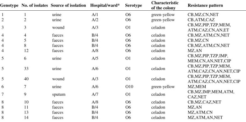
Table 1. Source of the isolation, hospital/ward, genotypes of clinical P. aeruginosa isolates and their phenotypic differentiation.
Genotype No. of isolates Source of isolation Hospital/ward* Serotype Characteristic
of the colony Resistance pattern
1 1 urine A/1 O6 green-yellow CB,MZ,CN,NET
2 2 urine A/2 O6 green-yellow CB,ATM,CAZ
3 3 wound A/3 O1 celadon CB,MZ,PIP,TZP,MEM,
ATM,CAZ,CN,AN,ET
4 4 faeces B/4 O6 celadon CB,MZ,ATM,CN,NET
4 5 faeces B/4 O6 celadon CB,MZ,CN
4 8 faeces B/4 O6 celadon CB,MZ,ATM,CN,NET
4 12 faeces A/8 O6 celadon MZ,AN
5 6 urine A/5 O1 celadon CB,MZ,PIP,TZP,IMP,
MEM,CN,AN,NET,CIP
5 33 urine A/6 O1 celadon CB,MZ,PIP,TZP,MEM,
ATM,CAZ,CN,AN,NET,CIP
5 40 wound A/3 O1 celadon CB,MZ,PIP,TZP,MEM,
ATM,CAZ,CN,AN,NET,CIP
6 7 urine A/6 O10 green-yellow MZ,MEM
7 9 sputum A/7 O1 celadon CB,MZ,IMP,MEM,ATM,
CAZ,NET
8 10 faeces A/8 O6 celadon CB,MZ,CAZ,NET
8 11 faeces B/4 O6 celadon MZ,AN
8 13 faeces B/4 O6 celadon MZ,ATM,CN
8 43 faeces A/8 O6 celadon CB,MZ,CAZ,AN,NET
8 45 faeces B/4 O6 blue MZ,CN
8 58 faeces A/8 O6 celadon CB,MZ,ATM,CN,NET
9 15 NCTC6749 - O6 green-blue CB,ATM,CAZ
10 16 wound A/9 PMF celadon CB,MZ,PIP,TZP,IMP,
MEM,CN,NET
11 17 wound A/3 O6 geen CB,MZ,PIP,TZP,CN, NET,CIP
11 18 wound A/2 O6 green-yellow CB,MZ,CN,AN,NET
11 55 wound A/2 O1 green CB,MZ,CN,AN,NET
12 19 bronchial
washings A/7 O15 blue
CB,MZ,PIP,TZP,IMP, MEM,ATM,CAZ,CN, AN,NET,CIP
13 20 faeces B/4 O6 celadon CB,PIP,ATM,CAZ,CN
13 21 faeces B/4 O6 green MZ,ATM
13 22 faeces B/4 O6 celadon CB,MZ,NET
13 25 faeces A/8 O6 celadon CB,MZ,CAZ
14 23 throat swab A/7 O6 celadon CB,MZ,NET
15 24 skin A/7 O6 celadon CB,MZ,IMP,MEM,NET
16 26 faeces A/8 O6 green MZ,MEM,ATM,CN,AN, NET
17 27 wound A/2 PMF celadon CB,MZ,CN,NET
18 28 bronchial
washings A/7 O6 celadon CB,MZ,CN,NET
19 29 wound A/9 O6 green-yellow MZ,CN,AN,NET
20 30 urine C O6 green-blue CB,MZ,MEM,ATM,
CAZ,CN,NET
21 31 faeces B/4 O6 celadon CB,MZ,IMP
21 36 faeces B/4 O6 celadon MZ,ATM,CN,NET
21 37 faeces A/8 O6 celadon MZ,ATM,AN
21 38 faeces B/4 O6 celadon sensitive to all
21 41 faeces B/4 O6 celadon CB,MZ
21 47 faeces B/4 O6 celadon MZ,ATM,CN
21 50 faeces B/4 O6 celadon CB,MZ,ATM,CAZ
21 52 faeces B/4 O6 celadon CB,MZ,ATM,CN
22 32 urine C O6 celadon CB,MZ,NET
23 34 bronchial
washings A/10 PMA green CB,MZ,PIP,CN,NET,CIP
23 48 bronchial
washings A/10 PMA green
CB,MZ,IMP,MEM,CN, AN,NET
23 51 bronchial
washings A/10 PMA green CB,MZ,CN,AN,NET,CIP
24 35 bronchial
washings A/7 O6 green MZ,IMP,MEM,CN,NET
25 39 urine A/10 O6 green CB,MZ,CN,AN,NET
26 42 throat swab A/7 PMC green CN,AN,NET,CIP
27 44 urine C O6 celadon CB,MZ,MEM,CN
28 46 wound A/9 O6 green MZ,CN,AN,NET
29 49 urine A/11 O9 green-blue MZ,PIP,NET
30 53 faeces A/8 O6 celadon CB,MZ,MEM,ATM,NET
31 54 ulceration A/9 PMA blue CB,MZ,NET,CIP
32 56 bronchial
washings A/7 O6 green-yellow CB, MZ, IMP, CN,NET
33 57 urine A/8 O6 celadon CN, AN,NET
34 59 faeces A/8 O6 celadon ATM,CN,AN,NET
35 60 blood A/12 O16 celadon CB,MZ,PIP,IMP,MEM, NET
36 61 bronchial
washings A/7 PMA green-yellow CB,MZ,CN
37 62 urine C O6 celadon CB,MZ
38 63 ear C PMA blue CB,MZ,PIP,TZP,CN, AN,NET
DISCUSSION
The hospital environment remarkably promotes selection
and quick distribution of resistant strains. One of the essential
steps leading to a reduction of nosocomial infections is a
constant monitoring of etiological agents and resistance of
intrahospital strains. It is of crucial importance to carry out
epidemiological surveys including a detailed characteristic and
relationship among strains isolated in particular environment
and time, as well as to become aware of risk factors, sources
and ways of infection distribution (1, 8, 9, 13). To obtain
reliable results the application of molecular methods seems to
be inevitable.
To differentiate precisely among P. aeruginosa isolated
from two hospitals in Siedlce (Poland), BOX-PCR typing was
carried out. PCR fingerprinting has shown 38 genetic patterns,
among them 7 main genotypes consisting of 3 to 8 strains and
31 other unique patterns. High number of genotypic patterns
pointed to marked intrahospital differentiation of P. aeruginosa
strains that are widely distributed in nature, especially in humid
environments. It indicated various sources of strains and their
constant exchange. Some strains were generally resistant to
tested antibiotics, what confirmed the development of
secondary resistance and their intrahospital selection. Based on
dates of strain isolation, and their resistance to antibiotics, it is
highly probable that selection of highly resistant isolates takes
place in ICU, Urologic and Orthopedic wards where P.
aeruginosa is one of the most frequent and severe causes of
infection, especially in patients with respiratory, urinary and
wound infections. Several studies have demonstrated
associations with a source of P. aeruginosa infection and
antibiotic resistance (1, 5, 18, 24). The other strains of
genotypes, especially those, which consisted of strains from
faeces (serotype O6) taken from patients hospitalized in the
Infectious Ward of the Municipal Hospital and the Paediatric
Ward of the Main Hospital, frequently expressed susceptibility
to tested antimicrobial agents. This proved incidence of
exogenous strains entering the hospital environment. Some of
the numerous genotypes were distributed in one, or more than
one unit. This may indicate that cross contamination among
patients lead to the spread of these genotypes among the
various units, possibly through transient hand carriage by
health care personnel due to contact with contaminated
surfaces, or by patient contact with contaminated surfaces or
medical equipment (19). The incidence of the same genotypes
of P. aeruginosa in two different hospitals drew attention to a
possibility of a long-distance strain transmission, which might
be linked to the movement of patients, visitors, medical and
paramedical staff. The importance of cross acquisition in the
epidemiology of nosocomial colonization and infection with P.
aerugionsa was reported by others (1, 8, 25). Fiett et al. (8)
demonstrated clonal relations within populations of P.
aeruginosa strains isolated in four different hospitals in Poland.
Bergmans et al. (1) who studied 100 patients admitted to an
ICU ward showed that cross colonization accounted for 50% of
all cases of acquired P. aeruginosa colonization, and the rest of
50% of patients were probably colonized from endogenous
sources. Cross transmission and treatment failure were also the
two main problems at Turkish medical centers (25).
This study demonstrated that BOX-PCR is a rapid, highly
discriminatory and reproducible assay that proved to be
powerful surveillance tools for typing as well as characterizing
clinical P. aeruginosa isolates. This is in agreement with the
studies of Syrmis et al. (22), in which the BOX-PCR method
showed the high discriminatory power. These authors reported
six major clonal groups, and 58 distinct clonal groups among
163 P. aeruginsa strains isolated from patients with cystic
fibrosis.
P. aeruginosa strains were also verified by classical typing
techniques. The studied strains showed poor differentiation of
phenotypic features, especially such as: serotypes and types of
growth on cetrimide agar. The total of 62 P. aeruginosa strains
were classified into 9 different serotypes. Most of them
(69.3%) belonged to O6 serotype, secondly to serotype O1
(9.7%) (the dominant type among MDR strains). The observed
strains demonstrated 5 types of growth on cetrimide agar
medium. The celadon type appeared most frequently (61.3%)
11.3%). The frequency of distribution of the O antigen types
differs considerably in various publications.
Czekajło-Kołodziej et. al. (4) demonstrated among over 50% of clinical
P. aeruginosa strains isolated from the lower respiratory tract
of patients admitted to ICU the production of green-yellow
colonies, typing by O11 sera, and resistance to many
antibiotics. Muller-Premru and Gubina (15) observed two O
serotypes 11 and 6 to be prevalent (36% and 14.4%
respectively) among clinical isolates. Antibiotic resistance of
strains was higher in serotype O11 than in serotype O6. In a
study of 73 P. aeruginosa strains from various clinical and
environmental sources, Pirnay et al. (17) reported the
predominant serotypes to be O11 (15.1%), O1 (12.3%), O6
(10.9%) and O12 (9.6%). Amongst 48 AFLP (amplified
fragment length polymorphism) types isolated from burns
patients, 58.3% were reported as serotypes O1, O6, O11 or
O12 (19). In a survey of 92 genetically distinct bacteraemia
isolates, O6 (25.0%) and O11 (18.0%) were reported to be the
most common serotypes (2). In a study of 23 isolates from
contact lens wearers, Thuruthyil et al. (23) reported O1
(30.0%), O6 (17.0%) and O11 (17.0%) as the most common
serotypes.
In conclusion, among all used methods in this work
BOX-PCR turned out to be a powerful tool for the study of clinical P.
aeruginosa isolates diversity. However, we suggest that
maximum discrimination can be best achieved by a
combination of phenotypic and genotypic methods.
ACKNOWLEDGEMENTS
We thank Mr. Henryk Matok for technical assistance. We
are also grateful to Mrs. Agnieszka Krechowska for the
provision of the bacterial strains. This work was supported by
grants from the Nature Faculty, University of Natural Sciences
and Humanities in Siedlce (Poland).
REFERENCES
1. Bergmans, D.C.; Bonten, M.J.; Vantiel, F.H. (1998). Cross colonization with Pseudomonas aeruginosa of patients in an intensive care unit.
Thorax. 53 (12), 1053-1058.
2. Berthelot, P.; Attree, I.; Plesiat, J.; Chabert, J.; de Bentzmann, S.; Pozzetto, B.; Grattard, F. (2003). Genotypic and phenotypic analysis of type III secretion system in a cohort of Pseudomonas aeruginosa bacteremia isolates: evidence for a possible association between O serotypes and exo genes. J. Infect. Dis. 188 (4), 512-518.
3. Clinical and Laboratory Standards Institute. Performance standards for antimicrobial testing (2006). 16th informational supplement M100-S16. Wayne, Pa: CLSI.
4. Czekajło-Kołodziej, U.; Giedrys-Kalemba, S.; M drala, D. (2006). Phenotypic and genotypic characteristics of Pseudomonas aeruginosa strains isolated from hospitals in the north-west region of Poland. Pol. J. Microbiol. 55 (2), 103-112.
5. Dambrauskien , A.; Adukauskien , D.; Jeroch, J.; Vitkauskien , A. (2009). Pseudomonas aeruginosa bacteremia: associations with a source of infection and antibiotic resistance. Medicina (Kaunas). 45 (1), 1-7. 6. Dawson, S.L.; Fry, J.C.; Dancer, B.N. (2002). A comparative evaluation
of five typing techniques for determining the diversity of fluorescent pseudomonads. J. Microbiol. Meth. 50, 9-22.
7. Deplano, A.; Denis, O.; Poirel, L.; Hocquet, D.; Nonhoff, C.; Byl, B.; Nordmann, P.; Vincent, J.L.; Struelens, M.J. (2005). Molecular characterization of an epidemic clone of panantibiotic-resistant Pseudomonas aeruginosa. J. Clin. Microbiol. 43 (3), 1198-1204. 8. 301.Fiett, J.; Trzci ski, K.; Hryniewicz, W.; Gniatkowski, M. (1998).
Molecular typing of Pseudomonas aeruginosa strains recovered from nosocomial infections caused by Pseudomonas aerugionsa. Przeg. Epid. 52 (4), 427-440 (in Polish).
9. Hauser, A.R.; Sriram, P. (2005). Severe Pseudomonas aeruginosa infections. Tackling the conundrum of drug resistance. Postgrad. Med. 117 (1),41-48.
10. Hoiby, N.; Pedersen, S.S.; Shand, G.H.; D ring, G.; Holder, I.A. (1989). Pseudomonas aeruginosa infection. Chemother. Basel. Karger. 42, 124-129.
11. Jones, R.N.; Croco, M.A.; Kugler, K.C.; Pfaller, M.A.; Beach, M.L. (2000). Respiratory tract pathogens isolated from patients hospitalized with suspected pneumonia: frequency of occurrence and susceptibility patterns from the Sentry Antimicrobial Surveillance Program. Diagn. Microbiol. Infect. Dis. 37 (2), 115-125.
12. Kingsford, N.M.; Raadsma, H.W. (1995). Detection of Pseudomonas aeruginosa from ovine fleece washings by PCR amplication of 16S ribosomal RNA. Vet. Microbiol. 47, 61-70.
13. Liu, Y.; Davin-Regli, A.; Bosi, C.; Charrel, R.N.; Bollet, C. (1996). Epidemiological investigation of Pseudomonas aeruginosa nosocomial bacteraemia isolates by PCR-based DNA fingerprinting analysis. J. Med. Microbiol. 45 (5), 359-365.
14. Morrison, A.J.; Wentzel, R.P. (1984). Epidemiology of infections due to Pseudomonas aeruginosa. Rev. Infect. Dis. 6, S627-S642.
university hospital. Zetralbl. Bacteriol. 289 (8), 857-867.
16. Olive, M.D.; Bean, P. 1999. Principles and applications of methods for DNA-based typing of microbial organisms. J. Clin. Microbiol. 37 (6), 1661-1669.
17. Pirnay, J.P.; De Vos, D.; Cochez, C.; Biloq, F.; Vanderkelen, A.; Zizi, M.; Ghyseles, B.; Cornelis, P. (2002). Pseudomonas aeruginosa displays an epidemic population structure. Environ. Microbiol. 4, 898-911.
18. Pirnay, J.P.; De Vos, D.; Cochez, C.; Biloq, F.; Pirson, J.; Struelens, M.; Duinslaeger, L.; Cornelis, P.; Zizi, M.; Vanderkelen, A. (2003). Molecular epidemiolology of Pseudomonas aeruginosa colonization in a burn unit: persistence of a multidrug resistant clone and a silver sulfadizine-resistant clone. J. Clin. Microbiol. 41 (3), 1192-1202. 19. Pittet, D.; Dharan, S.; Touveneau, S.; Sauvan, V.; Perneger, T.V. (1999).
Bacterial contamination of the hands of hospital staff during routine patient care. Arch. Intern. Med. 159 (8), 821-826.
20. Speert, D.P. (2002). Molecular epidemiology of Pseudomonas aeruginosa. Front. Biosci.1 (7), e354-361.
21. Speijer, H.; Savelkoul, P.H.M.; Bonten, M.J.; Stobberingh, E.E.; Tjhie, J.H.T. (1999). Application of different genotyping methods for
Pseudomonas aeruginosa in a setting of endemicity in an intensive care unit. J. Clin. Microbiol. 37 (11), 3654-3661.
22. Syrmis, M.W.; O´Carrol, M.R.; Sloots, T.P.; Coulter, C.; Wainwright, C.E.; Bell, S.C.; Nissen, M.D. (2004). Rapid genotyping of Pseudomonas aeruginosa isolates harboured by adult and paediatric patients with cystic fibrosis using repetitive-element-based PCR assays. J. Med. Microbiol. 53 (11), 1089-1096.
23. Thuruthyil, S.J.; Zhu, H.; Willcox, M.D. (2001). Serotype and adhesion of Pseudomonas aeruginosa isolated from contact lens wearers. Clin. Experiment. Ophthalmol. 29 (3), 147-149.
24. Yang, C.H.; Lee, S.; Su, P.W.; Yang, C.S.; Chuang, L.Y. (2008). Genotype and antibiotic susceptibility patterns of drug-resistant Pseudomonas aeruginosa and Acinetobacter baumannii isolates in Taiwan. Microb. DrugResist. 14 (4), 281-288.
25. Yetkin, G.; Otlu, B.; Cicek, A.; Kuzucu, C.; Durmaz, R. (2006). Clinical, microbiologic, and epidemiologic characteristics of Pseudomonas aeruginosa infections in a University Hospital, Malatya, Turkey. Am. J. Infect. Control. 34 (4), 188-192.