online | memorias.ioc.fiocruz.br
An overview of malaria transmission from
the perspective of Amazon Anopheles vectors
Paulo FP Pimenta1,2/+, Alessandra S Orfano1, Ana C Bahia3, Ana PM Duarte1,
Claudia M Ríos-Velásquez4, Fabrício F Melo1, Felipe AC Pessoa4, Giselle A Oliveira1,
Keillen MM Campos2, Luis Martínez Villegas1, Nilton Barnabé Rodrigues1, Rafael Nacif-Pimenta1,
Rejane C Simões5, Wuelton M Monteiro2, Rogerio Amino6, Yara M Traub-Cseko3, José BP Lima2,3,
Maria GV Barbosa2, Marcus VG Lacerda2,4, Wanderli P Tadei5, Nágila FC Secundino1
1Centro de Pesquisas René Rachou-Fiocruz, Belo Horizonte, MG, Brasil 2Fundação de Medicina Tropical Dr Heitor Vieira Dourado, Manaus,
AM, Brasil 3Instituto Oswaldo Cruz-Fiocruz, Rio de Janeiro, RJ, Brasil 4Instituto Leônidas e Maria Deane-Fiocruz, Manaus, AM, Brasil 5Instituto Nacional de Pesquisas da Amazônia, Manaus, AM, Brasil 6Unité de Biologie et Génétique du Paludisme, Institut Pasteur, Paris, France
In the Americas, areas with a high risk of malaria transmission are mainly located in the Amazon Forest, which extends across nine countries. One keystone step to understanding the Plasmodium life cycle in Anopheles species from the Amazon Region is to obtain experimentally infected mosquito vectors. Several attempts to colonise Ano- pheles species have been conducted, but with only short-lived success or no success at all. In this review, we review the literature on malaria transmission from the perspective of its Amazon vectors. Currently, it is possible to develop experimental Plasmodium vivax infection of the colonised and field-captured vectors in laboratories located close to Amazonian endemic areas. We are also reviewing studies related to the immune response to P. vivax infection of
Anopheles aquasalis, a coastal mosquito species. Finally, we discuss the importance of the modulation of Plasmo-dium infection by the vector microbiota and also consider the anopheline genomes. The establishment of experi-mental mosquito infections with Plasmodium falciparum, Plasmodium yoelii and Plasmodium berghei parasites that could provide interesting models for studying malaria in the Amazonian scenario is important. Understanding the molecular mechanisms involved in the development of the parasites in New World vectors is crucial in order to better determine the interaction process and vectorial competence.
Key words: Anopheles - Plasmodium - transmission - Amazon vectors
doi: 10.1590/0074-02760140266
Financial support: Bill & Melinda Gates Foundation (TransEpi Study), FIOCRUZ, PAPES, CNPq, CAPES, FAPEMIG, FAPERJ, FAPEAM NBR is a CAPES fellow (BEX 11603/13-5).
+ Corresponding author: [email protected] Received 22 July 2014
Accepted 18 December 2014
Malaria is an infectious disease that has a major im-pact on global public health and the economy, with an estimated 3.4 billion people at risk. Currently, malaria threatens almost one third of the world’s population in 104 tropical countries and territories where it is consid-ered an endemic disease. The World Health Organiza-tion (WHO) estimates that 207 million cases of malaria occurred globally in 2012 and led to 627,000 deaths. Africa, South-East Asia and the Eastern Mediterranean were the regions with the highest numbers of reported cases and deaths reported, mainly in children under five years of age (WHO 2013).
In the Americas, 22 countries are affected by malaria, with approximately 1.1 million cases and 1,100 deaths reg-istered in 2010. In this continent, 30% of the population is considered to be at risk and 8% are classified as being at
high risk. Areas with a high transmission risk are main-ly located in the Amazonian rainforest, which extends across nine countries including Brazil, Bolivia, Colombia, Ecuador, Peru, Venezuela, Guyana, Suriname and French Guiana. Brazil and Colombia accounted for 68% of the malaria cases in 2011 (PAHO 2011, WHO 2013).
In Brazil, approximately 241,000 clinical cases and 64 deaths were registered in 2012, most of them (99.88%) in the Amazon Region where malaria is endemic in nine states, namely, Acre, Amapá (AP), Amazonas (AM), Mato Grosso, Pará (PA), Rondônia, Roraima, Tocantins and Maranhão. PA and AM registered almost 70% of the cases in 2012; 14.4% were in urban areas, 25% in gold mine exploitation areas and the others were in rural settle-ments and indigenous areas (MS/SVS 2013, SVS 2013).
A gradual reduction in the overall number of cases has been observed over the last five years, but there has also been a significant increase in the number of cases in the Brazilian Amazon Region in 2012. Factors that contributed to the increased transmission of malaria include intensive and disorganised occupancy on the outskirts of cities, deforestation and artificial fishponds (MS/SVS 2013, SVS 2013).
Region or from the African continent, but a few were au-tochthonous from the endemic Atlantic Forest endemic region where few foci are maintained (Rezende et al. 2009, Duarte et al. 2013, Neves et al. 2013).
Malaria is due to infection by a parasitic protozoa of the Plasmodium genus. Several Plasmodium species in-fect humans and other animals, including birds, reptiles and rodents. In Brazil, three human Plasmodium parasites are prevalent. Plasmodium vivax is the predominant spe-cies (83.81%) and is responsible for cases associated with severe clinical complications and death (Alexandre et al. 2010, Costa et al. 2012, Lacerda et al. 2012). The preva-lence of Plasmodiumfalciparum (13.15%) has declined in the last decade, whilst Plasmodium malariae is the least prevalent species (0.037%). However, these numbers may be underestimated because the thick blood smear method that is used for routine malaria diagnosis may lead to mis-identification of the species (Cavasini et al. 2000).
Plasmodium cycle in the vector
Mosquitoes of the Anopheles genus are the vectors of the Plasmodium species, the causative agents of ma-larial disease. More than 400 species of the Anopheles
mosquito have been described and approximately 70 these species are potential vectorsof malaria that affect humans (Sinka et al. 2012). In the natural vector, the life cycle starts when the female Anopheles mosquito takes a blood meal from an infected vertebrate host and in-gests gametocytic forms of the parasite that are present in the blood (Smith et al. 2014).
One mosquito ingests an average of 103 gametocytes
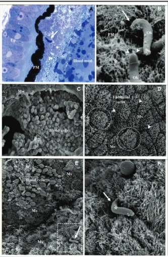
in an infected blood meal. Within minutes after the in-fective blood meal, these gametocytes undergo matu-ration inside the lumen of the midgut, which generates micro and macrogametocytes that will be fertilised and produce a diploid zygote (Sinden 1999). The mature zy-gote will differentiate into the mobile form of the para-site known as the ookinete via a process that can take up to 16-24 h, depending on the Plasmodium species (Ghosh et al. 2000, Dinglasan et al. 2009). This process starts with the exflagellation of the gametocytes in the mos-quito’s midgut after ingestion of the infected blood meal. Exflagellationwill lead to the formation of the micro and macrogametocytes and occurs mainly due to differences in temperature and pH and the production of xanturenic acid by the mosquito (Billker et al. 1997, 1998). The for-mation of the zygote occurs after fertilisation of the micro and macrogametocytes and will eventually differentiate into an ookinete. This development will only occur if the parasites are able to defeat the action of the digestive en-zymes that are secreted by the epithelium and are active throughout the midgut. It is believed that the ookinetes in the outer parts of the blood meal will die first from the actions of these digestive enzymes and the ookinetes that are closer to the interior of the blood meal and conse-quently farther away from the effects of the enzyme, will have a longer time in which to differentiate and survive the actions of the enzyme (Abraham & Jacobs-Lorena 2004). The ookinete, which is the mobile form of the parasite, will move and penetrate the peritrophic matrix (PM) and pass through the intestinal epithelium before transforming into an oocyst (Smith et al. 2014).
The PM is a layer comprised of chitin, proteins and proteoglycans that surround the blood meal that has been ingested (Fig. 1). Physical distension caused by the in-gestion of the blood and the blood meal itself are signals for the mosquito’s midgut to induce the formation of the PM. This matrix is seen as a physical barrier to many parasites as it prevents their contact with the insect gut (Ghosh et al. 2000). Several studies have suggested that
P. falciparum and Plasmodium gallinaceum may secrete chitinase additional to that already produced by the
sect which would allow the parasite to accomplish three crucial steps in the infection of the invertebrate host: (i) penetrate through the PM, (ii) escape the deadly action of digestive enzymes and (iii) successfully invade the epi-thelial cells of the intestine (Huber et al. 1991, Dessens et al. 1999, Vinetz et al. 1999, 2000). The details of the pen-etration of the PM by the ookinete are seen in Fig. 1A, B. The recently transformed ookinete moves in the direction of the mosquito epithelium (Fig. 1A) and penetrates the PM by introducing its anterior extremity into the fibrous layer of the internal side of the PM (Fig. 1B).
The penetration of the Plasmodium ookinete into the midgut epithelium is an important step in the infection of mosquitoes and has been thoroughly studied previously (Fig. 1B-F). The epithelial cells have polygonal shapes and their surfaces are covered with microvilli (Fig. 1D). The ookinete penetrates the microvilli clefts that exist among the epithelial cells toward their anterior extrem-ity (Fig. 1E, F) in order to initiate the invasion process.
Different theories have arisen regarding the ooki-nete’s strategies for penetration and invasion of the epithelial cells and escaping detection by the host’s im-mune system. After several years without any conclu-sive studies on how the ookinete invades the mosquito epithelium, Shahabuddin and Pimenta (1998) used an in vitro system to study the interaction of P. gallinaceum
with Aedes aegypti. The methodology consisting of the incubation of the parasites with dissected midgut was successfully applied to a study of the Leishmania-vector interaction (Pimenta et al. 1992, 1994). The result sug-gested the existence of specialised cells in the midgut epithelium of Ae. aegypti that the authors called Ross cells, which would serve as a specific entry point for the ookinete (Shahabuddin & Pimenta 1998). Subsequently, Han et al. (2000) proposed a time bomb theory in which parasites invade any epithelial cell in the midgut and this process of penetration triggers an immune response, causing this particular cell to begin apoptosis. However, a conclusive report from Barillas-Mury’s group at Na-tional Institute of Allergy and Infectious Diseases that was completed with our collaboration (Gupta et al. 2005) indicated that Ae. aegypti and Anopheles stephensi dif-fer in their mechanisms of epithelial repair after Plasmo-dium ookinete invasion. An. stephensi damaged cells via
an actin-mediated budding-off mechanism when invaded by either Plasmodium berghei or P. gallinaceum. In Ae. aegypti, the midgut epithelium is repaired by a unique actin cone zipper mechanism that involves the forma-tion of a cone-shaped actin aggregate at the base of the cell that closes sequentially, expelling the cellular con-tents into the midgut lumen as it brings together healthy neighbouring cells. This study had important findings: (i) it determined that the apparent target cells used by
P. gallinaceum to invade the vector epithelium were in fact an in vitro artifact; the Ross cells are believed to represent cells that have lost their integrity and some of their cytoplasmic contents after parasite invasion and (ii) these studies indicated that the epithelial responses of different mosquito vectors to Plasmodium depend on the vector-parasite combinations and are not universal.
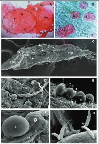
After crossing the epithelial layer of the gut, the ookinetes will remain between the intestinal epithelium
and the basal lamina, at which point the maturation of the oocyst will occur. A simple method of staining with mercurochrome (Merbromin) solution is useful for the identification of infected midguts. The rounded oocysts can be seen in bright red (Fig. 2A, B). Scanned electron microscope images of the external side of the infected midguts are valuable for showing the morphological aspects of the developing oocysts (Fig. 2C-F). These oocysts appear as protruding structures among the mus-cle fibres of the midgut wall (Fig. 2D). Some
cytes can be seen attached to oocysts (Fig. 2E). It is also possible to observe shrunken oocysts due to the rupture of the oocyst wall (Fig. 2F). Oocyst rupture and the sub-sequent release of sporozoites occur once the maturation is complete (usually within 10-24 days, depending on the
Plasmodium species). This leads to the release of any-where from hundreds to thousands of sporozoites into the mosquito haemocoel (Hillyer et al. 2007) (Fig. 1G). Before reaching the salivary gland, the sporozoites still need to overcome the other barriers that is produced by the immune system, including: (i) haemocytes (Fig. 2E), which are cells that are responsible for the internal de-fense system of the mosquito, (ii) antimicrobial peptides and (iii) other humoral factors (Dimopoulos et al. 2001).
In general, the process of invasion of the salivary gland by sporozoites is very inefficient; usually less than 20% of the total numbers of parasites produced are able to invade the organ (Korochkina et al. 2006, Hillyer et al. 2007). Those sporozoites that survive after overcom-ing various barriers to reachovercom-ing the salivary gland are finally able to invade the organ. By means of a specific recognition receptor present in the salivary gland of the vector, these parasites are able to adhere to and penetrate the basal lamina of the gland before penetrating the host
plasma membrane of the salivary cells. A number of par-asite ligands are necessary for the initial attachment of the sporozoites to the salivary glands, such as some re-gions of the circumsporozoite protein and thrombospon-din-related anonymous protein [see details in Sinden and Matuschewski (2005) and Aly et al. (2009)]. This process of invasion has been well described using the
P. gallinaceum/Ae. aegypti model (Pimenta et al. 1994). The penetration process appears to involve the forma-tion of membrane juncforma-tions. Once inside the host cells, the sporozoites are seen within vacuoles attached by their anterior end to the vacuolar membrane. Mitochon-dria surround and are closely associated with the invad-ing sporozoites. After the disruption of the membrane vacuole, the parasites traverse the cytoplasm, attach to and invade the secretory cavity through the apical plas-ma membrane of the cells. Inside the secretory cavity, the sporozoites are again seen inside the vacuoles. Upon escaping from these vacuoles, the sporozoites are posi-tioned in parallel arrays, forming large bundles attached by multilamellar membrane junctions. Several sporo-zoites are seen inside and around the secretory duct. Ex-cept for the penetration of the chitinous salivary duct, these observations have morphologically characterised
the entire process of sporozoite passage through the sali-vary gland (Pimenta et al. 1994). The sporozoites that are now inside the secretory duct of the salivary gland are ready to be injected by the mosquito bite into the skin of a new vertebrate host. An analysis of the amount of parasite that an infected mosquito could inject into the skin of a mouse varied between zero and approximately 1,300 and there appears to be a weak correlation of the number of injected sporozoites with the salivary gland load (Medica & Sinnis 2005).
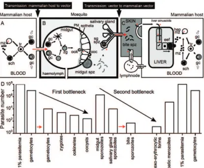
Considering the entire Plasmodium life cycle in the vector and in the vertebrate host, it is fascinating to ob-serve the complexity of distinct developmental forms and the parasite load during the course of infection. There is extraordinary adaptation of the Plasmodium parasite to its environment, which is reflected in morphological changes and the parasite load of distinct organs inside the vertebrate host and the mosquito vector (Baton & Ranford-Cartwright 2005, Medica & Sinnis 2005, Ami-no et al. 2006, Ma et al. 2010, Smith et al. 2014). During the stages that the Plasmodium moves from the mam-malian host to the vector and vice versa, two “bottle-necks” occur that are characterised by a small number of parasites. Fig. 3 shows an animated model that illustrates qualitative and quantitative views of the major steps of the life cycle of the P. berghei parasites infecting mice and An. stephensi mosquitoes. Murine-Plasmodium spp interaction studies are considered to be suitable experi-mental models to better understand the interaction be-tween malarial parasites and vectors.
The key Amazon Anopheles vectors
Among the Anopheles mosquito species that inhabit the Amazon, Anopheles darlingi, Anopheles albitarsis s.l. and Anopheles aquasalis are considered the principle mosquito vectors. Specifically, An. darlingi is the main vector in South America and has been associated with the dynamics of malaria transmission in the Amazonian regions of Bolivia, Colombia, French Guiana, Guyana, Peru, Suriname and Venezuela (Zimmerman 1992, Hi-wat et al. 2010). An. albitarsis s.l. inhabits regions of Venezuela (Rubio-Palis et al. 1992) and An. aquasalis
is found in Trinidad (Chadee & Kitron 1999), Guyana (Laubach et al. 2001) and Venezuela (Berti et al. 1993).
Other anopheline species can be secondary or oc-casional malaria vectors because of their population density, anthropophilic behaviour and natural infectiv-ity across their geographical distributions (Deane 1986, Zimmerman 1992, Sinka et al. 2010, 2012). Anopheles nuneztovari s.l. and Anopheles triannulatus s.l. are com-monly collected in the Amazon by researchers and they have been observed to be infected with P. vivax and P. falciparum, but their role as malaria vectors has yet to be elucidated (de Arruda et al. 1986, de Oliveira-Ferreira et al. 1990, Klein et al. 1991b, Tadei & Dutary 2000, da Silva-Vasconcelos et al. 2002, Póvoa et al. 2003, 2006, dos Santos et al. 2005, Galardo et al. 2007, da Rocha et al. 2008, Santos et al. 2009).
Recently, Foley et al. (2014) developed a study consid-ering the percentage of the area predicted to be suitable for mosquito habitation based on ecological niche
mod-els of Amazon vectors. They found that An. albitarsis
I, Anopheles janconnae and Anopheles marajoara had the highest percentage of their predicted suitable habi-tats overlapping the distribution models of P. falciparum
and P. vivax [see details in Foley et al. (2014)]. They also concluded that phylogenetic proximity might be related to malaria vectorial importance within the Albitarsis
group. The authors recognised that these findings would encourage additional studies of the transmission poten-tial of these Amazonian Anopheles species.
An. aquasalis is distributed predominantly along the Atlantic Coast because of its tolerance to saltwater environments, including in Venezuela, where it is con-sidered to be the primary coastal malaria vector of P. vivax (Galvão et al. 1942, Laubach et al. 2001, Póvoa et al. 2003, da Silva et al. 2006a).
Amazonian Anopheles species such as, Anopheles deaneorum, An. marajoara, Anopheles mattogrossensis, An. nuneztovari, Anopheles oswaldoi, Anopheles rondo-ni and An. triannulatus have been considered “naturally infected” with Plasmodium since they were captured with parasites in their blood meal (Galvão et al. 1942, Deane et al. 1948, de Arruda et al. 1986, Klein et al. 1991b, Branquinho et al. 1993, Tadei & Dutary 2000, Póvoa et al. 2001, 2003, 2006, da Silva-Vasconcelos et al. 2002, da Silva et al. 2006a, Galardo et al. 2007, da Rocha et al. 2008, Santos et al. 2009). However, their role as malaria vectors is not well defined.
Two crucial factors needed to label a mosquito a vec-tor are the demonstration that the species is anthropo-philic and identification of the same Plasmodium species or strain in patients from the same geographic region. In the field, the presence of Plasmodium oocysts in the mosquito midgut indicates parasite establishment in a susceptible vector. However, the discovery of only sporo-zoites in the dissected mosquito salivary gland can con-firm that the life cycle is complete and consequently that the Plasmodium parasite can be transmitted by a bite to human hosts. Moreover, recognition of the infection rate (i.e., the percentage of individuals in a mosquito popula-tion that carry Plasmodium) is an important parameter for defining vector competence and thus a key indicator in the description of malaria dynamics and transmission biology in a given geographic region. In contrast, the sole presence of an apparent abundance of a species along with parasites in the ingested blood meal is not sufficient to implicate a mosquito as a vector (Smith et al. 2014).
Colonisation of American anophelines
the major vector in several African countries, is the most well studied mosquito, including its interaction with hu-man and murine Plasmodium species that are considered causative agents of malaria (Moores 1953). Distinctly, the colonisation of An. darlingi, the major Amazon vec-tor,has proven to be difficult, as has that of other New World anopheline species.
Several attempts to colonise American species of
Anopheles under laboratory conditions have been con-ducted either unsuccessfully or with short-lived success. When describing the rationale for establishing a colony of Anophelesquadrimaculatus, Boyd et al. (1935) high-lighted two key starting points: (i) an abundant supply of food for the larvae and (ii) a stable and optimal tem-perature. Galvão et al. (1944) used Boyd’s technique with specifically sized cages (40 x 40 x 47 cm). They loaded approximately two thousand mosquitoes into each cage and the females started to lay eggs after seven days. Re-production led to An.albitarsis domesticus (An. mara-joara) mosquitoes reaching the seventh generation. Egg production in Anophelestarsimaculatus (An.aquasalis), however, was low and was maintained by only a few doz-en couples up to the fifth gdoz-eneration. The authors attrib-uted the colonisation problems to a lack of mating due to the space and type of food offered to the males. To begin a mosquito colony there are numerous factors that need to be controlled for, including the fact that several species do not undergo free copulation under laboratory condi-tions (Martinez-Palacios & Davidson 1967). Thus, for the establishment of the colony, the induced copulation ap-proach is often necessary. This method was developed by McDaniel and Horsfall (1957) for the Aedes spp and was later adapted by Baker et al. (1962) for Anopheles.
There are descriptions in the literature of various American Anopheles species that have been main-tained in insectaries for short periods of time, includ-ing Anophelespunctipennis, Anophelesmaculatus, An. aquasalis,An.albitarsis, An.deaneorum and An. mara-joara (Baker et al. 1962, Ow-Yang & Maria 1963, Baker 1964, Arruda et al. 1982, Klein et al. 1990, Horosko III et al. 1997). In the 2000s, the colonisation of Anopheles pseudopunctipennis, which is considered an important vector of human Plasmodium spp along the Andes in several countries, was noted to have occurred by means of free intercourse (Lardeux et al. 2007). The adult mosquitoes were exposed to a blue strobe light for 20 min for several nights, encouraging them to copulate naturally under laboratory conditions. After a few gen-erations, the researchers obtained a stable colony that reproduced by free mating. Corrêa et al. (1970) de-scribed some success in colonising and maintaining An. darlingi mosquitoesfor about two years. Subsequently, however, Buralli and Bergo (1988) failed to achieve suc-cessful results from the same laboratory and using the same methodology. More recently, Moreno et al. (2014) described a method for An. darlingi colonisation that also used the strobe light approach. They reported that
An. darlingi mosquitoes obtained after five generations were successfully infected with P. vivax by artificial membrane feeding similar to the previous work of Ríos-Velasquez et al. (2013) with field-captured mosquitoes.
One of the authors of this paper established colonies of two species of Neotropical anophelines 20 years ago.
An. albitarsis s.l. was colonised in 1993 by induced copu-lation. After about two years of colony maintenance with induced copulation, we noticed the successful occurrence of free copulation; we used large cages with a thousand adults and a sex ratio of approximately 1:1 (Horosko III et al. 1997). An.aquasalis was settled in 1995 from the beginning by the free coupling method. In 1998, a sec-ond American malaria vector was colonised, Anopheles albimanus, which is one of the main vectors of malaria in Central America and in the south of Mexico (Zerpa et al. 1998). The authors used a simple and efficient mainte-nance method for mosquito mating and laying eggs.
Today, to the best of our knowledge and according to the specialised literature related to Anopheles species, only two long-term colonised American malaria vec-tors, An. aquasalis and An. albimanus, are maintained in laboratories and have been used for experimental stud-ies, demonstrating that they are good models for study-ing the interaction of malaria vectors with Plasmodium
species. As examples of these types of studies in An. al-bimanus, there are reports showing the susceptibility of the vector to P. vivax (Herrera et al. 2011, Solarte et al. 2011) and to the murine P. berghei (Serrano-Pinto et al. 2010, Herrera-Ortiz et al. 2011). For An. aquasalis, there have been studies developed by our group related to their susceptibility to P. vivax infection, including those relat-ed to gene expression during parasitic infection (Bahia et al. 2010, 2011, 2013, Ríos-Velasquez et al. 2013).
Searching for a model to study the Plasmodium interaction with an American mosquito vector
An. aquasalis in nature: distribution, habitat and pop-ulation variability - An. aquasalis lives in sunny habitats with vegetation in fresh brackish water. It is believed that the mosquito prefers clean water such as that in stream pools, mangroves, ponds and ditches (Manguin et al. 1993, Grillet 2000). The demarcation of the An. aquasalis
territory to coastal regions and its tolerance to salt water could be evolutionary adaptations that have been selected to avoid competition for food with other Anopheles mos-quitoes (particularly during the larval phases), inserting the mosquito into the large and varied marine trophic chain (Sinka et al. 2010). The geographic distribution of
An. aquasalis covers the southern coastal region of Cen-tral America, the Caribbean Islands and South America, but this species can penetrate eight-10 miles inland from the coast because it has a flight capacity of up to 8 km. Its presence at the Atlantic Coast has been reported from SP to Nicaragua and at the Pacific Coast from Costa Rica to Ecuador, as well as in the Antilles and Trinidad and To-bago (Faran 1980, Chadee et al. 1992, Zimmerman 1992, Consoli & Lourenço-de-Oliveira 1994).
exam-ple, Giglioli (1963) reported the effect of mechanisation on a rice farm in Guyana, which led to the disappear-ance of buffalo in the region. This resulted in a change in the behaviour of An. aquasalis that had man as its main blood source. Nevertheless, this mosquito species has been associated with several outbreaks of malaria in several countries (Deane 1986, Berti et al. 1993, Lau-bach et al. 2001, Mouchet et al. 2008). In most of the territory it inhabits, this species is exophilic, zoophilic and crepuscular, but in the drier northeast area it is fre-quently endophilic and bites human hosts. The females are opportunists, feeding in both intra and peridomicili-ary areas of animals and humans. They begin to bite at sunset, reaching maximum activity in the early evening before decreasing later at night (Flores-Mendoza et al. 1996). Usually the mosquitoes rest in their peridomestic habitats before and after the blood meal.
Due to the importance of An. aquasalis as a vector of human malaria, it is necessary to perform studies to evaluate the genetic structure of diverse populations. In general, many Anopheles species are formed by complex-es of cryptic specicomplex-es. The taxonomic elucidation of thcomplex-ese complexes could reflect on the epidemiology and even on the control of malaria (Rosa-Freitas et al. 1998). To elu-cidate the dilemma of whether a given species is highly polymorphic or a complex of related species, an integrat-ed approach of performing several studies is necessary. These studies comprise taxonomic investigations apply-ing morphological, behavioural and molecular tools.
In its previous description, An. aquasalis was divided into two varieties: An. tarsimaculatus var. aquacaeles-tis, presenting the second hind tarsus with less than 1/6 of the length being black and An. tarsimaculatus var.
aquasalis, with nearly 1/2 of its length being black (Cur-ry 1932). Based on the morphological characters, many synonymous examples were proposed for this species. In 1941, Komp changed the name of the species known as An. tarsimaculatus var. aquacaelestis to Anopheles (Nyssorhynchus) emilianus by analysing egg character-istics. By studying the morphological characteristics of the eggs, larvae and adults, da Ramos (1942) renamed the same species An. (N.) oswaldoi guarujaensis. While working in Venezuela in 1948, Anduze (1948) found two different tonalities of mosquitoes and changed the name of the so-called An. aquacaelestis and An. aquasalis to var. guarauno and var. delta, respectively. Garcia et al. (1977) were working in Venezuela and studying sev-eral morphological characteristics in 1977 when they described An. aquasalis as a new species called Ano- pheles (Nyssorhynchus) deltaorinoquensis. While still working on Venezuelan mosquito populations in 1997, Maldonado et al. (1997) showed that the egg morphol-ogy of An. aquasalis varies within the species. More recently, a systematic studybased on the morphological characteristics supported the single species status for
An. aquasalis (Sallum et al. 2000). However, as a re-sult of these data using morphological tools, the species complex dilemma has yet to be resolved.
To elucidate the taxonomic relationships among An. aquasalis and An. emilianus in Venezuela, Perez and Conn (1992) conducted a chromosomal banding pattern
study on polytene chromosomes of different mosquito populations from endemic and non-endemic areas in that country. They observed that the banding patterns of the populations were identical to the standard chromo-some map of An. aquasalis from Brazil. In 1993, Conn et al. analysed populations of An. aquasalis from Ven-ezuela, Trinidad and Brazil using restriction enzyme digestion of mitochondrial DNA (mtDNA). The five enzymes surveyed yielded 12 mtDNA haplotypes. Es-timates of mtDNA sequence divergence between all the populations were within the range of interspecific dis-tances calculated for members of the anopheline species complexes. These results suggest a possible interspecific division in An. aquasalis populations north and south of the Amazon River delta (Conn et al. 1993, Linley et al. 1993). In 2002, examining variations in a fragment of the mitochondrial cytochrome oxidase I gene from five An. aquasalis Brazilian populations from PA and AP, Fairley et al. (2002) tested the hypothesis that the freshwater Amazon River acts as a barrier to gene flow in northeastern Brazil. Analytical results suggested that the localities within this region of northeastern Brazil constitute a single large population of An. aquasalis that spans the Amazon River delta.
To test the populations on either side of the Orinoco River (which is another potential freshwater barrier to gene flow for An. aquasalis), intragenomic heterogene-ity of the internal transcribed spacer (ITS)1 and ITS2 arrays were investigated by Fairley et al. (2005) in mos-quito populations from two geographic locations each in Brazil and in Venezuela and in a single location in Suriname. No sequences from either ITS had a diagnos-tic distribution or were informative for distinguishing between these populations, providing additional support for the status of An. aquasalis as a single species. In this same year, the relationship between An. aquasalis and other Amazonian malaria vectors was tested using the rDNA sequence ITS2. The results showed that this mark-er is compatible with the morphological taxonomic key established for Amazonian mosquitoes and that ITS2 sequence data has proven to be useful in species iden-tification and potentially to solve taxonomic problems (Marrelli et al. 2005). The same results were obtained in Colombia (Cienfuegos et al. 2011). Specifically, there were only five point mutations reported for ITS2 (Fairley et al. 2005). Two interesting questions that remain are how great is the morphological and genetic variability of An. aquasalis in endemic areas and are these factors related to vector competence for malarial parasites.
by the Plasmodium spp.All of these parasite species are cultured in the laboratory or maintained in experimental animals, making it easy to develop experimental research, but some combinations of parasite-mosquito do not occur in nature and might not resemble the real interactions seen between parasites and their vectors (Boete 2005).
In the past, experimental infection of mosquito vec-tors was initiated by direct placement of the mosqui-toes on the skin of malarial patients to encourage feed-ing (Klein et al. 1991a, c, da Silva et al. 2006b). Due to ethical issues, these types of studies are currently leaning towards the use of membrane-feeding assays instead in order to minimise the human interaction fac-tor. Several studies have confirmed that offering a blood meal through a membrane-feeding device is as efficient as direct feeding on human skin for the study of Plas-modium infection of mosquito vectors. A comparative study developed by Gouagna et al. (2013) compared the field-based xenodiagnoses and direct membrane feeding
assays evaluating the infectiousness to An. gambiae and concluded that the infection rates were similar with both methods. The membrane assay to infect mosquitoes is a simple method and can easily be applied in a laboratory without any sophisticated or complex devices.
From P. vivax infected patients to Amazon mosquito vectors - Today, it is possible to infect Amazon vectors in laboratories located in Manaus, the capital city of AM. The collaboration between three institutions, namely National Institute for Amazonian Research, Amazoni-an Oswaldo Cruz Foundation Amazoni-and Doctor Heitor Vieira Dourado Foundation for Tropical Medicine (FMT-HVD), has provided good conditions for developing important studies related to Plasmodium interaction with mosquito vectors. P. vivax is one of the most important causative agents of malaria in humans and is the most widespread and present parasite in America (Cruz et al. 2013); there-fore, we decided to focus on its interaction with mos-quito vectors. We used blood samples from adult
vol-Fig. 5: photographs showing details of the glass feeders for developing experimental infection of Anopheles aquasalis. A, B: images of the glass feeders filled with infected blood meals over black mesh cloth-ing for retaincloth-ing the mosquitoes inside the feedcloth-ing cages; A: the glass feeder is covered with an artificial membrane and piece of parafilm; B: a glass feeder covered with a natural membrane, dissected chick-en skin. The lateral side of the glass feeders (asterisks) are linked to aquarium tubings (not showing) for maintaining the circulating warm water. Inside the feeding cages, several mosquitoes are seen in the feeding activity (arrows in A and B).
unteers (ages >18 years) infected with P. vivax for our experiments and diagnosed malaria using thick blood smears stained with Giemsa stain. Approximately 3 mL of blood were collected from volunteers by venipunc-ture. After blood collection, all the patients were treated at the FMT-HVD or in the health posts where they were diagnosed, following ethical procedures determined by the Brazilian Health Ministry.
A simple experimental protocol was used to infect the mosquito vectors (Figs 4, 5). Briefly, adult mosquitoes were sugar-starved overnight prior to infection. Blood samples infected with P. vivax were offered to the mos-quitoes for a period of 45-90 min via a membrane-feed-ing assay through a glass feeder device (Figs 4B, 5A, B). A Parafilm® membrane was used to cover the glass
de-vice (Fig. 5A). Other natural membranes that can also be used for the experiments include the skin from two-three day-old chicks (Figs 4B, 5B) or from young mice or ham-sters. During the experimental infection, blood was held at 37-39ºC through a hose system connected to a thermal bath (Fig. 4A). Engorged mosquitoes were separated in rearing boxes. Five-eight days after ingesting infective blood meals, the midguts from the experimentally infect-ed mosquitoes were dissectinfect-ed in phosphate bufferinfect-ed sa-line (PBS), stained with 2% commercial mercurochrome (Merbromin), placed under a cover glass and examined for the presence of oocysts. Additionally, 12-14 days after infection, the mosquito salivary glands were dissected in PBS in order to observe the sporozoites.
Improving the knowledge of the vectorial compe-tence of Amazonian anopheline populations to Plasmo-dium is necessary to better understand the transmission of malaria in the region. At the end of 2013, our group published an article showing the characteristic aspects of the experimental P. vivax infection of key Anopheles spe-cies from the Brazilian Amazon and other surrounding South American countries (Ríos-Velasquez et al. 2013). This study compared the infection of four field-captured anophelines with the colonised An. aquasalis. The fol-lowing mosquito species were studied: (i) An. darlingi, the major malaria vector in all countries located in the Amazon Region, (ii) An. aquasalis and An. albitarsiss.l., also proven vectors, and (iii) An. nuneztovaris.l. and An. triannulatus s.l., which have been found to be infected, but their status as vectors is not yet well defined. Lar-vae from the anophelines were collected in the field and reared until the adult stages, except for An. aquasalis,
which was obtained from a well-established colony. All
Anopheles species tested were susceptible to experimen-tal P. vivax infection with the patient isolates. However, the proportion of infected mosquitoes and the infection intensity measured by oocyst number varied significant-ly among the species. Colonised An. aquasalis mosqui-toes showed the highest infection intensity. It was also observed that the components of the serum (by way of inactivation) could modify the infection rates, increas-ing the infection in An. darlingi and An. triannulatuss.l., but diminishing infection in An. albitarsiss.l. and An. aquasalis. The gametocyte density in the infected blood meal varied among the mosquito species. An. albitarsis s.l., An. aquasalis and An. nuneztovaris.l. had higher
in-fection rates than An. darlingi. This study was the first to characterise the experimental development of P. vivax
in Anopheles vectors from the Amazon. The data found enabled us to infer that the P. vivax-vector interaction presents variations depending on the species analysed (Ríos-Velasquez et al. 2013). This fact could have a direct impact on the vector competence of the anopheline spe-cies. Moreover, this comparative study demonstrated and endorsed An. aquasalis, the main vector in coastal South and Central America, as a feasible laboratory model. Both An. aquasalis, from an established colony, and P. vivax, from malarial patients, are now being used by our group as a model of human malaria transmission (Bahia et al. 2010, 2011, 2013, Ríos-Velasquez et al. 2013).
The cultivated P. falciparum parasite and mosquito vector interaction - P. falciparum is the human malaria parasite with the most devastating clinical consequences. In laboratories located close to the endemic regions, it is possible to study the interaction of P. falciparum with mos-quito vectors by feeding the mosmos-quito with collected in-fected blood from local patients (Harris et al. 2012). How-ever, with the introduction of the continuous culture of P. falciparum, it is now possible to study the factors involved in parasite-vector interactions in the laboratory far from the endemic areas. The first successful continuous culture was established and described by Trager and Jensen (1976).
The adaptation of several lines of P. falciparum -pro-ducing gametocytes in laboratories allowed the infection of colonised mosquito vectors (Trager & Jensen 1976, Carter & Miller 1979). Several studies have been performed by distinct research groups allowing the characteristics of
P. falciparum inside some important vectors from Africa and Asia, including the molecular aspects of the interac-tion and the immune response to the parasite infecinterac-tion to be understood (Rodrigues et al. 2012, Ramirez et al. 2014). Additionally, studies have shown that mosquito species exhibit a wide range of susceptibility to infection with a given P. falciparum line (Collins et al. 1986, Lambrechts et al. 2005) and different Plasmodium isolates also vary in their ability to infect a given mosquito strain (Niare et al. 2002, Lambrechts et al. 2005, Riehle et al. 2006).
A degree of adaptation was suggested between geo-graphically isolated populations of An. gambiae and P. falciparum when an An. gambiae colony was success-fully selected for resistance to New World P. falciparum
im-mune system and thioester-containing protein 1 (TEP1) (a complement like system) is correlated with parasite invasion (Molina-Cruz et al. 2012). Also of interest is an article demonstrating that P. falciparum development in a non-malaria vector, Culex quinquefasciatus, is blocked by the mosquito immune response after ookinetes have crossed the midgut epithelium and come in contact with the mosquito haemolymph (Molina-Cruz et al. 2013).
The identification of Brazilian P. falciparum lines that produce infective gametocytes will provide impor-tant information that will elucidate the parasite/vector interaction that is indispensable for future studies aimed at developing new strategies for blocking malaria trans-mission. The susceptibility of An. aquasalis and An. dar-lingi to this parasite under laboratory conditions needs to be further investigated.
Non-human Plasmodium species as a model for stud-ying the interaction with mosquito vectors - P.berghei,
P.yoelii and Plasmodium chabaudi are murine parasites that have been adapted in the laboratory and are con-sidered good models to investigate malaria in mammals and also to study parasite-mosquito interactions. These
Plasmodium species have been used in different labora-tories for several years to infect An.gambiae, Anopheles funestus, An. quadrimaculatus and An.stephensi, all of which are malaria vectors in Africa and Asia, mainly due to the vectors’ high susceptibility to infection with various malaria parasite species and strains (Yoeli et al. 1964, Vaughan et al. 1991, Sinden et al. 2002, Alavi et al. 2003, Akaki & Dvorak 2005, Frischknecht et al. 2006, Hume et al. 2007, Lo & Coetzee 2013, Xu et al. 2013).
There are several advantages of using an animal model of malaria andmany research groups worldwide have begun using murine Plasmodium-based experi-mental models to better understand the interaction be-tween malaria parasites and vectors. Essentially, these models have been helpfulin the evaluation of potential interventions for malaria control and to generate and test hypotheses about the biology of human malaria and drug tests (Killick-Kendrick 1978, Jaramillo-Gutierrez et al. 2009, Xu et al. 2013).
P. berghei was first found in the gut and salivary glands of Anopheles dureni (its natural invertebrate host) in Central Africa. Later, the parasite was isolated from the vertebrate host, the tree rat, Grammomys sur- daster, before being was passed on to white rats and re-sulting eventually in the K173 strain (Vincke 1954, Yoeli 1965, Sinden et al. 2002). P. berghei has largely been used as a reliable experimental model for malaria studies because of its relatively simple requirements for labo-ratory maintenance and the availability of permanent green fluorescent-labelled strains (Franke-Fayard et al. 2004).Consequently, P. berghei is one of the most com-monly studied Plasmodium species, particularly for elu-cidating the interactions between the parasites and their hosts (Anderson et al. 2004, Baldacci & Menard 2004, Ishino et al. 2004, Levashina 2004, Siden-Kiamos & Louis 2004). P. yoelii was originally found and isolated from rats in Central Africa. Three subspecies are recog-nised, namely P. yoelii yoelii, P. yoelii nigeriensis and
P. yoelii killicki, and they are widely used to study host immune responses and the genetic basis of parasite phe-notypes. P. chabaudi is a parasite of the African thicket rat, Thamnomys rutilans; it has been adapted to develop in the laboratory mouse and is one of the best laboratory models for the study of malaria. The species is one of the most common murine models that have been utilised within vaccine research. P. berghei and P. yoelii trans-genic lines that constitutively express green fluorescent protein (GFP) can develop throughout the entire life cycle in the vertebrate host and these mosquito vectors have been very useful in laboratorial experiments.
P. gallinaceum is an avian malaria parasite that is phylogenetically closer to P. falciparum than it is to many other malaria species (McCutchan et al. 1996, Roy & Irimia 2008) and has intriguingly become very useful in laboratories because it can be infected and complete its entire cycle in Ae. aegypti mosquitoes and in Aedes fluviatilis (Tason & Krettli 1978, de Camargo et al. 1983, Pimenta et al. 1994, Gupta et al. 2005). This model is now widely used for understanding the cell biology of parasitic infection and the routine chemotherapy test in chicks (Carvalho et al. 1992, Rocha et al. 1993a, b, Ram-irez et al. 1995, Krettli et al. 2001, da Rocha et al. 2004, Maciel et al. 2008, Rodrigues et al. 2008).
Few studies regarding New World vectors have been developed to date. An. albimanus, a Central America malaria vector, can be infected by P. yoelii, but cannot be effectively infected by P. berghei (Vaughan et al. 1994, Noden et al. 1995, Brucker & Bordenstein 2013). However, Frischknecht et al. (2006) demonstrated that a transformedGFP-P. berghei line can complete its life cycle in this North American vector. However, the sus-ceptibility of two important human malaria vectors of this parasite in South America, An. aquasalis and An. darlingi, requires further investigation under laboratory conditions. It was recently shown that An. funestus, an important vector in Sub-Saharan Africa, is permissive for
P. berghei development, which is in contrast with previ-ous reports (Xu et al. 2013). This kind of work highlights the importance of fully testing New World anopheline species for P. berghei experimental infections using dif-ferent parasite strains and mosquito populations.
The establishment of experimental infections using
An. aquasalis mosquitoes from colonies and P. yoelii
and P. berghei parasites could provide an interesting model for studying malaria in the Amazonian scenario. It could definitely be the first step in finally understand-ing the biology underlyunderstand-ing P. vivax and/or P. falciparum
infection of Brazilian vectors.
The immune response of the mosquito vector to Plasmodium infection
Understanding the molecular mechanisms involved in the development of the parasites in the vectors is an important step in determining the interaction proc-ess and vectorial competence. Mosquitoes, like other organisms, produce humoral and cellular immune re-sponses. A large range of molecules can be produced against pathogens such as bacteria, fungi, viruses and
or-gans and tissues as fat bodies, haemocytes and midgut cells (Yagi et al. 2004, Cirimotich et al. 2010). Recent studies using microarrays and transcriptome techniques have described how Plasmodium parasites can modulate the expression of immune genes in An. gambiae and An. stephensi (Dimopoulos et al. 2002, Xu et al. 2005, Dong et al. 2006, Baton et al. 2009). Actually, many studies have produced evidence supporting the fact that the vec-torial competence of a determined vector depends on the action of the mosquito immune system during the infec-tion process with Plasmodium species.
During several steps of the life cycle, mosquito im-mune defences can kill parasites, thereby controlling or eliminating the infection. Once Plasmodium parasites are ingested by female mosquitoes during blood feeding, they face the harsh environment of the digestive tract. It has been previously observed that these parasites can negatively or positively modulate the gene expression and activity of many of the mosquito’s digestive enzymes (Gass & Yeates 1979, Jahan et al. 1999, Somboon & Pra-panthadara 2002). There are several phenomena related to the mosquito vector’s defences that can occur. For ex-ample, the production of nitric oxide synthase (NOS) by the vector occurs from the period before the invasion of the intestinal epithelium to the time when the parasite crosses the epithelial cells. NOS is responsible for activa-tion of the producactiva-tion of the antimicrobial peptides that are responsible for the death of a large number of ooki-netes in the insect gut (Luckhart et al. 1998, Dimopoulos et al. 2001, Olayan et al. 2002, Herrera-Ortiz et al. 2011). Moreover, NOS is also an important component of the nitration process in Plasmodium-invaded midgut cells and targets parasites for complement activation through TEP1 protein (Oliveira et al. 2011). Additionally, due to this immune response (at least for the human Plasmo-dium), less than 10 ookinetes can successfully cross the intestinal epithelium and form viable oocysts (Ghosh et al. 2000). This means that only a small proportion of the ingested parasites will be able to successfully escape the interior of the intestine, cross over the PM and invade the epithelial cells of the intestine. Activation of the mela-nisation cascade may also occur during the crossing of the intestinal epithelium. A cascade of serine proteases which activates PPOs through a second cascade leads to the deposition of melanin and free radicals that are in-volved in the death of ookinetes (Luckhart et al. 1998, Hoffmann et al. 1999, Ghosh et al. 2000, Ligoxygakis et al. 2002, Cirimotich et al. 2010). The ookinetes that sur-vive the onslaught of the immune system will release the sporozoites. In the haemolymph, the phagocytosis of spo-rozoites by mosquito haemocytes has been described in
Ae. aegypti and An. gambiae (Hillyer et al. 2003, 2007). In addition to their phagocytic activity, these haemocytes are able to secrete substances that assist in promoting the death of the parasite (Blandin & Levashina 2007). Antimicrobial peptides that are rapidly produced by the fat body of the insect also represent an important step in fighting the infection. Actually, there is an intensive role that the mosquito’s immune system has to constantly undergo in order to fight back the infection.
The insect’s defense mechanisms are activated by intracellular immune signalling pathways. Toll, immu-nodeficiency (IMD) and JAK/STAT are the three major immune pathways, first described in Drosophila and then in Anopheles (Cirimotich et al. 2010). The Toll pathway activation by P. berghei is able to restrain parasite sur-vival in An. gambiae (Frolet et al. 2006). Over-activation of this pathway by silencing the negative regulator cactus dramatically reduced P. berghei loads in An. gambiae, An. stephensi and An. albimanus,but not P. falciparum num-bers in these same mosquito species (Garver et al. 2009). Interestingly, the IMD pathway plays an important role in limiting P. falciparum infection. Depletion of caspar, the negative regulator of the IMD pathway, promotes a
P. falciparum-refractoriness phenotype in An. gambiae
mosquitoes. However, the same phenotype is not achieved when P. berghei is used (Garver et al. 2009).
In An. gambiae, the JAK/STAT pathway medi-ates the killing of P. falciparum and P. berghei in the late infection phases after midgut invasion. Disruption of this pathway by silencing the transcription activa-tor, STAT-A, promotes P. berghei oocyst development. Meanwhile, the over-activation of the JAK/STAT path-way by depletion of the suppressors of cytokine signal-ling triggers NOS expression and decreases the infection levels (Gupta et al. 2009).
Reactive oxygen species (ROS) are generated by mitochondrial activity and/or activation of the immune system in mosquitoes (Kumar et al. 2003, Molina-Cruz et al. 2008, Gonçalves et al. 2012). In An. gambiae, the ROS-producing dual oxidase protein and an haemeper-oxidase (HPX2) are able to secrete a dityrosine network. This network prevents strong immune activation of the midgut by commensal gut bacteria. When Plasmodium
ookinetes invade epithelial cells, the dityrosine network is disrupted and a high level of NO, which has a strong negative effect on parasite survival, is produced (Kumar et al. 2010). In addition, the invasion of the An. gambiae
midgut epithelium by the P. berghei ookinetes induces the expression of a nicotinamide adenine dinucleotide phosphate (NADPH) oxidase, NADPH oxidase 5 and HPX2, which catalyses protein nitration leading to para-site opsonisation and killing through complement action in the mosquito’s haemolymph (Oliveira et al. 2011). Al-though ROS can promote parasite killing, they can also be hazardous to mosquito cells. Therefore, ROS production should be compartmentalised and their life-span must undergo fine regulation by the activation of detoxify-ing enzymes such as catalase and superoxide dismutase (SOD). In An. gambiae, catalase expression and activity is inhibited by P. berghei infection. The silencing of this enzyme decreases P. berghei survival (Molina-Cruz et al. 2008), emphasising that ROS are important immune effectors against Plasmodium parasites.
phenoloxi-dase (PO). Subsequent oxidation of phenols by PO leads to the production of quinones that polymerise to form melanin. Several serine proteases have been identified and characterised in the haemolymph of Anopheles in the presence of Plasmodium. Changes in the conformation of some membrane receptors activate a serine protease, which in turn triggers the activation of the PPO cascade that activates the melanisation immune response. PO is a very active enzyme and its activation intermediates are toxic both to invading microorganisms and for the insect itself. Therefore, its activation is limited to the site of in-fection and if not, it could lead to widespread and lethal melanisation for insects. In the plasma and haemocytes, inhibitory proteins such as serpins (SRPNs) can be found that regulate the activity of serine proteases (Volz et al. 2006). In mosquitoes, SRPNs regulate the cascade of PPO and determine whether or not malaria parasites are lysed, mainly via the activation of the Toll and IMD pathways (Gulley et al. 2013).
Many functional genetic studies have demonstrated in the An. gambiae/P. berghei system that melanisation can eliminate dead ookinetes (Blandin et al. 2004) or di-rectly mediate ookinete killing, based on the mosquito’s genetic background (Volz et al. 2006). The melanisa-tion response of Plasmodium has been particularly fol-lowed in refractory mosquitoes such as the An. gambiae
strain (L35), which melanises most Plasmodium spe-cies including the Brazilian P. falciparum 7G8 line; it is highly susceptible to some African P. falciparum strains such as LE5 and NF54 (Collins et al. 1986). Recently, Molina-Cruz et al. (2013) investigated whether these parasite lines differed in their ability to evade the mos-quito’s immune system. Silencing key components of the mosquito’s complement system (TEP1, LRIM1 or APL1) prevented melanisation of 7G8 parasites, reverting to the refractory phenotype. In contrast, it had no effect on the intensity of the infection with NF54, indicating that this line is able to evade the mosquito’s complement system. Furthermore, when L35 females were co-infected with a line that is melanised (7G8) and one that survives (3D7), this resulted in mixed infections with both live and en-capsulated parasites in individual midguts. The African 3D7 parasites were able to evade the mosquito comple-ment system even when 7G8 parasites were being melan-ised, indicating that immune evasion is parasite-specific and not systemic in nature. These findings suggest that evasion of the An. gambiae immune system by P. fal-ciparum may be a result of parasite adaptation to sym-patric mosquito vectors and may be an important factor driving malaria transmission (Molina-Cruz et al. 2012).
In the interaction studies of Plasmodium with their vector, more attention has been paid to the TEP1 that has a similar structure to that of vertebrate C3. Mosquito haemocytes synthesise and release TEP1 in the haemo-coel. TEP1 acts as an opsonin, promoting the phagocy-tosis of Gram-negative and Gram-positive bacteria in a thioester-dependent manner (Levashina et al. 2001). It was also observed that TEP1 can bind and mediate the killing of the midgut stages of P. berghei parasites (Blandin et al. 2004) and efficient binding of TEP1 to the ookinetes requires previous parasite targeting by
mid-gut protein nitration (Oliveira et al. 2011). Specifically, TEP1 binds to the surface of the P. berghei ookinetes escaping from the basal side of the mosquito midgut epithelium, mediating the death of the parasite (Blan-din et al. 2004). Moreover, TEP1-depleted susceptible and refractory (L35) An. gambiae mosquitoes showed enhanced development of Plasmodium oocysts, clearly demonstrating its anti-parasitic effect (Blandin et al. 2004) for P. berghei (Molina-Cruz et al. 2012) and for P. falciparum. Considering the LRIM1, LRR and APL1C cited in the above paragraph that also displayed a similar knock-down phenotype to that of TEP1 and increased P. berghei oocyst numbers in susceptible and L35 refracto-ry mosquitoes, as well as inhibiting ookinete melanisa-tion (Osta et al. 2004, Riehle et al. 2008, Povelones et al. 2009), there is a functional collaboration between these three proteins in mosquito anti-parasitic defence. Fur-ther studies of these complex molecules are necessary for a complete understanding of the innate immunity of these malarial vectors.
Haemocytes are the main players of the insect cel-lular response. The haemocyte types can vary greatly from flies to mosquitoes (Blandin & Levashina 2007). In An. gambiae, the main haemocyte populations are prohaemocytes, progenitor cells, granulocytes, phago-cytic cells and oenocytoids (Rodrigues et al. 2010). They are responsible for the melanisation and encapsulation of pathogens in the haemolymph. In addition, haemocytes can also produce humoral effectors that target Plasmodi-um parasites (Pinto et al. 2009). Recent studies have dem-onstrated that different Plasmodium species can trigger haemocyte differentiation in An. gambiae (Ramirez et al. 2014) and an increase in the granulocyte population is associated with immune protection towards subsequent
P. berghei infections (Rodrigues et al. 2010).
The Plasmodium life cycle is a complex process and one could argue that this complexity is due to the par-asite’s ability to alter itself on a cellular and molecular level. Recent studies have determined that the expression of Plasmodium surface proteins can control the vector in-fection. The P. falciparum gamete surface protein genes
Pfs48/45 and Pfs47 have been shown to have highly polymorphic regions (Conway et al. 2001, Anthony et al. 2007). Population studies have demonstrated an extreme geographical divergence of allele frequencies for both the
Pfs48/45 and Pfs47 genes. This strong population struc-ture is not observed in other P. falciparum genes. The
Pfs48/45 and Pfs47 genes have seven and 18 single nu-cleotide polymorphisms (SNPs), respectively, while other genes have fewer SNPs. The African lines had the most diverse combinations of these genes, whereas parasites from Brazil and Peru have the same SNP combination. Recently, Molina-Cruz et al. (2013) identified Pfs47 as an essential survival factor for P. falciparum that allows the parasite to evade the immune system of An. gambi-ae. Pfs47 suppresses midgut nitration responses that are critical in activating the complement-like system. Thus, the disruption of Pfs47 reduced parasite survival in the mosquito. These authors also provide evidence that Pfs47
outside of Africa. Understanding the molecular mecha-nisms involved in this step is crucial to interfering with the development of Plasmodium in mosquitoes.
Immune response of An. aquasalis to P. vivax infection
Because the genome sequence of this mosquito is still not available, differential subtraction mRNA librar-ies were generated to investigate how P. vivax infection modulates An. aquasalis gene expression (Bahia et al. 2010). Infection down-regulated the expression of the genes related to mosquito embryogenesis and energy metabolism, which was consistent with the notion that the activation of the immune system towards Plasmodi-um has a negative impact on reproductive fitness (Hop-wood et al. 2001, Ahmed & Hurd 2006). In contrast, only 3% of the obtained sequences were related to immunity. This weak immune activation could be associated with a high compatibility between P. vivax and An. aquasalis,
as demonstrated for other parasite-vector combinations (Jaramillo-Gutierrez et al. 2009).
Regarding the harsh environment of blood digestion in the P. vivax-An. aquasalis model, the expression of a chymotrypsin-like protease was heavily inhibited by infection 24 h after this infection occurred, showing that the parasite can negatively modulate this gene expres-sion. The same effect was not observed for a carboxy-peptidase A-like protein also found in this anopheline (Bahia et al. 2010). However, P. vivax infection induced the expression of a member of the SRPN family. These are classical inhibitors of serine proteases that partici-pate in blood digestion and the melanisation cascade (Dana et al. 2005, Michel et al. 2005). It is still unclear, however, whether these changes in digestive enzymes could have a protective effect on P. vivax development in An. aquasalis mosquitoes.
In P. vivax-infected An. aquasalis, catalase and SOD expression was induced 36 h post-infection (p.i.) in the whole mosquitoes. This induction was not observed in the infected midguts. However, midgut catalase and SOD activities were significantly lower 24 h after infection, in-dicating that P. vivax parasites can modulate the detoxi-fying response post-transcriptionally (Bahia et al. 2013). The silencing of catalase increased P. vivax infection and prevalence. These results are in contrast with previous reports for An. gambiae (Molina-Cruz et al. 2008) and suggest that ROS are necessary for P. vivax development in An. aquasalis mosquitoes, leading this parasite to ma-nipulate the detoxification system accordingly.
The role of IMD and Toll pathways on the P. vivax
-An. aquasalis interaction remains unclear. P. vivax can induce the expression of the antimicrobial peptide ce-cropin in An. aquasalis mosquitoes (Bahia et al. 2010) and cecropin production is under the control of IMD and Toll pathways in other mosquito species (Meister et al. 2005, Moon et al. 2011, Pan et al. 2012).
Bahia et al. (2011) showed that the JAK/STAT path-way is also activated in P. vivax-infected An. aquasalis
mosquitoes, but at an earlier stage than previously report-ed for An. gambiae (Gupta et al. 2009). The expression of STAT, the negative regulator protein inhibitor of activated
STAT1 and the immune effector NOS was induced by
Plasmodium at 24 and 36 h p.i. NOS is an important com-ponent of the nitration process that targets parasites for complement activation (Gonçalves et al. 2012). Besides to silencing of STAT promoted P. vivax development in An. aquasalis mosquitoes. The effect of the STAT pathway on
P. vivax infection at later stages is yet to be investigated. Consideration of anopheline genomes and those of New World vectors
The 2002 publication of the An. gambiae sensu stric-to (Holt et al. 2002) and the P. falciparum (Gardner et al. 2002) genomes marked a breaking point in the field of malaria vector biology research. The Anopheles project wrapped together decades of classic genetics knowledge, allowing us to better understand issues such as chromo-some and gene architecture. It also allowed vector biolo-gists to plunge into the area of comparative genomics through which the first comparisons made (Christophi-des et al. 2002, Zdobnov et al. 2002) addressed matters such as the composition of the immunity-related gene repertoire. In the post-genome era, several genetic en-gineering tools and strategies for vector control have arisen, have been implemented and have been assessed (Alphey et al. 2002, Lycett & Kafatos 2002, Scott et al. 2002, Benedict & Robinson 2003, Riehle et al. 2003, Tabachnick 2003, Toure et al. 2004, Sinkins & Gould 2006, Takken & Knols 2009, Isaacs et al. 2011, Sumitani et al. 2013). Nevertheless, the high diversity and plas-ticity that Plasmodium parasites have shown in verte-brate and inverteverte-brate hosts have led to the assumption that the parasites evolve faster and adapt rapidly, more so than human and anopheline hosts (Carius et al. 2001, Cohuet et al. 2010). As a consequence of this phenom-enon and with the experiences thus far accumulated, the vector biology community understood that sequencing the genomes of multiple mosquito and parasite species would be imperative to understanding and manipulating the vector-parasite interactions.
For this purpose, efforts were jointly channelled via
the Anopheles Genomes Cluster (AGC), which in 2008 formed the basis of what would become the first anoph-eline comparative genomics consortium (Besansky 2014). The committee identified and selected 16 mos-quito species whose genomes and transcriptomes were about to be published (Neafsey et al. 2013) and made available through the VectorBase (Megy et al. 2012). Unfortunately, An. albimanus is the only American vec-tor listed in the project and no attention was paid to the Amazon mosquitoes that are the vectors of the majority of the human cases on the continent.