(1) Nucleo de Doenças Infecciosas, Universidade Federal do Espírito Santo, Brazil. (2) Department of Medicine, Duke University Medical Center, Durham, NC, USA. (3) CIDM, the University of Sydney at Westmead Hospital; Westmead, NSW, Australia. (4) Faculty of Medicine Siriraj Hospital, Mahidol University, Bangkok, Thailand.
Correspondence to: Mariceli Araujo Ribeiro, PhD, Núcleo de Doenças Infecciosas, Universidade Federal do Espírito Santo, Av. Marechal Campos 1468, Maruípe, 29040-093 Vitória, ES,
MOLECULAR CHARACTERIZATION OF ENVIRONMENTAL
Cryptococcus neoformans
ISOLATED IN VITORIA, ES, BRAZIL
Mariceli Araujo RIBEIRO(1) & Popchai NGAMSKULRUNGROJ(2,3,4)
SUMMARY
Cryptococcus neoformans is the major cause of fungal meningitis, a potentially lethal mycosis. Bird excreta can be considered
a significant environmental reservoir of this species in urban areas, thirty-three samples of pigeon excreta were collected within the city of Vitoria, Brazil. Cryptococcus neoformans was isolated and identified using standard biochemical assays in ten samples. PCR
amplification with primer M13 and orotidine monophosphate pyrophosphorylase (URA5) gene-restriction fragment length polymorphism (RFLP) analysis discerned serotypes and genotypes within this species. All isolates were serotype A (C. neoformans var. grubii) and
genotype VNI. The two alternative alleles a and a at the mating type locus were determined by PCR amplification and mating assays performed on V8 medium. All isolates were MAT a mating type but only 50% were able to mate in vitro with the opposite mating
type MATa tester strains (JEC20, KN99a and Bt63). This study adds information on the ecology and molecular characterization of C. neoformans in the Southeast region of Brazil.
KEYWORDS: Cryptococcus neoformans; Epidemiology; Mating types; Genotyping; Serotypes.
INTRODUCTION
The basidiomycetous yeasts of the Cryptococcus neoformans species
complex (composed of C. neoformans var. neoformans, C. neoformans
var. grubii, and C. gattii) are the etiological agents of cryptococcosis, an opportunistic systemic mycosis that usually manifests as meningoencephalitis in both humans and animals. Other organs such as lungs and skin can also be infected4. Cryptococcal infections are believed to be acquired by inhalation
of airborne propagules from environmental source, assumed to be either by desiccated yeast or by basidiospore, deposited in the alveoli of the hosts12.
Under the revised taxonomy, the species C. neoformans consists of
two varieties: C. neoformans var. grubii (serotype A) and C. neoformans
var. neoformans (serotype D), and the hybrid of var. grubii and var. neoformans (serotype AD), whereas the species C. gattii comprises
the serotypes B and C17. The Cryptococcusneoformans complex has
been divided into eight major molecular types by M13 fingerprinting,
URA5-RFLP and AFLP analysis23,24. These include VNI and VNII (C.
neoformans var. grubii); VNIII (AD hybrid); VNIV (C. neoformans var. neoformans); VGI, VGII, VGIII and VGIV (C. gattii).
The distribution of serotypes around the world show that clinical and environmental isolates serotype A is present in a widespread distribution4,
whereas serotypes B and C were mostly limited to tropical and subtropical regions31. However, the ecological niche of C. gattii has expanded due
to discoveries of the yeast in temperate climate zone8,13. The occurrence
of serotype D is scarcely reported except insome European countries7
and areas of the United States33 and it was recently isolated in Southern
and South Eastern regions of Brazil from Eucalypt trees28. In Brazil, the
epidemiologyof serotype A in the southern and southeastern regions reproduces the picture observed worldwide. On the contrary, serotypeB is the most frequent agent of cryptococcosis in the northeasternregion23,27.
While C. gattii is found to be associated with trees, C. neoformans is
mostly recovered from bird excreta4.
Two mating types a and a are recognized in C. neoformans14. Over
95% of all clinical and environmental isolatesof C. neoformans are MATa serotype A isolates (Aa)11,37,38 and with worldwide distribution.
This bias in matingtype ratios has been postulated to be caused by wild-type haploid MATa cells of C. neoformans that could develop a hyphal phase under appropriate conditions, producing basidia with viable basidiospores16,37. Because of their small size, basidiospores are more
effectivelydispersed than the encapsulated vegetative yeast cells. Thus, haploid fruiting of MATa strains may explain the predominanceof this
mating type among environmental and clinical isolates.
The purposes of this study were to evaluate, by molecular methods, the serotypes, mating types and fertility of C. neoformans environmental
MATERIALS AND METHODS
Sampling: Thirty-three samples of pigeon excreta were collected from
outside and attic of public buildings, dockside warehouse, monuments and squares within the city of Vitoria, Brazil. A total of eight surveyed sites were selected because they were highly contaminated with pigeon excreta and have a significant flow of residents and tourists. The samples were treated as described by STAIB et al.32. after suspension of approximately
5 g of each pigeon excreta sample in sterile saline with chloramphenicol (0.05 g/L), 100 µL aliquots were spread on 10 plates with niger seed agar with 0.05 g/L chloramphenicol and 0.1 g/L biphenil. The plates were incubated at 27 ºC for two weeks. Dark brown colonies were confirmed as C. neoformans according to morphological characteristics and standard
biochemical assays.
DNA isolation: Genomic DNA was extracted based on the method
described by DEL POETA et al.6. Briefly one loop of the C. neoformans
grown on YEPD agar (1% yeast extract, 2% peptone, 2% dextrose and 2% agar) were suspended in 0.2 mL of 0.45 mm glass beads and 0.5 mL lysis buffer (50mM Tris, pH7.5; 20mM EDTA, pH8.0; 200mM NaCl; 2% triton; 1% SDS). The cells were disrupted by vigorous vortex, and then purified nucleic acids were extracted three times with phenol-chloroform-isoamyl alcohol (24:24:1). The aqueous phase was transferred to a fresh tube and genomic DNA was precipitated with absolute ethanol. Precipitated DNA was collected, resuspended in 0.5 mL TE buffer (10mM Tri-HCl, 1mM sodium EDTA, pH 8.0) and stored at -20 ºC.
PCR-fingerprinting: Oligonucleotides of the minisatellite-specific core
sequence of the wild-type phage M13 (5’-GAGGGTGGCGGTTCT-3’) were used as single primers for PCR amplification, according to MEYER
et al.23. PCR products were separated by electrophoresis in 1.4% agarose
gel in 1XTBE buffer stained with ethidium bromide at 0.5 ng mL-1 and
visualized under UV light. Reference isolates were used as comparison namely ATCC 34871 (serotype A)29, RV 45981 (serotype D)25, NIH312
(serotype C)9 and B4546 (serotype B)9.
PCR-RFLP: URA5 gene of each strain was amplified and digested as
previously described24. The digested amplicons were run on 3% agarose
gel against the reference strains [WM148 (VNI), WM626 (VNII), WM628 (VNIII), WM629 (VNIV), WM179 (VGI), WM178 (VGII), WM175 (VGIII), WM779 (VGIV)] to determine their molecular types.
Determination of mating type by PCR: The mating types were
determined by PCR with specific primer to the STE20aand STE20a
gene sequences, developed by LENGELER at al.19. The primers were
STE20a: JOHE 7264/KBL 5’AGC TGA TGC TGT GGA TTG AAT AC
3’, JOHE 7266/KBL 5’TGC AAT CAC AGC ACC TTA CAT AG 3’ and JOHE 7267/KBL 5’ATA GGC TGG TGC TGT GAA TTA AG 3’, JOHE
7269/KBL 5’TGC AGT CAC AGC ACC TTC TAT AC 3’ for STE20a.
Amplification reactions were performed as described by the authors and approximately 1Kb fragment was amplified from the mating type locus. The PCR amplicons were electrophoresed on 0.7% agarose gel in 1XTBE buffer at 100 V and then, stained in a solution of ethidium bromide at 0.5 ng mL-1. The gels were visualized by UV transilumination
and photographed. Two positive controls were used: JEC 21 (serotype D, MATa) and JEC 20 (serotype D, MAT a)20,26.
Mating experiments: Strains were pregrown on YEPD agar for two
days, and a little amount of cells was removed and patched onto solid
mating medium (5% V8 juice, 3 mM KH2PO4, 4% agar) pH 5.0 and pH
7.0, either alone or mixed with the mating type a strains. All the plates
were incubated in darkness at 25 ºC for up to four weeks15. The ability
to mate was tested against the serotype A mating type a strains KN99a and Bt63 and the serotype D mating type a JEC2021,26. The ability to
undergo same-sex mating were tested with the serotype A mating type
a strains KN99a and the serotype D mating type a JEC21. The haploid fruiting ability was tested by incubation of each strain alone. Plates were examined regularly for evidence of filamentation and basidiospore chains indicating mating reaction.
RESULTS
Sampling: Cryptococcus neoformans was isolated from 10/33 samples (30%) of positive pigeon excreta (Table 1). All of them were from public areas with great flow of people. All isolates were obtained from old, dry and withered pigeon dropping, not fresh, and the yeast
density in samples was above 1 x 10³ CFU/g. Only samples obtained
from places shielded from direct sunlight and rain were positive for
Cryptococcusneoformans.
PCR fingerprinting: The minisatellite-specific primer M13 was able
to generate individual strain-specific DNA polymorphism, allowing the easy differentiation among the two serotypes of C. neoformans (A and D) as well as serotypes B/C of C. gattii (Fig. 1). PCR fingerprinting
profile revealed a high level of homogeneity among strains and gave identical patterns for serotype A when compared with reference strain ATCC 34871. The URA5 RFLP confirmed all the strains to be VNI
molecular type (Fig. 2).
Determination of mating type: Studies revealed that all environmental Cryptococcus neoformans serotype A strains were MATa mating type
as determined by PCR amplification of the STE20a gene sequence,
producing an expected DNA fragment of 1 Kb for each strain. The primers STE20a did not amplify any DNA fragment from serotype A strains indicating that all strains A had only MATa alleles (Fig. 3).
Mating experiments: Mating studies showed that 50% of 10 isolates
were able to mate with their opposite mating type MATa tester isolates (JEC20, KN99a and Bt63). It confirmed the MATa mating type detected
by PCR, since none of the isolates mated with MATa mating type tester
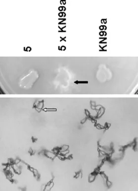
strains JEC21 and H99, and, thus, no same-sex mating. We graded the mating typing experiment results into three categories: sterile, fertile and robustly fertile and observed that only strain number 5 fit the third category (Fig. 4). Interestingly, this strain was not able to grow at the temperature of 37 ºC (data not shown).
DISCUSSION
Our results support the molecular application for typing on serotypes of C. neoformans as shown in a previous study23. MEYER et al.24 showed
predominance of serotype A (C.neoformans var. grubii) with an identical
or similar PCR fingerprinting and RAPD profiles, indicating a conserved molecular pattern in this serotype.
This method does not only substitute the immunological serotyping in
C. neoformans, but also shows a high discriminatory power. Genotyping
of environmental isolates with primer M13 allowed the differentiation between the two varieties types of C. neoformans according to MEYER et
Table 1
Isolation of Cryptococcus neoformans according to habitat
Local Samples Sheltered Isolation
Public building P1A1 No Negative
P1A2 Yes Positive
P1A3 Yes Positive
P1A4 No Negative
P1A5 No Negative
P1A 6 Yes Positive
Public building P2A1 No Negative
P2A 2 No Negative
P2A3 Yes Negative
Public building P3A1 Yes Positive
P3A2 Yes Negative
P3A3 Yes Positive
P3A4 No Negative
P3A5 No Negative
Public building P4A1 No Negative
P4A2 Yes Positive
P4A3 Yes Negative
P4A4 No Negative
Squares P5A1 No Negative
P5A2 No Negative
P5A3 No Negative
P5A4 No Negative
Monuments P6A1 No Negative
P6A2 No Negative
P6A3 No Negative
P6A4 Yes Positive
Dockside warehouse P7A1 Yes Negative
P7A2 No Negative
P7A3 Yes Positive
P7A4 Yes Negative
Dockside warehouse P8A1 Yes Positive
P8A2 Yes Positive
P8A3 Yes Negative
Fig. 1 - The PCR fingerprints using the M13 primer reveal the similar pattern between C. neoformans in this study and the reference serotype A strain (ATCC 34871). Lines 1-10: environmental C. neoformans strains; lines 11-14: reference strains: serotypes A, B, C and D. M: molecular marker (1Kb+ DNA ladder, Invitrogen®)
Fig. 2 - The URA5-RFLP of C. neoformans. Lines 1-10: environmental C. neofomans strains from this study; lines 11-14: reference strains VNI, VNII, VNIII, VNIV; lines 15-18: C. gattii reference strains VGI, VGII, VGIII, VGIV. M: molecular marker (1Kb+ DNA ladder, Invitrogen®). All strains collected in this study belong to molecular type VNI.
al.23. However, a problem occurs with primer M13: it does not differentiate
serotypes D and AD24. In this study we did not detect any hybrid strains,
molecular type VNIII, confirmed with URA5 RFLP (Fig. 2).
GARCIA-HERMOSO et al.10 showed that the distribution of clinical
isolates from African patients diagnosed with cryptococcosis in France was significantly different from that of clinical isolates recovered from the European patients, suggesting that African patients might have been
infected in their native countries but not in France. TINTELNOT et al.34
also studying patients with cryptococosis among European residents, at the beginning of the illness, informed that 22% of these patients originally came from Asia.
The city of Vitoria, Espirito Santo State, built on an area of 45 km2
in area consists of colonial buildings, ports and beaches. The climate is tropical and humid, with an average maximum monthly temperature of 30.4 °C (86.72 °F) and minimum of 24 °C (75.2 °F), and a heavy rainy season mainly in the months of October to January. It lies on the Southeast part of the Brazilian coast and this study revealed a comparable result as environmental surveys in the same geographic and climatic area, namely Rio de Janeiro18 and Sao Paulo30, where C. neoformans serotype A was
predominantly recovered from pigeon excreta.
Only a few Brazilian studies have investigated the molecular types of environmental Cryptococcus1,2,3,22,24,28,35. The distribution of molecular types
using the specific primer M13 shows the predominance of VNI (serotype A) in surveyed regions. It is worthwhile to note the presence of VNIV (serotype D) in eucalypt trees in the deep south of Brazil, a subtropical region2,28, and
of VGI (C. gattii serotype B) in excreta of psittaciformes in the same region1.
Study of clinical isolates in São Paulo showed that all of them were serotype A and the majority belonged to the molecular type VNI22.
The sexual cycle of C. neoformans involves fusion of a and a cells determined by two alleles within the single MAT locus, designated MATa or MATa14. In this study, all the environmental isolates produced
amplicons only of STE20a (Fig. 3) and, it was observed that the PCR
method was more successful in determining mating type alleles than mating experiments as previously shown21,33,34, since mating in the
laboratory with MATatester strains (JEC20, KN99a and Bt63) identified
only 50% of the MATa strains. Among them, same-sex mating of the
isolates was ruled out because none of the MATa strains mated with
opposite mating type MATa taster strains (JEC21 and H99).
No MATastrainwas recovered in this study. This emphasizes the
sexual reproduction by opposite mating type which does not appear to be dominant in C.neoformans var. grubii. An imbalanced ratioof a/a
spores generation is frequently observed and the rare MATa serotype A isolates (Aa) accounted with only 3 of >2,000 isolates examined21 and
theywere discovered confined in specific geographical regions of Africa, such as Tanzania19,Botswana21, and Italy36.
This study shows the environmental predominance of Cryptococcus
neoformans serotype A, molecular type VNI and mating-type a in the
city of Vitoria and adds information on the ecology and molecular characterization of C. neoformans in the Southeast region of Brazil.
RESUMO
Caracterização molecular de cepas ambientais de Cryptococcus neoformans isoladas em Vitória, ES, Brasil
O “complexo Cryptococcus neoformans” é constituído por C.
neoformans var. neoformans, C. neoformans var. grubii, e C. gattii. Trinta
por cento de amostras de excrementos de pombos coletados dentro da cidade de Vitória, Brasil, foram positivas para Cryptococcus neoformans,
espécie identificada por testes bioquímicos convencionais. Amplificação
Fig. 4 - Example of mating typing reaction between strain number 5 and its opposite mating type MATa tester isolate KN99a. The black and white arrow represents filamentation and basidiospores chains on V8 medium, respectively.
por PCR com primer M13 e análise por orotidine monophosphate
pyrophosphorylase (URA5) gene-“restriction fragment length
polymorphism” (RFLP) distinguiram sorotipos e genotipos dentro desta espécie. Todos os isolados ambientais foram sorotipo A (C. neoformans
var. grubii) e genotipo VNI. Os dois alelos alternativos a e a do locus “mating type” foram determinados por PCR e por testes de “mating” em meio V8. Todos os isolados foram “mating type” tipo MAT a mas somente
50% foram capazes de conjugar in vitro com cepas MATa,de “mating
type” oposto(JEC20, KN99a e Bt63). Este estudo adiciona informações
sobre a ecologia e caracterização molecular de cepas ambientais de C. neoformans, isoladas na região sudeste do Brasil.
ACKNOWLEDGMENTS
We thank Dr. Gary Cox and Dr. John Perfect from Duke University, USA, and Dr. Wieland Meyer, Australia for laboratory’s supplies and advices. The reference isolates were kindly provided by Dr. Wiley A. Schell, Dr. Andrew Alspaugh from Duke University and Dr. Wieland Meyer. Part of this study was supported by Grant from Foundation for Research of State of Espirito Santo (FAPES), Brazil.
REFERENCES
1. ABEGG, M.A.; CELLA, F.L.; FAGANELLO, J. et al. - Cryptococcus neoformans
and Cryptococcus gattii isolated from the excreta of psittaciformes in a southern Brazilian zoological garden. Mycopathologia, 161: 83-91, 2006.
2. CASALI, A.K.; GOULART, L.; ROSA, E. et al. - Molecular typing of clinical and
environmental Cryptococcus neoformans isolates in the Brazilian State Rio Grande do Sul. FEMS Yeast Res., 3: 405-415, 2003.
3. CARVALHO, V.G.; TERCETI, M.S.; DIAS. A.L. et al. - Serotype and mating type
characterization of Cryptococcus neoformans by multiplex PCR. Rev. Inst. Med. trop. S. Paulo, 49: 207-210, 2007.
4. CASADEVALL, A. & PERFECT, J.R. - Cryptococcus neoformans. Washington,
ASM, 1998.
5. CHATURVEDI, S.; RODEGHIER, B.; FAN, J. et al. - Direct PCR of Cryptococcus neoformans MATalpha and MATa pheromones to determine mating type, ploidy,
and variety: a tool for epidemiological and molecular pathogenesis studies. J. clin. Microbiol., 38: 2007-2009, 2000.
6. DEL POETA, M.; TOFFALETTI, D.L.; RUDE, T.H. et al. - Topoisomerase I is
essential in Cryptococcus neoformans: role in pathobiology and as an antifungal target. Genetics, 152: 167-178, 1999.
7. DROMER, F.; MATHOULIN. S.; DUPONT, B.; LETENNEUR, L. & RONIN, O. - Individual and environmental factors associated with infection due to Cryptococcus neoformans
serotype D. French Cryptococcosis Study Group. Clin. infect. Dis., 23: 91-96, 1996. 8. ESCANDÓN, P.; SÁNCHEZ. A.; MARTINEZ, M.; MEYER, W. & CASTAÑEDA, E.
- Molecular epidemiology of clinical and environmental isolates of the Cryptococcus neoformans species complex reveals a high genetic diversity and the presence of the
molecular type VGII mating type a in Colombia. FEMS Yeast Res., 6: 625-635, 2006.
9. FRASER, J.A.; SUBARAN, R.L.; NICHOLS, C.B. & HEITMAN, J. - Recapitulation of the sexual cycle of the primary fungal pathogen Cryptococcus neoformans var.
gattii: implications for an outbreak on Vancouver Island, Canada. Eukaryot Cell,
2: 1036-1045, 2003.
10. GARCIA-HERMOSO, D.; JANBON, G. & DROMER, F. - Epidemiological evidence for dormant Cryptococcus neoformans infection. J. clin. Microbiol., 37: 3204-3209,
1999.
11. HALLIDAY, C.L.; BUI, T.; KROCKENBERGER, M. et al. - Presence of alpha and a mating types in environmental and clinical collections of Cryptococcus neoformans
var. gattii strains from Australia. J. clin. Microbiol., 37: 2920-2926, 1999. 12. HULL, C.M. & HEITMAN, J. - Genetics of Cryptococcus neoformans. Ann. Rev.
Genet., 36: 557-615, 2002.
13. KIDD, S.E.; HAGEN, F.; TSCHARKE, R.L. et al. - A rare genotype of Cryptococcus gattii caused the cryptococcosis outbreak on Vancouver Island (British Columbia, Canada). Proc. nat. Acad. Sci. (Wash.), 101: 17258-17263, 2004.
14. KWON-CHUNG, K.J. - Morphogenesis of Filobasidiella neoformans, the sexual state of Cryptococcus neoformans. Mycologia, 68: 821-833, 1976
15. KWON-CHUNG, K.J.; BENNETT, J.E. & RHODES, J.C. - Taxonomic studies on
Filobasidiella species and their anamorphs. Antonie v. Leeuwenhoek, 48: 25-38, 1982.
16. KWON-CHUNG, K.J; EDMAN, J.C. & WICKES, B.L. - Genetic association of mating types and virulence in Cryptococcus neoformans. Infect. Immun., 60: 602-605, 1992.
17. KWON-CHUNG, K.J.; BOEKHOUT, T.; FELL, J.W. & DIAZ, M. - (1557) Proposal to conserve the name Cryptococcus gattii against C. hondurianus and C. basillisporus
(Basidiomycota, Hymenomycetes, Tremellomycetidae). Taxon, 51: 804-806, 2002. 18. LAZERA, M.S.; WANKE, B. & NISHIKAWA, M.M. - Isolation of both varieties
of Cryptococcus neoformans from saprophytic sources in the city of Rio de Janeiro, Brazil. J. med. vet. Mycol., 31: 449-454, 1993.
19. LENGELER, K.B.; WANG, P.; COX, G.M.; PERFECT, J.R. & HEITMAN, J. - Identification of the MATa mating-type locus of Cryptococcus neoformans reveals a serotype A MATa strain thought to have been extinct. Proc. nat. Acad. Sci. (Wash.), 97: 14455-14460, 2000.
20. LENGELER, K.B.; FOX, D.S.; FRASER, J.A. et al. - Mating-type locus of
Cryptococcus neoformans: a step in the evolution of sex chromosomes. Eukaryot. Cell, 1: 704-718, 2002.
21. LITVINTSEVA, A.P.; MARRA, R.E.; NIELSEN, K. et al. - Evidence of sexual recombination among Cryptococcus neoformans serotype A isolates in sub-Saharan Africa. Eukaryot. Cell, 2: 1162-1168, 2003.
22. MATSUMOTO, M.T.; FUSCO-ALMEIDA, A.M.; BAEZA, L.C.; MELHEM, M.S. & MENDES-GIANNINI, M.J. - Genotyping, serotyping and determination of mating-type of Cryptococcus neoformans clinical isolates from São Paulo State, Brazil. Rev. Inst. Med. trop. S. Paulo, 49: 41-47, 2007.
23. MEYER, W.; MARSZEWSKA, K.; AMIRMOSTOFIAN, M. et al. - Molecular typing of global isolates of Cryptococcus neoformans var. neoformans by polymerase chain reaction fingerprinting and randomly amplified polymorphic DNA-a pilot study to standardize techniques on which to base a detailed epidemiological survey. Electrophoresis, 20: 1790-1799, 1999.
24. MEYER, W.; CASTAÑEDA, A.; JACKSON, S. et al. - Molecular typing of Ibero American Cryptococcus neoformans isolates. Emerg. infect. Dis., 9: 189-195, 2003.
25. N G A M W O N G S AT I T, P. ; S U K RO O N G R E U N G , S . ; N I L A K U L , C . ; PRACHAYASITTIKUL, V. & TANTIMAVANICH, S. - Electrophoretic karyotypes of
C. neoformans serotype A recovered from Thai patients with AIDS. Mycopathologia, 159: 189-197, 2005.
26. NIELSEN, K.; COX, G.M.; WANG, P. et al. - Sexual cycle of Cryptococcus neoformans var. grubii and virulence of congenic a and alpha isolates. Infect. Immun., 71: 4831-4841, 2003.
27. NISHIKAWA, M.M.; LAZERA, M.S.; BARBOSA, G.G. et al. - Serotyping of 467
28. RIBEIRO, A.M.; SILVA, L.K.; SCHRANK, I.S. et al. - Isolation of Cryptococcus neoformans var. neoformans serotype D from Eucalypts in South Brazil. Med. Mycol., 44: 707-713, 2006.
29. SCHMEDING, K.A.; JONG, S.C. & HUGH, R. - Biochemical variation of
Cryptococcus neoformans. Mycopathologia, 84: 121-131, 1984.
30. SOARES, M.C.; PAULA, C.R.; DIAS, A.L.; CASEIRO, M.M. & COSTA, S.O. - Environmental strains of Cryptococcus neoformans variety grubii in the city of Santos, SP, Brazil. Rev. Inst. Med. trop. S. Paulo, 47: 31-36, 2005.
31. SORRELL, T.C.; CHEN, S.C.; RUMA, P. et al. - Concordance of clinical and environmental isolates of Cryptococcus neoformans var. gattii by random amplification of polymorphic DNA analysis and PCR fingerprinting. J. clin. Microbiol., 34: 1253-1260, 1996.
32. STAIB, F.; SEIBOLD, M.; ANTWEILER, E. et al. - The brown colour effect (BCE) of Cryptococcus neoformans in the diagnosis, control and epidemiology of
C. neoformans infections in AIDS patients. Zbl. Bakt. Mikrobiol. Hyg. [A]., 266: 167-177, 1987.
33. STEENBERGEN, J.N. & CASADEVALL, A. - Prevalence of Cryptococcus neoformans var. neoformans (Serotype D) and Cryptococcus neoformans var. grubii
(Serotype A) isolates in New York City. J. clin. Microbiol., 38: 1974-1976, 2000.
34. TINTELNOT, K.; LEMMER, K.; LOSERT, H.; SCHAR, G. & POLAK, A. - Follow-up of epidemiological data of cryptococcosis in Austria, Germany and Switzerland with special focus on the characterization of clinical isolates. Mycoses, 47: 455-464, 2004.
35. TRILLES, L.; LAZERA, M.; WANKE, B.; THEELEN, B. & BOEKHOUT, T. - Genetic characterization of environmental isolates of the Cryptococcus neoformans
species complex from Brazil. Med. Mycol., 41: 383-390, 2003.
36. VIVIANI, M.A.; ESPOSTO, M.C.; COGLIATI, M.; MONTAGNA, M.T. & WICKES, B.L. - Isolation of a Cryptococcus neoformans serotype A MATa strain from the Italian environment. Med. Mycol., 39: 383-386, 2001.
37. WICKES, B.L.; MAYORGA, M.E.; EDMAN, U. & EDMAN, J.C. - Dimorphism and haploid fruiting in Cryptococcus neoformans: association with the alpha-mating type. Proc. nat. Acad. Sci. (Wash.), 93: 7327-7331, 1996.
38. YAN, Z.; LI, X. & XU, J. - Geographic distribution of mating type alleles of
Cryptococcus neoformans in four areas of the United States. J. clin. Microbiol., 40: 965-972, 2002.