online | memorias.ioc.fiocruz.br
A potent trypanocidal component from the fungus Lentinus strigosus
inhibits trypanothione reductase and modulates PBMC proliferation
Betania Barros Cota, Luiz Henrique Rosa/++, Elaine Maria Souza Fagundes/+++,
Olindo Assis Martins-Filho1, Rodrigo Correa-Oliveira2, Alvaro José Romanha3,
Carlos Augusto Rosa4, Carlos Leomar Zani/+
Laboratório de Química de Produtos Naturais 1Laboratório de Biomarcadores de Diagnóstico e Monitoração 2Laboratório de Imunologia
Celular e Molecular 3Laboratório de Parasitologia Celular e Molecular, Instituto René Rachou-Fiocruz, Av Augusto de Lima 1715, 30190-002
Belo Horizonte, MG, Brasil 4Departamento de Microbiologia, Instituto de Ciências Biológicas, Universidade Federal de Minas Gerais,
Belo Horizonte, MG, Brasil
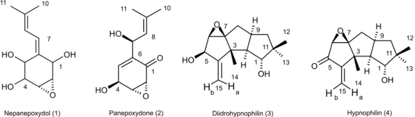
The fungus Lentinus strigosus (Pegler 1983) (Polyporaceae, basidiomycete) was selected in a screen for inhibitory activity on Trypanosoma cruzi trypanothione reductase (TR). The crude extract of L. strigosus was able to completely inhibit TR at 20 µg/ml. Two triquinane sesquiterpenoids (dihydrohypnophilin and hypnophilin), in addition to two panepoxydol derivatives (neopanepoxydol and panepoxydone), were isolated using a bioassay-guided fractionation protocol. Hypnophilin and panepoxydone displayed IC50 values of 0.8 and 38.9 µM in the TR assay, respectively, while the other two compounds were inactive. The activity of hypnophilin was confirmed in a secondary assay with the intracellular amastigote forms of T. cruzi, in which it presented an IC50 value of 2.5 µM. Quantitative flow cytometry experiments demonstrated that hypnophilin at 4 µM also reduced the proliferation of human peripheral blood monocluear cells (PBMC) stimulated with phytohemaglutinin, without any apparent interference on the viability of lymphocytes and monocytes. As the host immune response plays a pivotal role in the adverse events triggered by antigen release during treatment with trypanocidal drugs, the ability of hypnophilin to kill the intracellular forms of T. cruzi while modulating human PBMC proliferation suggests that this terpenoid may be a promising prototype for the development of new chemotherapeutical agents for Chagas disease.
Key words: fungal natural products - Chagas disease - drug discovery - immunomodulators - Basidiomycota
Chagas disease is caused by the protozoan parasite
Trypanosoma cruzi and affects millions of people in Latin America, having an enormous economic and social impact in the endemic areas. These patients rely on treat-ment with nitrofuran (nifurtimox; Bayer) or nitroimida-zol (benznidanitroimida-zole; Roche), medicines that display little or no activity in chronic infections, in spite of their beneficial effect during the acute phase of the disease (Cançado 2002, Coura & Castro 2002). However, signifi-cant differences in the therapeutic effectiveness are ob-served between these two drugs, especially when consid-ering distinct geographical areas, which is probably due to the occurrence of naturally resistant and susceptible
T. cruzi strains (Filardi & Brener 1987, Toledo et al. 2003). Furthermore, both drugs cause several side effects that contribute to their reduced use in clinical medicine (Can-çado 1985). Thus, new compounds are needed to develop more effective medicines to treat Chagas disease,
espe-Financial support: Fapemig, Capes, CNPq, Fiocruz + Corresponding author: [email protected]
++ Current address: Laboratório de Microbiologia, Instituto de Ciências Exatas e Biológicas, Universidade Federal de Ouro Preto, Ouro Preto, MG, Brasil
+++ Current address: Departamento de Fisiologia e Biofísica, Univer-sidade Federal de Minas Gerais, Belo Horizonte, MG, Brasil Received 12 December 2007
Accepted 9 May 2008
cially in its chronic phase, is needed (Schmidt & Krauth-Siegel 2002, Nwaka & Ridley 2003). However, due to low profit prospects, the development of new trypanocidal drugs is not attractive to the pharmaceutical industry (Nwaka & Ridley 2003). Thus, this endeavor is being con-ducted mainly in academic laboratories (Fairlamb 1999).
Among the many different strategies for drug discov-ery, screening the local biodiversity for bioactive natural products using appropriate bioassays is an interesting alternative for researchers in affected areas, especially in countries with a rich biodiversity. There is a vast lit-erature describing that natural products produced by plants, fungi and other organisms are the direct source of or inspiration for many of the currently available medicines (Newman & Cragg 2007). In this regard, our group screens the Brazilian biodiversity for new bioac-tive natural products using in vitro bioassays related to neglected diseases, especially Chagas disease and leish-maniasis (Ribeiro et al.1997, Rosa et al. 2005, 2006). We evaluated more than 3,000 extracts from plant and fungi using an inhibition assay with trypanothione re-ductase (TR) (unpublished results). This enzyme is a val-idated molecular target in Trypanosoma and Leishmania species (Fairlamb et al.1985, Dumas et al. 1997, Tovar et al. 1998, Krieger et al. 2000). As a result of the screening, we found that the ethyl acetate extract of the culture of
Species from the genus Lentinus are normally sapro-phytic, lamellate wood-decaying basidiomycetes (Kirk et al. 2001). Most of its 40 species grow in the tropics, although some can be found in the boreal region (Pegler 1983). Previous investigations on the chemical composi-tion of Lentinus species identified several sesquiterpenes from L. lepideus (Hanssen1982, 1985) and L. connatus
(Rukachaisirikul et al. 2005) and benzoquinones from
L. degener (Anchel et al. 1948) and L. adhaerens (Lauer et al. 1991). However, to the best of our knowledge, no in-vestigations studying TR inhibitors were performed with any species from this genus, and this is the first study on the species L. strigosus.
Here, we describe the bioassay-guided isolation and identification of the active compounds present in
L. strigosus extract. We also show that one of these com-pounds, the terpenoid hypnophilin, has an interesting biological activity profile, making it an attractive lead for further investigations of drugs to treat Chagas disease.
MAtERiALS And MEtHOdS
General experimental procedures - The optical rota-tions were measured on a Perkin-Elmer 341 polarimeter (25°C, Na lamp, 589 nm). Infrared spectra were obtained on a Shimadzu FTIR-8400, with the samples in KBr pel-lets. 1H (400 MHz), 13CNMR (100 MHz), DEPT, HMQC,
and HMBC NMR spectra were measured on a Brucker DRX 400 spectrometer. Electron impact (70 eV) low-resolution mass spectra (EI-MS) were measured with a Shimadzu QP5050A spectrometer, equipped with a direct insertion probe. Electrospray ionization mass spectra (ESI-MS) were measured on a Thermo Finnigan LCQ-Advantage spectrometer.
Biological material - The L.strigosus (Schwein) Fr. isolate was deposited in the UFMGCB collection (Uni-versidade Federal de Minas Gerais, Brazil) and received the code UFMGCB975. The specimen was identified us-ing classical taxonomic methods (Pegler 1983).
Fungus fermentation - The fungus was grown on malt extract agar (MEA, Difco, US) and transferred (three 5 mm discs) to ten 250 ml Erlenmeyer flasks containing 25 ml of MEC medium (malt extract 2%, peptone 0.1%, glucose 1.5%). The flasks were shaken at 150 rpm and 28°C for five days. The contents of these culture flasks were then transferred to an 8 l bioreactor and cultivated for nine days at 28°C.
Extract preparation - The mycelium was separated from the culture by vacuum filtration, and the filtrate was extracted with ethyl acetate (5 x 300 ml for each 1,000 ml of broth). The organic phase was separated and dried over anhydrous sodium sulfate. After filtration, the solution was concentrated in a rotary evaporator at temperatures below 45°C. The extract was then dried in a vacuum centrifuge at 40°C to yield 2.29 g of a brown gum. An aliquot was used to prepare a stock solution at 20 mg/ml in dimethylsulphoxide (DMSO), which was used in all biological assays. Both the crude extract and stock solution were stored at -40°C.
Isolation and purification - Initially, 100 µg of the crude extract was separated in an analytical HPLC col-umn (Shim-pack ODS, 5 µm, 4.6 x 250 mm), using a linear gradient from 10 to 100% CH3OH in 25 min at 1.0 ml/min. The effluent was monitored with a photo-diode array UV detector and collected in a 96-well plate (220 µl per well, 80 wells). The plates were dried in an oven at 40°C and then subjected to the TR bioassay to iden-tify the fractions containing metabolites with inhibitory activity (Fig. 1). A semi-preparative fractionation was then performed in a 20 x 250 mm reverse-phase HPLC column (Shim-pack prep-ODS, 5 µm), using a 34 min linear gradient from 15 to 100% CH3CN in H2O. Five aliquots of approximately 100 mg of crude extract were fractionated at a flow rate of 10 ml/min, with UV detec-tion at 210 and 254 nm. One fracdetec-tion afforded a white amorphous solid that, upon crystallization with a mix-ture of CH2Cl2-Hex, yielded 32 mg of dihydrohypnophi-lin (3). The remaining fractions were further fractionated by semi-preparative isocratic normal-phase HPLC (Shim-pack prep-Si, 5 µm, column 20 x 250mm), eluted with CH2Cl2-MeOH (95:5 or 97:3) at 10 ml/min. This procedure yielded 70 mg of panepoxydone (2), 3.8 mg of neopanep-oxydol (1), and 14 mg of hypnophilin (4).
Neopanepoxydol (1) - White amorphous powder; IR
(KBr): νmax = 3308 (OH), 2962 (aliph.), 2862 (aliph.), 1600
(C = C), 1038 (C = C) cm-1. EI-MS (70 eV), m/z 212 [M]+●
(C11H16O4). 1H NMR (CDCl
3, 400 MHz): δ 6.62 (1H, m,
H-7), 6.53 (1H, m, H-8), 4.56 (1H, broad singlet, H-1), 4.21 (1H, d, J = 5.5 Hz, H-5), 3.96 (1H, dd, J = 1.1; 5.5 Hz, H-4), 3.38 (1H, ddd, J = 0.6; 2.5; 3.5 Hz, H-2), 3.28 (1H, dd,
J = 0.6; 2.0; 4.0 Hz, H-3), 1.81 (3H, s, H-10/-11), 1.79 (3H,
s, H-10 or H-11). 13C NMR (CDCl
3,100 MHz): δ 138.08
(C-9), 132.92 (C-6), 128.34 (C-7), 121.86 (C-8), 73.20 (C-5), 71.00 (C-1 or C-4), 70.90 (C-1 or C-4), 57.81 (C-2), 56.66 (C-3), 26.74 (C-10 or C-11), 18.04 (C-10 or C-11).
Panepoxydone (2) - Pale yellow oil. [α]25
D -61° (c 0.4,
CH2Cl2). IR (KBr) νmax = 3381 (OH), 2974 (aliph.), 2880 (aliph.), 1682 (C = O), 1383 (C-H), 1043 (C-O) cm-1.
ESI-MS m/z 209 [M - H]- (C
11H14O4).
1H NMR (400 MHz,
CDCl3): δ 6.70 (1H, ddd, J = 1.3, 2.5, 3.6 Hz, H-5), 5.29 (1H, qd, J = 1.3, 8.8 Hz, H-7), 5.02 (1H, d, J = 8.8 Hz, H-8), 4.68 (1H, sl, H-4), 3.80 (1H, dq, J = 1.3; 2.5; 3.6 Hz, H-3), 3.46 (1H, dd, J = 1.0; 3.6 Hz, H-2), 1.72 (6H, s, H-10 or H-11). 13C NMR (100 MHz, CDCl
3): δ 194.47 (C-1),
138.98 (C-6), 138.26 (C-9), 137.78 (C-5), 123.68 (C-8), 65.35 (C-7), 63.16 (C-4), 57.71 (C-3), 53.88 (C-2), 25.90 (C-10 or C-11), 18.42 (C-10 or C-11).
Dihydrohypnophilin (3) - White crystals from CH2Cl2
-Hex. [α]25
D +135° (c 0.5, CHCl3). IR (KBr) νmax = 3391
(OH), 2939 (aliph.), 2858 (aliph.), 1666 (C = C), 1458 (ali-ph.), 1392 (C-H), 1222 (C-O), 1045 (C-O), 957 (C-H), 883 (C-H) cm-1. EI-MS (70 eV), m/z 250 [M]+● (C
15H22O3). 1H NMR (400 MHz, CDCl
3): δ 5.32 (1H, d, J = 2.2 Hz,
H-15a), 5.15 (1H, d, J = 2.2 Hz, H-15b), 4.62 (1H, q,
J = 2.2 Hz, H-5), 3.80 (1H, d, J = 8.4 Hz, H-1), 3.47 (1H,
d, J = 2.2 Hz, H-6), 2.60 (1H, m, H-9), 2.03 (1H, dd,
(2H, d, J = 9.0 Hz, H-8), 1.17 (3H, s, H-14), 1.13 (1H, m,
J = 12.0 Hz, H-10β), 1.05 (3H, s, H-12), 0.89 (3H, s, H-13). 13C NMR (100 MHz, CDCl
3): δ 159.64 (C-4), 112.53
(C-15), 80.96 (C-1), 75.03, (C-7), 74.19, (C-5), 63.73 (C-6), 55.25 (C-2), 47.55 (C-3), 46.36 (C-10), 44.22 (C-11), 34.81 (C-9), 30.74 (C-8), 26.48 (C-12), 19.78 (C-13), 17.51 (C-14).
Hypnophilin (4) - Colorless oil. [α]25
D -74° (c 0.9,
CHCl3). IR (KBr) νmax = 3443 (OH), 2937 (aliph.), 2870 (aliph.), 1728 (C = O), 1636 (C = C), 1464 (C-H), 1375 (C-H), 1115 (C-O) cm-1. EI-MS (70 eV), m/z 248 [M]+●
(C15H20O3). 1H NMR (400 MHz, CDCl
3): δ 6.13 (1H,
s, H-15a), 5.45 (1H, s, H-15b), 3.87 (1H, d, J = 9.0 Hz, H-1), 3.43 (1H, s, H-6), 2.64 (1H, m, H-9), 2.14 (1H, dd,
J = 9.0, 12.0 Hz, H-2), 1.95 (2H, m, H-8), 1.90 (1H, m, H-10α), 1.30 (3H, s, H-14), 1.23 (1H, m, H-10β), 1.07 (3H, s, H-12), 0.89 (3H, s, H-13). 13C NMR (100 MHz, CDCl
3): δ 197.60 (C-5), 153.73 (C-4), 121.61 (C-15), 81.23 (C-1), 76.04 (C-7), 61.14 (C-6), 56.13 (C-2), 46.12 (C-10), 45.54 (C-3), 44.14 (C-11), 34.52 (C-9); 30.75 (C-8), 26.43 (C-12), 19.66 (C-13), 17.75 (C-14).
Cell preparations - Venous blood from healthy adult volunteers was collected in heparinized tubes and cen-trifuged over a Ficoll-Hypaque cushion (Histopaque, Sigma, St Louis, MO). Peripheral blood mononuclear cells (PBMC) were collected from the Ficoll/Hypaque interphase and washed three times in RPMI prior to fur-ther processing. The cell suspensions were adjusted to 1.5 x 10⁶ cells/ml.
Viability assays - PBMC viability was determined using a modification of the protocol described by Souza-Fagundes et al. (2002). Briefly, the cells were treated with pure compounds at different concentrations for 72 h at 37ºC in a 95% relative humidity atmosphere with 5%
CO2. After this period, the cells were incubated for 10 min with trypan blue (0.4% in NaCl 0.9%). In the flow cytometry experiments, trypan blue fluorescence was measured in the FL2 channel. Fluorescence intensity was used to discriminate viable (non-fluorescent) and non-viable (fluorescent) lymphocytes. Controls with 0.1% DMSO run in parallel did not demonstrate any dif-ferences in cell viability (data not shown).
PBMC culture - All cultures were performed in RPMI-1640 medium (GIBCO, Grand Island, NY), sup-plemented with 5% (v/v) heat-inactivated, pooled human sera type AB (Flow Laboratories, Royaune, UN) and L-glutamine (2 mM, GIBCO, Grand Island, NY). An antibiotic/antimicotic solution containing 1,000 U/ml penicillin, 1,000 µg/ml streptomycin and 25 µg/ml fungi-sone (SIGMA, St. Louis, MO) was added to control fungal and bacterial contamination.
Lymphocyte proliferation assay - In vitro cellular proliferation (blastogenesis) was assessed as previously described (Gazzinelli et al. 1983). Briefly, 1 x 10⁶ cells/ml
(1.5 x 10⁵ cells per well) were cultured in complete
RPMI-1640 in flat-bottomed microtiter plates (Costar, tissue culture treated polystyrene # 3512, Corning, NY, USA). The cultures were stimulated with 2.5 µg/ml of PHA (SIGMA, St Louis, MO) and incubated for 72 h at 37°C in a humidified atmosphere containing 5% CO2. The cell proliferation and viability were determined using an MTT (methyl thiazolyl tetrazolium)-based colorimetric assay (Jiang & Xu 2003). The results were expressed as percent inhibition of the lymphocyte proliferation in relation to the control (without drugs).
al. (2003) in 96-well plates (Costar 9017, Corning, USA) us-ing Hepes buffer (40 mM pH 7.5) with 1 mM EDTA. Each assay well (250 µl) contained enzyme (6 mU), trypano-thione (0.25 nmol) and NADPH (50 nmol). The extracts, fractions or pure compounds were added to the above mixture and incubated at 30°C during 30 min. After this period, 17.5 nmol of DTNB [5,5’-dithiobis(2-nitrobenzoic acid), Ellman’s reagent] was added, and the absorbance (Abs) was measured at 412 nm in the kinetic mode every
10 s for 5 min. The resulting slope of the δAbs/δt plot is
proportional to DTNB reduction and the enzyme activity (Hamilton et al. 2003). The enzyme inhibition was
calcu-lated as the ratio between (δAbs/δt) of the experimental
wells and that of the controls without drug, that is, percent
inhibition = (1 - (δAbs)exp/(δAbs)contr) x 100.
Assay with T. cruzi amastigotes - This assay was performed as described previously by Buckner et al. (1996) using a T. cruzi (Tulahuen strain) expressing the
Escherichia coli β-galactosidase gene. Infective trypo -mastigote forms were obtained through culture in mono-layers of mouse L929 fibroblasts in RPMI-1640 medi-um (pH 7.2-7.4) without phenol red (Gibco BRL) plus 10% fetal bovine serum, 2mM glutamine and 40 µg/ml gentamycin. For the bioassay, 4,000 L929 cells in 80 µl of supplemented medium were added to each well of a 96-well microtiter plate. After an overnight incubation, 40,000 trypomastigotes in 20 µl were added to the cells and incubated for 2 h. The medium containing extrace-lullar parasites was replaced with 200 µl of fresh medi-um and the plate was incubated for an additional 48 h to establish the infection. The medium was then replaced with solutions of the compounds at different concen-trations in fresh medium (200 µl), and the plate was incubated for 96 h. After this period, 50 µl of 500 µM chlorophenol red glycoside in 0.5% Nonidet P40 was added to each well, and the plate was incubated for 24 h, after which the absorbance at 570 nm was measured. Controls with uninfected cells, untreated infected cells, and medium only were run in parallel. The re-sults are expressed as percent reduction of the absor-bance in the experimental wells in comparison with the control wells with untreated infected cells (percent inhibition = (1 - Absexp/Abscontr) x 100). Triplicates were run in the same plate, and the experiments were re-peated at least once.
RESULtS
L.strigosus (Schwein) Fr. was grown in MEC medi-um, and the culture extracted with ethyl acetate yielded 2.29 g of a crude extract that was able toinhibit TR ac-tivity by 100% at 20 µg/ml. In order to identify the active components of the extract, 100 µg of the crude extract was fractionated in an analytical HPLC column, and the effluent was collected in a 96-well microplate (250 µl/ well) while recording the UV chromatogram (at 210 nm). After solvent elimination, the dry films in the plate were dissolved with the assay buffer, and the solutions were subjected to the TR inhibition assay. The results of this experiment are presented as an overlay plot of the chro-matogram and as the percentage of TR inhibition (Fig. 1), clearly showing that the inhibitory activity was con-centrated on compounds eluting around 13 and 19 min. The HPLC method was then scaled up to isolate the com-pounds in sufficient amounts for structural elucidation and IC50 determinations. Four compounds were isolated, either by crystallization of the collected fractions or after further chromatographic steps, as described in the Mate-rials and Methods section. Based on the analysis of their UV, IV, Mass, and NMR spectra and on comparison with data reported in the literature, the compounds were iden-tified as neopanepoxydol (1), panepoxydone (2), dihydro-hypnophilin (3) and dihydro-hypnophilin (4), as shown in Fig. 2. Detailed analysis of the HMQC and HMBC NMR data allowed the unambiguous assignments of C-12 and C-14 chemical shifts in dihydrohypnophilinand hypnophilin, requiring the correction of those proposed by Abate and Abraham (1994). Thus, the attributions for C-12/C-14 in
dihydrohypnophilin should be corrected from 17.5/26.5 δ to 26.48/17.51 δ, and in hypnophilin from 17.6/26.4 δ to 26.43/17.45 δ.
All pure compounds were tested at 20 µg/ml in the TR, amastigote, and PBMC assays (Table I). The results show that panepoxydone and hypnophilin were active in all bioassays while neopanepoxydol and dihydro-hypnophilin were inactive. Dose response experiments with the active compounds confirmed these preliminary results, with hypnophilin and panepoxydone disclosing IC50 values around 1 and 39 µM, respectively (Table II). Hypnophilin was also more active than panepoxydone against intracellular amastigotes, having an IC50 value of 2.5 µM. Panepoxydone, however, was more effective in inhibiting PBMC proliferation, having an IC50 of 1.3 µM.
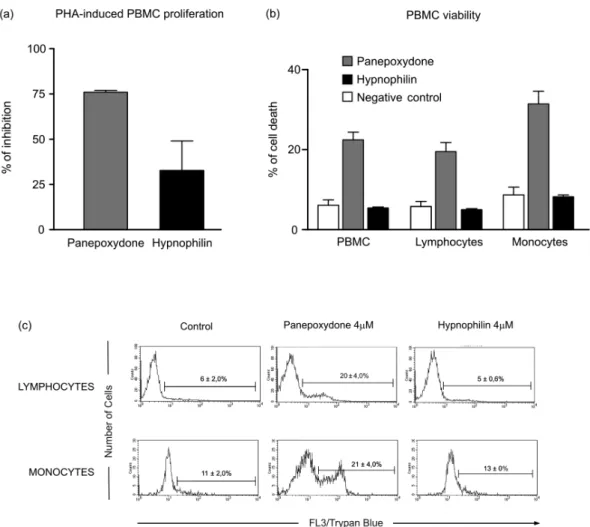
From the experimental data used to construct the dose response curves (data not shown), it was estimated that 4 µM hypnophilin is able to block 100% of TR activity and inhibit 80% of the growth of intracellu-lar amastigotes, with only minimal inhibitory activity (20%) on the proliferation of PHA-stimulated PBMC (Fig. 3). According to the data, at this concentration, panepoxydone would be almost inactive against amas-tigotes and still present some toxicity to PBMC. To in-vestigate this hypothesis, the impact of panepoxydone and hypnophilin at 4 µM on lymphocyte and monocyte subpopulations of the PBMC was evaluated.
The results (Fig. 4a, b) show that panepoxydone in-duced cell death on the lymphocyte and monocyte popu-lations compared with PBMC control cultures while, as expected, hypnophilin was not cytotoxic. These results were corroborated by quantitative analyses by flow cy-tometry (Fig. 4c), which showed that panepoxydone re-duced the lymphocytes population by 20%, while hyp-nophilin showed only a 5% reduction, which is similar to the control culture without drug. Similar results were observed for the monocytes population, where panep-oxydone significantly induced cell death (21%), while the percentage of death caused by hypnophilin mirrored the control without treatment (11%).
diSCUSSiOn
A bioassay-guided procedure based on the inhibition of TR was used to detect and isolate neopanepoxydol (1), panepoxydone (2), dihydrohypnophilin (3) and
hypno-philin (4) from L. strigosus. Our results confirm the effi-ciency of the method based on analytical HPLC and off-line bioassay to identify the retention time of the active components (panepoxydone and hypnophilin) present in the extract, facilitating their isolation.
Hypnophilin (4) was previously isolated from the cul-ture of Pleurotellus hypnophilus (Gianetti et al. 1986),
Lentinus critinus (Abate & Abraham1994) and L. con-natus (Rukachaisirikul et al. 2005). Dihydrohypnophilin (3) was isolated from a culture of L. crinitus (Abate & Abraham1994) and L. conatus (Rukachaisirikul et al. 2005). Panepoxydone and neopanepoxydol were obtained from fermentations of Panus rudis and Panus conchatus
(Kis et al. 1970),while panepoxydone was isolated with hypnophilin from cultures of the L. connatus (Rukachai-sirikul et al. 2005) and L. crinitus (Erkel et al. 1996).
Panepoxydone and hypnophilin inhibited TR in the same concentration range of a series of synthesized polyaminoguanidines and polyaminobiguanides (IC50 values from 0.95 to 69.47 µM)(Bi et al. 2006). Under our assay conditions, hypnophilin inhibits TR in the low micromolar range (IC50 = 1 µM), making it about three times more active than the reference compound clomip-ramine (IC50 = 3 µM), a known inhibitor of TR. It is also more active than the macrocyclic spermidine alkaloid lunarine (IC50 = 65 µM, Hamilton et al. 2005), which was isolated from Lunaria biennis. These results indi-cate that hypnophilin can be considered a good starting point for a medicinal chemistry program aimed at deter-mining derivatives with higher TR inhibitory activity.
Concerning the mechanism by which panepoxydone (2) and hypnophilin (4) inhibit TR, we speculate that it may be similar to that proposed for the alkaloid lunarine (Hamilton et al. 2005), in which the presence of con-jugated carbonyl groups make it susceptible to nucleo-philic attack by the thiol groups present in the active site of TR. The lack of activity by neopanepoxydol (1) and dihydrohypnophilin (3), which are closely related to two TABLE I
Activity of compounds isolated from Lentinus strigosus
on trypanothione reductase (TR), intracellular amastigotes (AMA) of Trypanosoma cruzi, and human lymphocyte
proliferation (PBMC) assays
Compounds % inhibitiona at 20 µg/mL
TR AMA PBMC
Panepoxydone 95 ± 7 95 ± 7 100 ± 12 Hypnophilin 100 ± 1 80 ± 9 100 ± 17 Neopanepoxydol 0 ± 8 0 ± 16 8 ± 13 Dihydrohypnophilin 0 ± 7 0 ± 11 15 ± 11
a: mean value ± S.D. of at least two independent experments; PBMC: peripheral blood monoclear cell.
TABLE II
IC50 values of panepoxydone and hypnophilin on TR, AMA and PBMC assays
Compoundsa IC
50 (µM)
b
TR AMA PBMC
Panepoxydone 38.9 ± 1.4 8.7 ± 0.1 1.3 ± 0.1 Hypnophilin 0.8 ± 0.1 2.5 ± 0.7 8.9 ± 1.4
a: IC50 (µM) of the standard drugs: TR assay, clomipramine, 2.9 ± 0.1; AMA assay, benznidazole, 1.5 ± 0.7; b: mean value ± S.D. of three independent experiments.
and fourbut do not carry the conjugated carbonyl moi-ety, reinforces this hypothesis.
Besides its activity in the biochemical assay, hypno-philin was also active against the intracellular amastig-otes of the Tulahuen strain of T. cruzi infecting L929 cells. Inhibition of the multiplication of T. cruzi intracellular amastigotes is clinically relevant because these are the proliferating forms of the parasite. These parasites ma-ture into trypomastigote forms that cause the rupma-ture of the host cells, thus promoting tissue damage in the in-fected organs, especially the esophagus, colon and heart of chronic patients of Chagas disease. Although circu-lating parasites cannot be observed by blood inspection in this phase, T. cruzi antigens may trigger the immune system by multiple mechanisms resulting in inflamma-tion and fibrosis. An inflammatory response may result in accumulation and activation of monocytes in addition to increasing the concentration of macrophage-derived cytokines in the plasma (Aliberti et al. 1996). These cy-tokines can promote an immediate response and also af-fect organs and tissues.
There is a general consensus that the host immune response plays a pivotal role in the adverse events caused upon the massive antigen release triggered by trypanocidal agents during treatment (Sathler-Avelar et al. 2006). With this in mind, our results show that hypnophilin display a non-cytotoxic antiproliferative activity on PBMC stimu-lated with PHA, suggesting that, besides its trypanocidal activity, hypnophilin can eventually reduce tissue damage by interfering with the proliferation of cells involved with the immune response.
the majority of the 18 million patients with T. cruzi are currently in the chronic phase (Prata 2001). In this con-nection, our findings indicate that hypnophilin is a lead candidate for development of new drugs to treat chronic Chagas disease. Additional studies are needed to charac-terize the molecular basis of hypnophilin-induced immu-nomodulatory activity. We speculate that it may involve the up-regulation of IL-10 synthesis, as is the case with benznidazole (Pascutti et al. 2004), or the engagement of other cytokines and/or apoptosis mechanisms that would allow the effective parasite killing in the absence of im-munomediated tissue damage.
Two metabolites, panepoxydone and hypnophilin, with significant TR inhibitory activity were isolated from L. strigosus, a tropical basidiomycete investigated for the first time. Hypnophilin disclosed significant
anti-T. cruzi activity associated with minor non-cytotoxic immunomodulatory activity on human leukocytes, sug-gesting that it is a good lead candidate for developing new drugs against Chagas disease.
ACknOwLEdgEMEntS
To Rosana Alves, Rodrigo Leite and Aline Vaz for their technical assistance. To Prof. Alan Fairlamb (Univ. of Dundee, Scotland) for for providing the recombinant trypanothione re-ductase, and to Prof. Frederick Buckner (University of Washing-ton, USA) for the Tulahuen strain of T. cruzi expressing the E. coli β-galactosidase gene.
REFEREnCES
Abate D, Abraham WR 1994. Antimicrobial metabolites from Lenti-nus crinitus. J Antibiot47: 1348-1350.
Aliberti JC, Cardoso MA, Martins GA, Gazzinelli RT, Vieira LQ, Silva JS 1996. Interleukin-12 mediates resistance to Trypano-soma cruzi in mice and is produced by murine macrophages in response to live trypomastigotes. Infect Immun 64: 1961-1967.
Anchel M, Hervey A, Kavanagh F, Polatnick J, Robbins WJ 1948. Antibiotic substances from basidiomycetes III. Coprinus similes and Lentinus degener. Proc Natl Acad Sci U S A 34: 498-499.
Bi X, Lopez C, Bacchi CJ, Rattendi D, Woster PW 2006. Novel alkyl-polyaminoguanidines and alkylpolyaminobiguanides with potent antitrypanosomal activity. Bioorg Med Chem Lett 16: 3229-3232.
Buckner FS, Verlinde CLMJ, La Flamme AC, van Voorhis WC 1996. Efficient technique for screening drugs for activity against Try-panosoma cruzi using parasites expressing beta-galactosidase. Antimicrob Agents Chemother 40: 2592-2597.
Cançado JR 1985. Tratamento específico. In JR Cançado, Cardiopatia Chagásica, Fundação Carlos Chagas, Belo Horizonte, p 327-355.
Cançado JR2002. Long term evaluation of etiological treatment of Chagas disease with benznidazole. Rev Inst Met Trop Sao Paulo 44: 29-37.
Coura JR, Castro SL 2002. A critical review on Chagas disease che-motherapy. Mem Inst Oswaldo Cruz 97: 3-24.
Dumas C, Ouellette M, Tovar J, Cunningham ML, Fairlamb AH, Tamar S, Olivier M, Papadopoulou B 1997.Disruption of the trypanothi-one reductase gene of Leishmania decreases its ability to survive oxidative stress in macrophages. EMBO J 16: 2590-2598.
Erkel G, Anke T, Sterner O 1996. Inhibition of NF-κB activation by panepoxydone. Biophys Res Commun 226: 214-221.
Fairlamb AH 1999. Future prospects for the chemotherapy of Cha-gas’s disease. Medicina (B Aires)59: 179-187.
Fairlamb AH, Blackburn P, Ulrich P 1985. Trypanothione-A novel bis(glutationyl)spermidine cofator for glutathione-reductase in trypanosomatids. Science 227: 1485-1487.
Filardi LS, Brener Z 1987. Susceptibility and natural resistance of Trypanosoma cruzi strains to drugs used clinically in Chagas disease. Trans R Soc Trop Med Hyg 81: 755-759.
Gazzinelli G, Katz N, Rocha RS, Colley DG 1983. Immune response during human schistosomiasis mansoni X. Production and stan-dartization of an antigen-induced mitogenic activity by periph-eral blood mononuclear cells from treated but not active cases of schistosomiasis. J Immunol130: 2891-2895.
Gianetti BM, Steffan B, Steglich W, Kupka J, Anke T 1986. Antibiot- Antibiot-ics from basidiomycetes. Part 24. AntibiotAntibiot-ics with a rearranged hirsutane skeleton from Pleurotellus hypnophilus (Agaricales). Tetrahedron 42: 3587-3593.
Hamilton CJ, Saravanamuthu A, Eggleston IM, Fairlamb AH 2003. Ellman’s-reagent-mediated regeneration of trypnothione in situ: substrate-economical microplate and time-dependent inhibition assays for trypanothione reductase. Biochem J 369: 529-537.
Hamilton CJ, Saravanamuthu A, Poupat C, Fairlamb AH, Eggleston IM 2005. Time-dependent inhibitors of trypanothione reductase: Analogues of the spermidine alkaloid lunarine and related natu-ral products. Bioorg Med Chem14: 2266-2278.
Hanssen HP 1982. Sesquiterpene hydrocarbons from Lentinus lepi-deus. Phytochemystry 21: 1159-1160.
Hanssen HP 1985. Sesquiterpene alcohols from Lentinus lepideus. Phytochemystry 24: 1293-1294.
Jiang J, Xu Q 2003. Immunomodulatory activity of the aqueous ex-Immunomodulatory activity of the aqueous ex-tract from rhizome of Smilax glabra in the later phase of adju-vant-induced arthritis in rats. J Ethnopharmacol 85: 53-59.
Kirk PM, Cannon PF, David JC, Stalpers JA 2001. Ainsworth & Bisby’s dictionary of the fungi, 9th ed, CABI Publishing, Britain, 655 pp.
Kis Z, Glosse A, Sigg HP, Hruban L, Snatzke G 1970. Die struktur von panepoxydon und verwandten pilzmetaboliten. Helv Chim Acta 53: 1577-1597.
Krieger S, Schwarz W, Ariyanayagam MR, Fairlamb AH, Krauth-Siegel RL, Clayton C 2000. Trypanosomes lacking trypanothi-Trypanosomes lacking trypanothi-one reductase are avirulent and show increased sensitivity to oxidative stress. Mol Microbiol 35: 542-552.
Lauer U, Anke T, Hansske F 1991. Antibiotics from basidiomycetes. XXXVIII. 2-methoxy-5-methyl-1,4-benzoquinone, a tromboxa-ne A2 receptor antagonist from L adherens. J Antibiot 44: 59-65.
Muelas-Serrano S, Le-Senne A, Fernandez-Portillo C, Nogal JJ, Ochoa C, Gomez-Barrio A 2002. In vitro and in vivo anti- Try-panosoma cruzi activity of a novel nitro-derivative. Mem Inst Oswaldo Cruz 97: 553-557.
Newman DJ, Cragg GM 2007. Natural products as sources of new drugs over the last 25 years. J Nat Prod 70: 461-477.
Nwaka S, Ridley RG 2003. Virtual drug discovery and development for neglected diseases through public-private partnerships. Natu- Natu-re Rev Drug Discov 2: 919-928.
Pascutti MF, Pitashny M, Nocito AL, Guermonprez P, Amigorena S, Wietzerbin J, Serra E, Bottasso O, Revelli S 2004. Benznidazole, a drug used in Chagas’ disease, ameliorates LPS-induced inflam-matory response in mice. Life Sci76: 685-697.
Pegler DN 1983. The genus Lentinus: a world monograph,Kew Bul-letin Additional Series X, HMSO, London, 281 pp.
Ribeiro A, Piló-Veloso D, Romanha AJ, Zani CL 1997. Trypanocidal flavonoids from Trixis vauthieri. J Nat Prod 60: 836-838.
Rosa LH, Cota BB, Machado KMG, Rosa CA, Zani CL 2005. Anti- Anti-fungal compound produced by Oudemansiella canarii (Basidi-omycota). World J Microbiol Biotechnol 21: 983-987.
Rosa LH, Souza-Fagundes EM, Machado KMG, Alves TMA, Ro-manha AJ, Oliveira RC, Rosa CA, Zani CL 2006. Cytotoxic, immunosuppressive and tripanocidal activities of agrocybin, a polyacetylene produced by Agrocybe perfecta (Basidiomycota). World J Microbiol Biotechnol 22: 539-545.
Rukachaisirikul V, Tansakul C, Saithong S, Pakawatchai C, Isaka M, Suvannakad R 2005. Hirsutane sesquiterpenes from the fungus Lentinus connatus BCC 8996. J Nat Prod 68: 1674-1676.
Sathler-Avelar R, Vitelli-Avelar DM, Massara RL, Borges JD, Lana M, Teixeira-Carvalho A, Dias JC, Eloi-Santos SM, Martins-Filho OA 2006. Benznidazole treatment during early-indeterminate Chagas disease shifted the cytokine expression by innate and adaptive immunity cells toward a type 1-modulated immune pro-file. Scand J Immunol64: 554-563.
Schmidt A, Krauth-Siegel R 2002. Enzymes of the trypanothione me-tabolism as targets for antitrypanosomal drug development. Curr Top Med Chem 2: 1239-1259.
Souza-Fagundes E, Queiroz ABR, Martins-Filho O, Gazzinelli G, Corrêa-Oliveira R, Alves TMA, Zani CL 2002. Screening and fractionation of plant extracts with antiproliferative activity on human peripheral blood mononuclear cells. Mem Inst Oswaldo Cruz 97: 1207-1212.
Tanowitz HB, Kirchhoff LV, Simon D, Morris SA, Weiss LM, Wittner M 1992. Chagas disease. Clin Microbiol Rev 5: 400-419.
Toledo MJ, Bahia MT, Carneiro CM, Martins-Filho O, Tibayrenc M, Barnabe C, Tafuri WL, Lana M 2003. Chemotherapy with ben-Chemotherapy with ben-znidazole and itraconazole for mice infected with different Try-panosoma cruzi clonal genotypes. Antimicrob Agents Chemother 47: 223-230.