online | memorias.ioc.fiocruz.br
Dengue-2 infection and the induction of apoptosis
in human primary monocytes
Amanda Torrentes-Carvalho, Elzinandes L Azeredo, Sonia RI Reis, Alessandro S Miranda, Mariana Gandini, Luciana S Barbosa, Claire F Kubelka/+
Laboratório de Imunologia Viral, Instituto Oswaldo Cruz-Fiocruz, Av. Brasil 4365, 21045-900 Rio de Janeiro, RJ, Brasil
Monocytes/macrophages are important targets for dengue virus (DENV) replication; they induce inflammatory mediators and are sources of viral dissemination in the initial phase of the disease. Apoptosis is an active process of cellular destruction genetically regulated, in which a complex enzymatic pathway is activated and may be trigged by many viral infections. Since the mechanisms of apoptotic induction in DENV-infected target cells are not yet defined, we investigated the virus-cell interaction using a model of primary human monocyte infection with DENV-2 with the aim of identifying apoptotic markers. Cultures analyzed by flow cytometry and confocal microscopy yielded DENV antigen positive cells with rates that peaked at the second day post infection (p.i.), decayed afterwards and produced the apoptosis-related cytokines TNF-α and IL-10. Phosphatidylserine, an early marker for apoptosis, was increased at the cell surface and the Fas death receptor was upregulated at the second day p.i. at significantly higher rates in DENV infected cell cultures than controls. However, no detectable changes were observed in the expression of the anti-apoptotic protein Bcl-2 in infected cultures. Our data support virus modulation of extrinsic apoptotic factors in the in vitro model of human monocyte DENV-2 infection. DENV may be interfering in activation and death mecha-nisms by inducing apoptosis in target cells.
Key words: dengue - apoptosis - monocytes - human
Financial support: IOC-FIOCRUZ, DECICT/ CNPq, FAPERJ, ICGEB + Corresponding author: claire@ioc.fiocruz.br
Received 28 June 2009 Accepted 19 October 2009
The dengue fever emergency has become one of the most dramatic public health issues in Latin America. Epidemics are presenting increased severity and fre-quency and are continuously expanding geographically (Gubler 2002). In Brazil, the disease is endemic and during the last five years the region has accounted for 70% reported cases in the Americas (Nogueira et al. 2007). Infection with the Dengue virus (DENV) causes a disease spectrum ranging from asymptomatic to shock and hemorrhages, with a risk of lethality. Acute vascu-lar permeability may be followed by abnormal vascuvascu-lar homeostasis. Clinical manifestations include plasmatic leakage, bleeding tendency and liver commitment fol-lowed by mild seric transaminase elevation. The phys-iopathology of the disease may be explained as result of infected monocyte activation by the production of fac-tors that act to increase the vascular permeability and disturb the coagulation system. Pleural effusion, as-cites and hemoconcentration are signs of intravascular volume loss. Such factors promote rapid progression to shock (Gibbons & Vaughn 2002).
Mononuclear phagocytes are considered the main targets for viral replication (Jessie et al. 2004, Neves-Souza et al. 2005). Cells such as B lymphocytes, mono-cytes, hepatocytes and dendritic cells are described as
potential virus targets and may undergo programmed cell death when infected in culture. Reports show apop-totic induction in DENV infected endothelial cells elevates IL-8 and RANTES production, which are chemokines related to inflammatory processes during cell migration (Avirutnan et al. 1998).
Monocytes/macrophages and dendritic cells are responsible for the dissemination of the virus after its entrance into the dermis by mosquito bite (Kou et al. 2008). Monocytes and T lymphocytes are activated during human infection, producing several pro-inflam-matory mediators and may have amplified activation during secondary infection to become sources of in-tense cytokine production. Several investigations have followed patients during acute infection to monitor
their plasma cytokine content and found that TNF-α is
among the first detected cytokines in patients as well as in peripheral blood mononuclear cell (PBMC) cultures (Bethell et al. 1998, Braga et al. 2001, Reis et al. 2007). In general, these mediators may promote platelet and endothelial activation, culminating in thrombocytope-nia, vascular permeability, hypotension and shock, as observed in severe cases of dengue (Fink et al. 2006). The TNF superfamily cytokines mediate their effects
by activating transcription factors, including NF-κB,
which culminate in apoptotic processes and/or cellular proliferation (Karin & Lin 2002).
death (Bruchhaus et al. 2007). It is a physiologic mecha-nism that may exert pathogenic effects as well. Cells with damaged DNA can be removed without harm via the ac-tivation of a complex enzymatic chain of intracellular signaling systems that lead to the destruction of compo-nents that are essential to cell survival. These events oc-cur in response to a variety of signals and stimuli, both internal and external, including growth factor deficiency, hormones, cytokines and suicidal cells that activate death receptors and endogenous proteases (Lemasters 2005). During the apoptotic process, changes in the cellular membrane expose phosphatidylserine to the external cell surface; subsequently, there is cleavage of the DNA double helix by endogenous endonucleases, among other changes (Vermes et al. 1995, Merchant et al. 2001).
Apoptosis is usually induced by an extrinsic signal transduction triggered by a death receptor such as TNF receptor (TNF-RI), TRAIL-RI/II and/or Fas (CD95/
APO-1) binding to TNF-α, TRAIL or Fas ligand. After
signaling, an enzymatic cascade leads to the activation of a series of cysteinyl aspartate proteases known as cas-pases (Feig & Peter 2007) and then to cell degredation. However, the apoptotic process can be triggered by en-dogenous events as well. It is well-known that mitochon-dria play a central role in mediating intrinsic apoptotic signals. Changes in the external membrane mitochondri-al potentimitochondri-al and the production of reactive oxygen spe-cies are related to initial apoptotic events. When death signals reach the mitochondria, they lead to a collapse in the membrane potential that results in organelle rup-ture and release of pro-apoptotic proteins to cytoplasm, inducing caspase activation (Loeffler & Kroemer 2000, Gupta 2003). The mitochondrial apoptotic program is also regulated by several Bcl-2 family members that are known to have an active role in inducting/inhibiting cell death (Gross 2001, Dejean et al. 2006).
DENV may be inducing apoptosis and disseminat-ing its viral progenies to neighbor cells through phago-cytized apoptotic bodies; alternatively, it may be acting to inhibit cell death in order to ensure virus survival and replication. Considering that phagocytosis of apoptotic cells is essential in preventing a deregulated inflamma-tory process and inducing specific immunological re-sponses, the induction of apoptosis may be an attempt by the host immune system to limit the extent of the infection. Since monocytes/macrophages represent the main targets for infection by DENV and have an es-sential role in developing disease severity, it would be extremely useful to reveal processes that modulate the virus-cell interaction effect, with an emphasis on cell death mechanisms. To that end, we here studied an in vitro infection model in which primary human
mono-cytes are infected with DENV-2, produce TNF-α, IL-10
and are expressing apoptotic markers such as phosphati-dylserine exposition and Fas upregulation, indicating that apoptotic processes are occurring.
MATERIALS AND METHODS
Virus strain, cell cultures, virus stock preparation and titration - DENV, serotype 2, strain 16681 was
pro-vided by Dr. SB Halstead (Naval Medical Research
Cen-ter, USA). For virus stock preparation, an Aedes albopic-tus C6/36 cell clone was grown as a monolayer culture, as previously described by Reis et al. (2007). Virus stock was titrated by serial dilution cultures in microtiter plates and detected by immunofluorescence, as previously de-scribed (Miagostovich et al. 1993). Virus titer was calcu-lated as 50% tissue culture infectious dose (TCID50/mL). The inactivated virus was prepared by incubating the in-oculum for 30 min/56°C. Titrated virus stock was used in culture at a final concentration of 1.37 x 10⁸ TCID
50/mL. In vitro purification of PBMC - Human peripheral
blood was obtained from the Hospital Universitário Cle-mentino Fraga, Rio de Janeiro. PBMCs were isolated from healthy donors’ buffy coats by centrifugation on a density gradient (350 g/30 min in Ficoll-Paque Plus, Amersham Biosciences Corp) according to standard procedures. Cell suspensions (1 x 10⁶ cells/mL) in RPMI
1640 medium containing 200 mM glutamine, 100 U/mL penicillin and 10 mg/mL streptomycin, were incubated at 37°C under humid atmosphere with 5% CO2 and al-lowed to adhere on uncoated polystyrene flasks (150 cm3)
during 90 min for monocyte enrichment. Non-adherent cells were gently removed by washing after which the adherent cells were detached by mechanical cell harvest-ing with cell scrapers in cold cell culture medium. Cell viability was verified in culture by Trypan blue
exclu-sion and presented ≥ 95% viability. Enriched monocytes
were suspended in RPMI 1640 medium supplemented with 10% FCS and seeded at 1 x 10⁶ cells/mL on 24-well
plates or 16-well cell culture chambers (Labtech, Nunc).
Infection of adherent PBMCs - After an overnight in-cubation, cell cultures were infected using a DENV-2
di-luted inoculum (300 μL), inactivated virus or maintained
with culture medium. After 2 h incubation for adsorp-tion, culture supernatants were replaced with medium containing 2% FCS and further incubated for 24-120 h. Wells were set in triplicates for each different parameter in culture. After 48 h, supernatants were collected and stocked at -20ºC until cytokine measurement and cells were recovered for viral antigen (Ag), co-stimulatory, intrinsic and extrinsic apoptotic marker determination by flow cytometry.
Determination of viral Ag and specific co-stimulato-ry marker by flow cytometco-stimulato-ry in human monocytes - Cells were recovered by scratching with a plastic microtip us-ing cold medium and were set at 1 x 10⁶cells/microtube;
centrifugation, membrane permeabilization was carried out with 1 mL 0.1% saponine in PBS with FCS and NaN3. Cell surface unspecific receptors were blocked with 5% inactivated human plasma in PBS/BSA at 4ºC/30 min. Primary monoclonal antibodies reactive to Dengue Com-plex (Chemicon) or isotype anti-mouse IgG2a diluted in PBS with saponine, FCS and NaN3 were added to cells for a 60 min incubation. Cells were washed once with 1 mL PBS containing FCS and NaN3 and further incu-bated with a secondary anti-mouse IgG1 labeled with Alexa 488 (BD Pharmingen) for 30 min. Alternatively, for double staining with CD14 and DENV, cells were first surface labeled with PE-labeled antibodies to CD14 (DAKO) and then intracellularly labeled with monoclo-nal antibody to Dengue Complex-Alexa 647 (coupled with Molecular Probes labeling kit). Cells were then ac-quired (5,000 events for gated monocytes) on a FACS-Calibur flow cytometer (Beckon & Dickinson). Analysis was performed using FlowJo Software (TreeStar Inc).
Assessment of apoptosis - Apoptotic detection assays were carried out by surface labeling with the Ca2+
depen-dent phosphatidylserine-binding protein annexin V. Unin-fected and inUnin-fected cells were recovered by scratching with plastic microtips using cold medium and were set at 1 x 10⁶/
microtube; they were then centrifuged (350 g, 5 min) and washed once with PBS 1X. Cells were labeled by incuba-tion with Annexin V-FITC and propidium iodide (PI) in a binding buffer (Apoptosis detection Kit I # 556547, BD Pharmingen) for 15 min at RT in the dark, according to the manufacturer’s instruction. For extrinsic (surface) apoptotic molecules labeling, cells were blocked and incubated with FITC-labeled antibodies to Fas (Southern Bioteck), a death receptor. Intracellular staining was performed for detection of Bcl-2, an intrinsic anti-apoptotic protein, using PE-labeled antibodies to Bcl-2 (Caltag) as described above. Specific isotypes were used as negative controls. For an apoptotic positive control, culture wells were treated with Brefeldin A (Sigma), a fungal macrocyclic lactone that is a potent in-hibitor of membrane trafficking and vesicular transport and which therefore induces apoptosis when incubated with cul-tures at low concentrations for long time periods (Shao et al. 1996, Guo et al. 1998). Stained cells were acquired (5,000 for gated monocytes) on a FACSCalibur flow cytometer. Analysis was performed using FlowJo Software.
Confocal microscopy - Cells were isolated and cultured in Labtech microchambers (Nunc). Two days after infec-tion, as described above, supernatants were recovered and adherent cells were washed once with PBS 1X and fixed with 0.05 mL cold paraformaldehyde at 2% and Triton 0.2% (Sigma) at 4ºC/30 min. After fixation, wells were washed with PBS/BSA and blocked with 5% inactivated plasma in PBS/saponine at 4ºC/30 min. Subsequently, cells were incubated with Dengue Complex-reactive primary mono-clonal antibodies or isotype anti-mouse IgG2a (Chemicon) for 37ºC/60 min in a humid chamber. Cells were washed twice with PBS/BSA and further labelled with a secondary anti-mouse IgG1 coupled to Alexa 488 (BD Pharmingen) for 37ºC/60 min in a humid chamber. The microchambers were washed with distillated water and the chambers were removed from slides. Slides were dried and mounted in
glycerol with 2% anti-fading agent. Cells were observed under a fluorescence microscope LSM510-META (Zeiss) using excitation/emission (488 nm/ 540 nm - green fluo-rescence dye) for DENV. Digital pictures of nuclei and vi-rus were obtained for each stain using a CCD camera.
Ethics - Procedures performed during this work were approved by the Ethical Committee of the Fundação Os-waldo Cruz, Brazilian Health Ministry (recognized by the Brazilian National Ethical Committee) (111/00).
Statistical analyses - Data were first tested for nor-mality with the GraphPad Prism version 5 for Windows GraphPad Software (http://www.graphpad.com) in order to determine the significance of differences in dengue Ag, Annexin V and Fas death receptor positive cell num-bers under several culture treatment conditions. Data values passed the Kolmogorow-Smirnov normality test and were evaluated for significance using the one-way ANOVA (more than two groups). Altered parameters were considered significant at p < 0.05.
RESULTS
DENV Ags are expressed after monocyte infection; proinflammatory cytokine TNF-a production peaked at two days and IL-10 at three days post infection (p.i.) - Monocytes infected with DENV-2 (strain 16681) were cultured for five days in order to evaluate cell infection and cytokine production. Both DENV intracellular Ag and secreted cytokines present in the cell culture su-pernatant were determined daily. Cytoplasmatic virus localization was observed by confocal microscopy with immunofluorescent labeling (Fig. 1A). Infected cells are CD14+ monocytes as represented by contour plots
dur-ing flow cytometry analysis within the monocyte gate (Fig. 1B). Increased DENV Ag+ cell rates were detected
by flow cytometry until the second day p.i., when peak values were reached. After this time point, DENV Ag+
cell percentages decreased (Fig. 1C). Monocytes from 12 PBMC healthy donors showed mean virus infection percentage of 40 ± 4% on the second day p.i. with a min-imum of 18% and a maxmin-imum of 58% DENV Ag+ cells.
TNF-α is a pro-inflammatory cytokine that followed
the same kinetic pattern as cell infection, with maximum levels at day two and a subsequent gradual decay from 3-5 days after infection (Fig. 2). IL-10 is an
anti-inflam-matory cytokine that may be induced by TNF-α as a reg -ulatory feedback mechanism and reached its peak a day later. Both are detected in cell culture supernatants and are most likely produced as a result of cell activation by the virus and stay in culture longer than viral Ags detect-ed intracellularly. Most experiments performdetect-ed showdetect-ed that intracellular virus was already high by the first day, peaked at the second day and then decayed abruptly and from the third day onwards was either undetected or de-tected at low rates. In contrast, cytokines displayed statis-tically significant differences in their levels for 3-5 days.
Changes related to programed cell death in mono-cytes infected with DENV-2
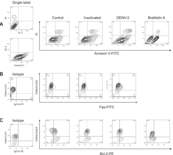
was inducing apoptosis in human monocytes, we searched for an early marker of cell death, the display of phosphati-dylserine at the cell membrane, by labeling with annexin V and PI. The presence of annexin V without PI labeling is used to identify apoptotic and autophagic cells at early stages of cell death, when annexin V binding is exclusive. Double-labeling occurs at later stages of apoptosis and in subsequent necrotic processes. Brefeldin A, a lactone an-tibiotic produced by fungal organisms that induces apop-tosis, was added to cell cultures as positive control.
Two days after infection of monocytes with DENV-2, cells were recovered for flow cytometry analysis. Fig. 3A
shows contour plots representing annexin V and PI label-ing on monocytes incubated with cell culture medium, inactivated virus, infectious DENV-2 or brefeldin A. In-fected cells presented an increase in single-labeled an-nexin V+ cells as compared to cultures incubated with
inactivated virus or medium. No significant differences were observed in the frequency of cells that underwent apoptosis followed by necrosis (annexin-V+ PI+) when
inactivated was compared to infectious virus. Cells in-cubated only with inactivated virus displayed equivalent percentages as those incubated with medium, indicating that physiological culture conditions are permitting cells to die spontaneously or recovery procedures may damage some cells. Nevertheless, the virus enhanced the apoptot-ic process that was occurring in culture. Data from five different PBMC donors for annexin V+PI- cells are plotted
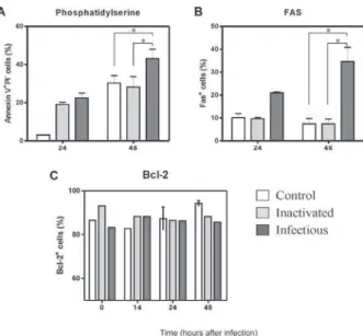
in Fig. 4A, showing the significant difference between cultures exposed to infectious DENV-2 and controls. Differences in early apoptotic markers between infected and uninfected cells were not significant until 48 h.
Fas receptor expression is detected on infected mono-cytes - The Fas cell surface receptor (CD95) can be
stim-Fig. 1: Dengue virus (DENV)- antigen (Ag) detection in human mono-cytes after in vitro DENV-2 infection:monocyte-enriched human pe-ripheral blood mononuclear cell (PBMC) were incubated for 2-5 days with infectious DENV-2 (strain 16681) and negative controls (heat-in-activated virus and culture medium). Cells were labeled with antibody to DENV-complex and anti-mouse IgG Alexa-488 or -647. DENV Ag was detected in infected monocytes by (A) confocal microscopy in two different fields or by (B) flow cytometry observed in contour plots. Time dependent DENV Ag detection was determined by FACS in three different PBMC donors (C) (average ± standard error is plotted).
Fig. 2:proinflammatory cytokine TNF-α and Il-10 production after mono -cyte infection by Dengue virus (DENV)-2: mono-cyte-enriched human peripheral blood mononuclear cell (PBMC) was infected with DENV-2 (strain 16681). Medium and inactivated DENV were used as controls and
at 1-5 day incubation time cell culture supernatants were tested for TNF-α
ulated by agonists or the natural ligand FasL, recruiting proteins with distinct functions such as cytosolic adaptor proteins that ultimately bind to the initiating caspase (cas-pase 8) and trigger apoptosis (Limjindaporn et al. 2007). DENV-2 infected monocytes were also analyzed in order to detect the Fas expression on cell surface. By the first day after infection, Fas+ cells were already more common
in DENV-2 exposed cells than in controls. At two days after infection, Fas+ cell frequencies were significantly
increased in the presence of the infectious virus, as de-tected in six different PBMC donors (Fig. 4B).
Bcl-2 oncoprotein expression is not altered in in-fected monocytes - The proto-oncogene Bcl-2 works as an important repressor of programmed cell death. We investigated whether DENV-2 infected monocytes, ap-parently more susceptible to death, might have altered Bcl-2 expression and therefore an altered degree of in-hibition of apoptosis. We detected Bcl-2 by labeling monocytes from 1-5 days after DENV-2 infection and analysed the cells by flow cytometry (data not shown). Bcl-2 expression remained unchanged during this
peri-od, although alternative treatment with brefeldin A was able to downregulate this protein, as can be observed in Fig. 3C (showing data from the first 48 h). In order to ascertain whether cells were infected or not with DENV, we performed double-labeling with antibodies directed against Bcl-2 and DENV. We detected double labeling in 39 ± 4% cells at 24 h and 50 ± 2% at 48 h, in contrast to negative double label in controls (< 1%), indicating that the presence of the DENV-2 does not interfere with Bcl-2 expression; most infected cells are still expressing this protein and the apoptosic mechanisms appear to act independently from Bcl-2 regulation.
DISCUSSION
Mononuclear phagocytes such as monocytes and den-dritic cells are the main DENV target cells, both in vitro and in vivo and their interaction with the virus has been shown to be relevant to the host response to infection (Ha- lstead & O’Rourke 1977, Wu et al. 2000, Neves-Souza et al. 2005). After signaling pathway activation by viruses, these cells have upregulated surface activation molecules
and begin producing cytokines and chemokines. Modula-tion of the innate and adaptative immunity interface oc-curs as a result (Frankenberger et al. 1996, Bosch et al. 2002, Strauss-Ayali et al. 2007), likely influencing the protection and/or pathology onset during infection.
DENV replicates in human primary peripheral mono-cytes (Halstead & O’Rourke 1977, Chen & Wang 2002). We have previously infected human monocyte cultures with DENV-2 Asian strain 16681 (Sydow et al. 2000) and other DENV strains (Neves-Souza et al. 2005). Here we show a time-dependent detection of DENV Ags starting as early as the first day after infection, with a detection peak at 48 h and a subsequent decay. These results re-semble other reports using this virus strain in dendritic cells, although peak virus production may be related not only to the strain type but also with the inoculum concen-tration (Cologna & Rico-Hesse 2003). Intracellular virus
detection was related to TNF-α extracellular production, with a maximum production at the second day. TNF-α
is probably produced as a result of virus replication and remains in culture, being detected at significant levels at five days, by which point intracellular virus is found at low levels. It is likely that non-infected monocytes are
activated and become important targets for TNF-α pro -duction at the later stage of infection.
Programmed cell death is an essential regulatory process in the immunological system. During immune responses to viral infections, apoptosis may occur as a direct pathogenic mechanism of viral escape, allowing apoptotic bodies containing virus to disseminate
infec-Fig. 4: Dengue virus (DENV)-2 infection enhanced apoptosis of hu-man monocytes as observed by Fas and phosphatidilserine detection but no Bcl-2 change: monocyte-enriched human peripheral blood mononuclear cell (PBMC) was incubated for 14-48 h with infectious DENV-2 (strain 16681) or negative control (heat-inactivated virus or culture medium). Cells were labeled with (A) annexin V- FITC to detect early apoptotic cells and with propidium iodide (PI) to detect necrosis (n = 2 at 24 h; n = 6 at 48 h) and (B) anti-Fas-FITC (n = 1 at 24 h; n = 5 at 48 h) or (C) Bcl-2 PE (n = 4 all time points). Monocyte gated cells were analyzed. Asterisks means p < 0.05 in Wilcoxon test.
tion to neighbor cells. Alternatively, apoptosis may be used as an immune mechanism to induce opposed ef-fects, becoming deleterious to viral progenies and re- presenting an appropriate response from host to limit viral replication (Servet-Delprat et al. 2000). Apoptosis can be either Ag-dependent, initiated by death recep-tor signaling and/or activating intracellular pathways, or it may occur by negligence when Ag-specific recep-tors are not stimulated. When the cellular microenvi-ronment is depleted of survival factors, another type of programmed cell death occurs, a phenomenon known as authophagy (Lemasters 2005, Feig & Peter 2007). Moreover, modulation of programmed cell death plays an important role in several pathologies related to viral infections such as AIDS and measles (Oyaizu & Pahwa 1995, Servet-Delprat et al. 2000). DENV replication seems to be essential to induce both phagocytosis and apoptosis in human monocytes, as reported by Espina et al. (2003). However, these authors showed no quantita-tive evidence about apoptotic events. Considering that monocytes are among the main target cells for DENV, we investigated markers of programmed cell death dur-ing human primary monocyte infection, as well as the production of related cytokines.
During phagocytosis, the virus-cell interaction may lead to production of cytoplasmatic lysosomal compo-nents that can result in deleterious effects for both the virus and host cells. Labeling cells with annexin V, which binds to the exposed phosphatidylserine found on apoptotic cells and the simultaneous exclusion with propidium iodine evaluated by flow cytometry allowed us to quantify early apoptosis after two days of DENV infection in monocytes. This observation matches with
the peak production of TNF-α, which is a proinflamma -tory cytokine that plays an important role in the induc-tion of apoptosis. We suggest that the maturainduc-tion process and the cytokine production by these cells are activation pathways involved in apoptotic processes. We also ob-served that some monocytes cultured without infectious virus die, although at lower rates. It is possible that they are undergoing cell death processes by mechanisms such as authophagy if survival factors are not present at suf-ficient concentrations in cultures (Yu et al. 2006).
Among TNF family receptor series, the Fas receptor is related to the pathenogenesis of viral infection such as measles and hepatitis B (Servet-Delprat et al. 2000, Wang et al. 2004). Dendritic cells infected by measles virus present upregulated expression of Fas, becoming more susceptible to apoptosis mediated by the Fas/FasL system. Also, a protein from the DENV capsid induces apoptosis in HepG2 cells in a Fas-dependent fashion (Limjindaporn et al. 2007). Fas signaling induces IL-8,
TNF-α and IL-1β production in mononuclear phago -cytes, modulating pro-inflammatory responses by a cas- pase-dependent pathway (Park et al. 2003).
been detected in patients with dengue fever and often have been found to be associated with increased severity (Green et al. 1999, Azeredo et al. 2001).
We demonstrated here for the first time that human monocytes infected in vitro by DENV have upregulated Fas expression concomitant with the viral peak, indi-cating that programmed cell death induced by DENV in our study model may be influenced by this extrinsic apoptotic pathway. It is possible that this process leads to extracellular release of virus, facilitating neighbor cell infection, in addition to inducing simultaneously monocyte maturation. Fas and DENV Ag double cell labeling in DENV-2 infected cultures shows that Fas ex-pression may occur in DENV-Ag positive and negative
cells. DENV-Ag is detected in Fas-negative cells (data not shown). Although our data indicate that apoptosis is induced in monocytes during Fas expression, we
can-not exclude the possibility that TNF-α/TNFR receptor,
cytokines and/or other mechanisms are participating in apoptotic processes mediated by DENV. Azeredo at al.
(2001) reported elevated circulating TNF-α and TNF sol -uble receptors in patients with acute dengue fever. The cytoplasmatic expression of the proto-oncogene Bcl-2 plays a role in increasing cell survival and has contrary effects to Fas during apoptosis (Boudet et al. 1996, Mo- lica et al. 1996). However, no significant changes were detected with regard to Bcl-2 expression in monocytes infected with DENV-2, in contrast to what we observed
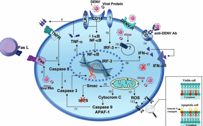
Fig. 5:hypothesis for signaling pathways involved in cell death and cellular activation during monocyte infection by dengue virus (DENV). After DENV inoculation by Aedes aegypti mosquito into the human host skin, the virus would infect one of the main target cells - circulating monocytes - through their co-receptors such as TLR-2 (CD14-TLR-2) or by other not yet known cellular receptor (1). The recognition and/or viral endocytosis mediated by these receptors would lead to the activation of transcription factors in charged of the cytokine induction and other
mediators involved in viral clearance and in the survival/death from the infected cell. Among transcription factors, the IκB-NF-κB complex is dissociated, permitting the NF-κB translocation to the nucleus (2) and, consequently, inducing proinflammatory cytokine expression such as TNF-α (3). TNF-α interaction with its transmembrane receptors, mainly TNFR1, induces recruitment from another series of cytoplasmatic
signaling proteins (4) that lead initially to caspase-8 activation (5). Within a sequence of well orientated events, the caspase-8 induces effector caspase-3 activation (6) that is involved in inhibition and/or activation of cytoplasmatic constituents in charged of maintaining cell survival (7). During infection it would be also possible to observe the increase in the Fas receptor expression (8), which interacts with its ligand - FasL, present in neighbor cells, triggers the apoptosis signaling cascade similarly to the TNF/TNFR pathway. The virus infection would also activate the intrinsic cell death pathway, in which mitochondria has a critical role (9). Indeed, several reports have been demonstrating that an alteration in mitochondrial membrane permeability promotes the organelle dysfunction (10) and secretion of apoptotic proteins that activate caspase-9, which in a sequence, activates caspase-3 (11). The mitochondrial membrane potential and permeability alterations lead to free radical secretion (12) that, e. g., would contribute to the cell death processes by inducing phosphatidylserine exposition on external membrane from the apoptotic cell, facilitating in this way, their recognition by phagocytic cells (13). The cell death activation may be associated with monocyte activation
events, such as the increase from CD16 (FCγIII), characteristic of monocytes with high proinflammatory capacity (14). On the other hand, the infection by DENV would induce the transcription factor IRF-3 (15), involved in the increase of anti-viral cytokines such as IFN-α (16). The
in CD8 positive T-lymphocytes from dengue patients, which presented low levels of Bcl-2 (Azeredo et al. 2006). The present data suggest that other anti-apoptotic proteins, such as Bcl-xL, might be negatively regulated during infection in monocytes.
According to our data and other relevant studies, we present here a model for DENV infection in monocytes in which complex interactions between the virus and host cell occur, leading to the activation of transcription factors, cytokines and enzymatic factors. These interac-tions may induce not only pathologic proinflammatory responses that influence the severity and progression of the disease, but also virus clearance and apoptotic path-ways that could be controlling infection by anti-viral mechanisms [(Chen et al. 1999, Diamond et al. 2000, Bielefeldt-Ohmann et al. 2001, Grandvaux et al. 2002, Navarro-Sanchez et al. 2003, Neves-Souza et al. 2005, Azeredo et al. 2006, 2009, Seth et al. 2006, Chareon-sirisuthigul et al. 2007, Reis et al. 2007, Durbin et al. 2008, Kou et al. 2008 (see details in Fig. 5)]. The present study may, therefore, contribute to a better understand-ing of the cell activation mechanisms triggered durunderstand-ing DENV infections occurring in human target cells. Fur-ther investigations will be required in order to define in detail the apoptotic pathways involved in dengue fever and to determine whether or not there is a potential be-nefic role from apoptosis in the resolution of infection.
ACKNOWLEDGMENTS
To Bernardo M Pascarelli, by helping in confocal image analysis and uptake at the Confocal Microscopy Technological Platform, Fiocruz, and to Luzia Maria de Oliveira Pinto, for suggestions and encouragement. Amanda Torrentes de Car-valho was a fellowship from CNPq.
REFERENCES
Avirutnan P, Malasit P, Seliger B, Bhakdi S, Husmann M 1998. Den-gue virus infection of human endothelial cells leads to chemokine production, complement activation and apoptosis. J Immunol 161: 6338-6346.
Azeredo EL, Neves-Souza PC, Alvarenga AR, Reis SRNI, Torrentes-Carvalho A, Zagne SMO, Nogueira RMR, Oliveira-Pinto LM, Kubelka CF 2009. TLR-2, TLR-4 and CD16 are upregulated in human monocytes after dengue virus in vitro infection and are associated with mild dengue in patients, while HLA-DR is down-regulated during severe dengue fever. Immunology, in press.
Azeredo EL, Zagne SMO, Alvarenga AR, Nogueira RMR, Kubelka CF, Oliveira-Pinto LM 2006. Activated peripheral lymphocytes with increased expression of cell adhesion molecules and cyto-toxic markers are associated with dengue fever disease. Mem Inst Oswaldo Cruz 101: 437-449.
Azeredo EL, Zagne SM, Santiago MA, Gouvea AS, Santana AA, Neves-Souza PC, Nogueira RM, Miagostovich MP, Kubelka CF 2001. Characterisation of lymphocyte response and cy-tokine patterns in patients with dengue fever. Immunobiology 204: 494-507.
Bethell DB, Flobbe K, Cao XT, Day NP, Pham TP, Buurman WA, Cardosa MJ, White NJ, Kwiatkowski D 1998. Pathophysiologic and prognostic role of cytokines in dengue hemorrhagic fever.
J Infect Dis 177: 778-782.
Bielefeldt-Ohmann H, Meyer M, Fitzpatrick DR, Mackenzie JS 2001. Dengue virus binding to human leukocyte cell lines:
re-ceptor usage differs between cell types and virus strains. Virus Res 73: 81-89.
Bosch I, Xhaja K, Estevez L, Raines G, Melichar H, Warke RV, Fournier MV, Ennis FA, Rothman AL 2002. Increased produc-tion of interleukin-8 in primary human monocytes and in human epithelial and endothelial cell lines after dengue virus challenge.
J Virol 76: 5588-5597.
Boudet F, Lecoeur H, Gougeon ML 1996. Apoptosis associated with
ex vivo down-regulation of Bcl-2 and up-regulation of Fas in potential cytotoxic CD8+ T lymphocytes during HIV infection.
J Immunol 156: 2282-2293.
Braga EL, Moura P, Pinto LM, Ignacio SR, Oliveira MJ, Cordeiro MT, Kubelka CF 2001. Detection of circulant tumor necrosis factor-alpha, soluble tumor necrosis factor p75 and interferon-gamma in Brazilian patients with dengue fever and dengue hemorrhagic fever. Mem Inst Oswaldo Cruz 96: 229-232.
Bruchhaus I, Roeder T, Rennenberg A, Heussler VT 2007. Protozoan parasites: programmed cell death as a mechanism of parasitism.
Trends Parasitol 23: 376-383.
Bzowska M, Guzik K, Barczyk K, Ernst M, Flad HD, Pryjma J 2002. Increased IL-10 production during spontaneous apoptosis of monocytes. Eur J Immunol 32: 2011-2020.
Chareonsirisuthigul T, Kalayanarooj S, Ubol S 2007. Dengue virus (DENV) antibody-dependent enhancement of infection upregu-lates the production of anti-inflammatory cytokines, but sup-presses anti-DENV free radical and pro-inflammatory cytokine production in THP-1 cells. J Gen Virol 88: 365-375.
Chen YC, Wang SY 2002. Activation of terminally differentiated human monocytes/macrophages by dengue virus: productive infection, hi-erarchical production of innate cytokines and chemokines and the synergistic effect of lipopolysaccharide. J Virol 76: 9877-9887.
Chen YC, Wang SY, King CC 1999. Bacterial lipopolysaccharide inhibits dengue virus infection of primary human monocytes/ macrophages by blockade of virus entry via a CD14-dependent mechanism. J Virol 73: 2650-2657.
Cologna R, Rico-Hesse R 2003. American genotype structures de-crease dengue virus output from human monocytes and dendritic cells. J Virol 77: 3929-3938.
Dejean LM, Martinez-Caballero S, Manon S, Kinnally KW 2006. Reg-ulation of the mitochondrial apoptosis-induced channel, MAC, by BCL-2 family proteins. Biochim Biophys Acta 1762: 191-201.
Diamond MS, Roberts TG, Edgil D, Lu B, Ernst J, Harris E 2000. Modulation of dengue virus infection in human cells by alpha, beta and gamma interferons. J Virol 74: 4957-4966.
Durbin AP, Vargas MJ, Wanionek K, Hammond SN, Gordon A, Rocha C, Balmaseda A, Harris E 2008. Phenotyping of peripheral blood mononuclear cells during acute dengue illness demonstrates in-fection and increased activation of monocytes in severe cases compared to classic dengue fever. Virology 376: 429-435.
Espina LM, Valero NJ, Hernandez JM, Mosquera JA 2003. Increased apoptosis and expression of tumor necrosis factor-alpha caused by infection of cultured human monocytes with dengue virus. Am J Trop Med Hyg 68: 48-53.
Feig C, Peter ME 2007. How apoptosis got the immune system in shape. Eur J Immunol 37 (Suppl 1): S61-70.
Fink J, Gu F, Vasudevan SG 2006. Role of T cells, cytokines and an-tibody in dengue fever and dengue haemorrhagic fever. Rev Med Virol 16: 263-275.
monocyte subpopulations: a polymerase chain reaction analysis.
Blood 87: 373-377.
Gibbons RV, Vaughn DW 2002. Dengue: an escalating problem. BMJ 324: 1563-1566.
Grandvaux N, Ten Oever BR, Servant MJ, Hiscott J 2002. The in-terferon antiviral response: from viral invasion to evasion. Curr Opin Infect Dis 15: 259-267.
Green S, Vaughn DW, Kalayanarooj S, Nimmannitya S, Suntayakorn S, Nisalak A, Rothman AL, Ennis FA 1999. Elevated plasma in-terleukin-10 levels in acute dengue correlate with disease sever-ity. J Med Virol 59: 329-334.
Gross A 2001. BCL-2 proteins: regulators of the mitochondrial apop-totic program. IUBMB Life 52: 231-236.
Gubler DJ 2002. The global emergence/resurgence of arboviral dis-eases as public health problems. Arch Med Res 33: 330-342.
Guo H, Tittle TV, Allen H, Maziarz RT 1998. Brefeldin A-mediated apoptosis requires the activation of caspases and is inhibited by Bcl-2. Exp Cell Res 245: 57-68.
Gupta S 2003. Molecular signaling in death receptor and mitochon-drial pathways of apoptosis - Review. Int J Oncol 22: 15-20.
Halstead SB, O’Rourke EJ 1977. Dengue viruses and mononuclear phagocytes. I. Infection enhancement by non-neutralizing anti-body. J Exp Med 146: 201-217.
Jessie K, Fong MY, Devi S, Lam SK, Wong KT 2004. Localization of dengue virus in naturally infected human tissues, by immunohis-tochemistry and in situ hybridization. J Infect Dis 189: 1411-1418.
Karin M, Lin A 2002. NF-kappaB at the crossroads of life and death.
Nat Immunol 3: 221-227.
Kou Z, Quinn M, Chen H, Rodrigo WW, Rose RC, Schlesinger JJ, Jin X 2008. Monocytes, but not T or B cells, are the principal target cells for dengue virus (DV) infection among human peripheral blood mononuclear cells. J Med Virol 80: 134-146.
Lemasters JJ 2005. Dying a thousand deaths: redundant pathways from different organelles to apoptosis and necrosis. Gastroenter-ology 129: 351-360.
Limjindaporn T, Netsawang J, Noisakran S, Thiemmeca S, Wongwi-wat W, Sudsaward S, Avirutnan P, Puttikhunt C, Kasinrerk W, Sriburi R, Sittisombut N, Yenchitsomanus PT, Malasit P 2007. Sensitization to Fas-mediated apoptosis by dengue virus capsid protein. Biochem Biophys Res Commun 362: 334-339.
Loeffler M, Kroemer G 2000. The mitochondrion in cell death con-trol: certainties and incognita. Exp Cell Res 256: 19-26.
Merchant SH, Gonchoroff NJ, Hutchison RE 2001. Apoptotic index by Annexin V flow cytometry: adjunct to morphologic and cytogenet-ic diagnosis of myelodysplastcytogenet-ic syndromes. Cytometry 46: 28-32.
Miagostovich MP, Nogueira RM, Cavalcanti SM, Marzochi KB, Schatzmayr HG 1993. Dengue epidemic in the state of Rio de Janeiro, Brazil: virological and epidemiological aspects. Rev Inst Med Trop Sao Paulo 35: 149-154.
Molica S, Mannella A, Dattilo A, Levato D, Iuliano F, Peta A, Con-sarino C, Magro S 1996. Differential expression of BCL-2 onco-protein and Fas antigen on normal peripheral blood and leukemic bone marrow cells. A flow cytometric analysis. Haematologica 81: 302-309.
Navarro-Sanchez E, Altmeyer R, Amara A, Schwartz O, Fieschi F, Virelizier JL, Arenzana-Seisdedos F, Despres P 2003. Dendritic-cell-specific ICAM3-grabbing non-integrin is essential for the
productive infection of human dendritic cells by mosquito-cell-derived dengue viruses. EMBO Rep 4: 723-728.
Neves-Souza PC, Azeredo EL, Zagne SM, Valls-de-Souza R, Reis SR, Cerqueira DI, Nogueira RM, Kubelka CF 2005. Inducible nitric ox-ide synthase (iNOS) expression in monocytes during acute dengue fever in patients and during in vitro infection. BMC Infect Dis 5: 64.
Nogueira RMR, de Araújo JMG, Schatzmayr HG 2007. Dengue virus-es in Brazil, 1986-2006. Rev Panam Salud Publica22: 358-363.
Oyaizu N, Pahwa S 1995. Role of apoptosis in HIV disease pathogen-esis. J Clin Immunol 15: 217-231.
Park DR, Thomsen AR, Frevert CW, Pham U, Skerrett SJ, Kiener PA, Liles WC (2003). Fas (CD95) induces proinflammatory cy-tokine responses by human monocytes and monocyte-derived-macrophages. J Immunol 170: 6209-6216.
Reis SR, Sampaio AL, Henriques MG, Gandini M, Azeredo EL, Kubelka CF 2007. An in vitro model for dengue virus infec-tion that exhibits human monocyte infecinfec-tion, multiple cytokine production and dexamethasone immunomodulation. Mem Inst Oswaldo Cruz 102: 983-990.
Schmidt T, Karsunky H, Frass B, Baum W, Denzel A, Moroy T 2000. A novel protein (Fbf-1) that binds to CD95/APO-1/FAS and shows sequence similarity to trichohyalin and plectin. Biochim Biophys Acta 1493: 249-254.
Servet-Delprat C, Vidalain PO, Azocar O, Le Deist F, Fischer A, Rabour-din-Combe C 2000. Consequences of Fas-mediated human dendritic cell apoptosis induced by measles virus. J Virol 74: 4387-4393.
Seth RB, Sun L, Chen ZJ 2006. Antiviral innate immunity pathways.
Cell Res 16: 141-147.
Shao RG, Shimizu T, Pommier Y 1996. Brefeldin A is a potent in-ducer of apoptosis in human cancer cells independently of p53.
Exp Cell Res 227: 190-196.
Strauss-Ayali D, Conrad SM, Mosser DM 2007. Monocyte subpopula-tions and their differentiation patterns during infection. J Leukoc Biol 82: 244-252.
Sydow FF, Santiago MA, Neves-Souza PC, Cerqueira DI, Gouvea AS, Lavatori MF, Bertho AL, Kubelka CF 2000. Comparison of dengue infection in human mononuclear leukocytes with mos-quito C6/36 and mammalian Vero cells using f low cytometry to detect virus antigen. Mem Inst Oswaldo Cruz 95: 483-489.
Vermes I, Haanen C, Steffens-Nakken H, Reutelingsperger C 1995. A novel assay for apoptosis. Flow cytometric detection of phos-phatidylserine expression on early apoptotic cells using fluores-cein labelled Annexin V. J Immunol Methods 184: 39-51.
Wang WH, Gregori G, Hullinger RL, Andrisani OM 2004. Sus-tained activation of p38 mitogen-activated protein kinase and c-Jun N-terminal kinase pathways by hepatitis B virus X pro-tein mediates apoptosis via induction of Fas/FasL and tumor ne-crosis factor (TNF) receptor 1/TNF-alpha expression. Mol Cell Biol 24: 10352-10365.
Wu SJ, Grouard-Vogel G, Sun W, Mascola JR, Brachtel E, Putvatana R, Louder MK, Filgueira L, Marovich MA, Wong HK, Blauvelt A, Murphy GS, Robb ML, Innes BL, Birx DL, Hayes CG, Fran-kel SS 2000. Human skin Langerhans cells are targets of dengue virus infection. Nat Med 6: 816-820.