In vivo
evaluation of the antimutagenic and antigenotoxic effects of
b
-glucan
extracted from
Saccharomyces cerevisiae
in acute treatment with multiple
doses
Rodrigo Juliano Oliveira
1,2,3, Maria José Sparça Salles
4, Ariane Fernanda da Silva
4,
Tatiane Yumi Nakamura Kanno
4, Ana Carolina dos Santos Lourenço
4, Véssia da Silva Leite
4,
Hevenilton José Matiazi
5, João Renato Pesarini
1,6, Lúcia Regina Ribeiro
6and Mário Sérgio Mantovani
41
Centro de Estudos em Célula Tronco, Terapia Celular e Genética Toxicológica, Núcleo de Hospital
Universitário, Universidade Federal de Mato Grosso do Sul, Campo Grande, MS, Brazil.
2
Programa de Pós-graduação em Saúde em Desenvolvimento na Região Centro-Oeste, Faculdade de
Medicina “Dr. Hélio Mandetta”, Universidade Federal de Mato Grosso do Sul, Campo Grande, MS, Brazil.
3
Programa de Mestrado em Farmácia, Centro de Ciências Biológicas e da Saúde,
Universidade Federal de Mato Grosso do Sul, Campo Grande, MS, Brazil.
4
Departamento de Biologia Geral, Universidade Estadual de Londrina, Londrina, PR, Brazil.
5
Laboratório de Tecnologia em Alimentos e Medicamentos, Universidade Estadual de Londrina,
Londrina, PR, Brazil.
6
Programa de Pós-graduação em Biologia Celular e Molecular, Instituto de Biociências,
Universidade Estadual Paulista “Julio de Mesquita Filho”, Rio Claro, SP, Brazil.
Abstract
Ample evidence suggests that cancer is triggered by mutagenic damage and diets or supplements capable of reduc-ing such incidences can be related to the prevention of neoplasy development or to an improvement in life quality of patients who undergo chemotherapy. This research aimed to evaluate the antimutagenic and antigenotoxic activity ofb-glucan. We set up 8 experimental groups: control (Group 1), cyclophosphamide (Group 2), Groups 3-5 to assess the effect ofb-glucan administration, and Groups 6-8 to evaluate the association between cyclophosphamide and
b-glucan. The intraperitonial concentrations ofb-glucan used were 100, 150 and 200 mg/kg. Micronucleus and
comet assays showed that within the first week of treatmentb-glucan presented a damage reduction rate between 100-62.04% and 94.34-59.52% for mutagenic and genotoxic damages, respectively. This activity decreased as the treatment was extended. During the sixth week of treatment antimutagenicity rates were reduced to 59.51-39.83% and antigenotoxicity was not effective. This leads to the conclusion that the efficacy ofb-glucan in preventing DNA damage is limited when treatment is extended, and that its use as a chemotherapeutic adjuvant need to be better clarified.
Keywords:b-glucan, cyclophosphamide, antimutagenicity, antigenotoxicity, mice.
Received: September 6, 2012; Accepted: April 27, 2013.
Introduction
Experimental studies on the interaction between diet, nutrition and cancer can be developed in animal models and/or cell culture, and most of the epidemiological and trial studies on animal models and cell culture focus on the observation, control and analysis of cancer development steps. Thus, trial studies can provide support to
epidemio-logical findings, as testing is done under controlled condi-tions. Furthermore, they can provide strong evidence on the relationship of diet, nutrition and cancer development and/or treatment (Torrinhaset al., 2006). Correlations be-tween diet, cancer prevention andb-glucan are described in two major reviews (Mantovaniet al., 2008; Kumaret al., 2012) which indicate important health implications for hu-mans.
Basically, in studies involving nutrition and cancer, the induction of tumor development in animals can be done by: (I) using different carcinogenic agents: mainly chemi-cal ones or, incertain cases, by irradiation exposure or
par-www.sbg.org.br
Send correspondence to Rodrigo Juliano Oliveira. Faculdade de Medicina, Universidade Federal do Mato Grosso do Sul, Cidade Universitária, S/N. 79070-900 Campo Grande, MS, Brazil. E-mail: [email protected].
ticular strains of viruses; and (II) through tumor cell trans-plants which are able to grow and turn into tumors when implanted into the host (Torrinhaset al., 2006).
In general, carcinogenicity induced by chemical agents, radiation and viruses is based on the concept of car-cinogenicity via multiple steps. This concept implies that cancer development is a process that occurs in sequential steps, named: initiation, promotion, progression and mani-festation. Carcinogenesis is an experimentally well-cate-gorized process (Pitot and Dragan, 1991; Pitot, 1993), and it also probable occurs in human beings (Pinkerton and Dubé, 1991; Fearon and Jones, 1992; Sugimura, 1992).
Initiation is associated with a DNA alteration,e.g. in-duced by exposure to a carcinogenic agent. This alteration is considered a genotoxic activity (Camargoet al., 1994). Consequently, diets or dietetic compounds that can dimin-ish the risk of genotoxic or mutagenic effects can be associ-ated indirectly to cancer prevention by decreasing the cancer initiation risk of cells.
b-glucan, a sugar polymer, is currently widely studied for the prevention of DNA damage related to cancer devel-opment. This sugar can be extracted from the cell wall of certain cereals, fungi and yeast (Cisneros et al., 1996; Zimmerman et al., 1998; Turnbull et al., 1999; Masihi, 2000).b-glucan of yeast shows is composed of D-glucose molecules linked in theb-(1®3) position and containing lateral chainsb-(1®6) (Di Luzioet al., 1979) with great potential for the biotechnological production of drugs.
The chemopreventive efficacy of this polysaccharide duringin vitrotrials on the prevention of mutagenic dam-age to the cell linedam-ages V79, CHO-k1, CHO-xrs5, HTC and to human lymphocytes was assessed in several studies (Slamenová et al., 2003; Angeli et al., 2006, 2009a,b; Oliveiraet al., 2006, 2007). Other reports are on the pre-vention against damage induced by doxorubicin, cisplatin and cyclophosphamide in vivo (Chorvatovicová et al., 1996, 1998; Tohamyet al., 2003; Linet al., 2004; Oliveira
et al., 2009a).
Notwithstanding such reports concerning chemopre-ventive effects of this molecule in acute or subchronic tri-als, there is still the need of studies to show the usefulness ofb-glucan in chronic and acute treatments with multiple doses. Thus, the aim of this study was to investigate the antimutagenic and antigenotoxic activity ofb-glucan ex-tracted from Saccharomyces cerevisiae in damages in-duced by cyclophosphamide in acute treatments with multiple doses.
Material and Methods
DNA damage-inducing agent
For inducing DNA damage we used the alkylating agent cyclophosphamide (Fosfaseron®), diluted in Ca+2 -and Mg+2-free, phosphate-buffered saline (PBS) (NaCl
137 mM; KCl 2.7 mM; Na2HPO4 3.9 mM; KH2PO4
1.8 mM, pH 7.4) at a final concentration of 50 mg/kg of body weight (b.w.). This was administrated intraperito-nially (i.p.).
Extraction and preparation ofb-glucan
Theb-glucan molecules tested in this study were ex-tracted fromSaccharomyces cerevisiae. Theseb-glucans have their main chains composed of D-glucose molecules linked by theb- type (1®3) with lateral branches in which there are links ofb- type (1®6). They were extracted by autolysis ofSaccharomyces cerevisiae. The cell wall was separated by means of centrifugation at 6500g for 8 min followed by heat treatment (70 °C for 5 h) in NaOH (10%), washed and centrifuged three times and finally dried in an incubator at 40 °C. Nuclear magnetic resonance (NMR) analysis showed the presence of (1.3 and 1.6)b-D-glucan with purity of 85%. Subsequently, the glucan was solu-bilized using DMSO (dimethyl sulfoxide) and urea 8 M in the proportion of 100 mL (DMSO): 60 g (Urea). In a warm water bath, 100 mL of DMSO with 10 mL of concentrated sulfuric acid was added, and the mixture was stirred for 4 h at 100 °C. Dialysis was then performed with approximately 100 L of ultrapure water (Milli-Q) and then concentrated in a rotary evaporator at 40 °C followed by lyophilization.
Solutions ofb-glucan were prepared in sterile Ca+2 -and Mg+2-free PBS (NaCl 137 mM; KCl 2.7 mM; Na2HPO4
3.9 mM; KH2PO41.8 mM, pH 7.4) at concentrations of
100, 150 and 200 mg/kg (b.w.). The doses were determined in pilot trials according to indications by Tohamyet al.
(2003) and Oliveiraet al.(2009a) (data not shown).
Animals
Experimental design and analysis methods
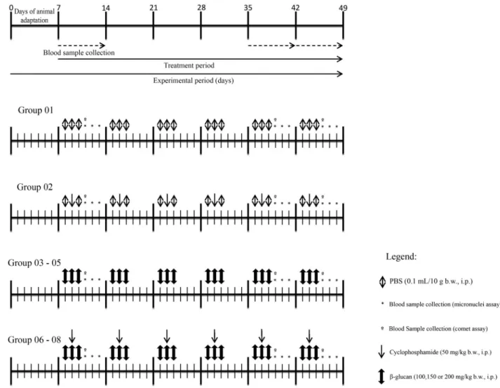
The animals were randomly divided into eight experi-mental groups (n = 6) as follows: animals from the control group (Group 01) received sterile Ca+2- and Mg+2-free PBS (NaCl 137 mM; KCl 2.7 mM; Na2HPO43.9 mM; KH2PO4
1.8 mM, pH 7.4) at a volume of 0.1 mL/10 g (b.w.; i.p.) dur-ing 3 consecutive days. Animals belongdur-ing to the cyclo-phosphamide group (Group 02) received this chemother-apy agent at a concentration of 50 mg/kg (b.w.; i.p.) on the second day of the treatment and PBS during the next couple of days. For the mutagenicity evaluation, Groups 03-05 re-ceived b-glucan (i.p.) for three consecutive days, at the doses of 100, 150 and 200 mg/kg (b.w.), respectively. For the antimutagenicity evaluation, Groups 06-08 received
b-glucan (i.p.) for three consecutive days at the doses men-tioned before and one dose of cyclophosphamide on the second day of treatment (Figure 1).
Peripheral blood was sampled from the experimental groups by puncturing the tail vein at three different time points to evaluate the mutagenic and/or antimutagenic po-tential by means of micronucleus testing in peripheral
blood. Blood sample designated as time points T0 and T1 were always taken before the administration of treatment, within intervals of 24 hours. Time point T2 corresponds to a blood sample taken at 48 hours after the last administra-tion of cyclophosphamide. At this time point, an amount of 30mL was also collected to evaluate the genotoxicity and antigenotoxicity using the comet test.
Animals were treated during three consecutive days per week, for six consecutive weeks. Blood samples were taken in the first, fifth and sixth week.
Micronucleus assay in peripheral blood
The micronucleus assay was originally described by Hayashi et al.(1990), with certain changes proposed by Oliveiraet al.(2009a). The slides were warmed to 70 °C and covered with a layer of 20mL of Acridine Orange in an aqueous solution (1.0 mg/mL). After the preparation of the slides, a drop of peripheral blood was deposited on the slide and covered by a coverslip. Analyses were performed with a fluorescence microscope (Bioval®) at 40X magnifica-tion, with a 420-490 nm excitation and a 520 nm barrier
ter. A total of 2,000 cells were analyzed per animal, and the statistical analysis was performed using an unpaired Stu-dent’st-test (p < 0.05).
Comet assay
The alkaline Comet assay proposed by Singhet al.
(1988) was performed with modifications and under indi-rect light. Briefly, 20mL of a blood cell suspension was em-bedded into 120mL of 0.5% low melting point agarose and layered onto a pre-coated slide with a thin layer of normal melting point agarose. The slide was covered with a glass coverslip and cooled to 4 °C for 20 min and immersed in lysis solution for 1 h. Next, the slides were transferred to an electrophoresis chamber containing a pH > 13.0 buffer (300 mM NaOH and 1 mM EDTA, prepared from a stock solution of 10 N NaOH and 200 mM EDTA, pH 10.0) at 4 °C for 20 min to denature DNA. Electrophoresis was run at 25 V and 300 mA (1.25 V/cm). Subsequently, the slides were neutralized with pH 7.5 buffer (0.4 M Tris-HCl) with three changes of 5 min, air-dried, fixed in absolute ethanol for 10 min and stored for later scoring.
For coloration, the slides were stained with 100mL of ethidium bromide (20mg/mL) and evaluated using a fluo-rescence microscope (Bioval®) at 40X, using a 420-490 nm excitation and a 520 nm barrier filter.
Three independent repetitions were done, and 100 cells were scored per treatment, classifying the comets as follows: (class 0) cells without a comet tail; (class 1) cells with a tail smaller than the diameter of the nucleus; (class 2) cells with a tail 1 to 2 times the diameter of the
nu-cleus; (class 3) cells with a tail greater than 2 times the diameter of the nucleus. Apoptotic cells that showed a com-pletely fragmented nucleus were not counted (Kobayashiet al., 1995).
The total score was calculated by adding the resulting values after the multiplication of the total cells observed in each class of lesion by the number of the class. Statistical analysis was performed using an unpairedStudent’st-test (p < 0.05).
Calculation of the damage reduction percentage
The cyclophosphamide damage reduction percentage (DR%) byb-glucan administration was calculated as the mean of Group 2 minus the mean of an associated group (Groups 6-8) divided by the mean of Group 2 minus the mean of Group l. The result was multiplied by 100 and ex-pressed as DR% (Manoharan and Banerjee, 1985; Waters
et al., 1990).
Results
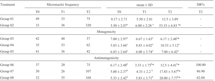
Table 1 shows the frequency, average, standard devi-ation and DR% related to the comet assay in peripheral blood on the first week of the trial. The baseline micro-nuclei frequency for all animals did not show a statistically significant difference at the beginning of the experiment.
During mutagenicity assessment, no such activity was confirmed for b-glucan. The animals of Group 3, which received only b-glucan in lower concentrations, showed a statistically significant reduction in the baseline micronuclei frequency at time T2.
Table 1- Frequency, mean, standard deviation and damage reduction percentage related to the micronucleus test in peripheral blood of mice during the first week of treatment.
Treatment Micronuclei frequency mean±SD DR%
T0 T1 T2 T0 T1 T2 T2
Group 01 49 33 75 8.17±2.71 5.50±2.81 12.5±3.89 -Group 02 33 36 320 5.50±2.07a 6.00±2.28a 53.33±6.83a*
-Mutagenicity
Group 03 42 40 37 7.00±2.97a 6.67±1.63a 6.17±2.48a* -Group 04 35 53 62 5.83±3.66a 8.83±4.02a 10.33±5.12a -Group 05 41 36 42 6.83±2.64a 6.00±3.74a 7.00±6.42a
-Antimutagenicity
Group 06 37 20 75 6.17±2.48b 3.33±1.75b* 12.5±4.41b* 100.00 Group 07 30 26 107 5.00±2.37b 4.33±2.2b 17.83±5.67b* 86.90
Group 08 32 35 168 5.33±2.42b 5.83±3.71b 28.00±7.77b* 62.04
Group 01 - control (PBS - 0.1 mL/10.0 g), Group 02 - cyclophosphamide (50 mg/kg), Group 03 -b-glucan (100 mg/kg), Group 04 -b-glucan (150 mg/kg), Group 05 -b-glucan (200 mg/kg), Group 06 -b-glucan (100 mg/kg) + cyclophosphamide (50 mg/kg), Group 07 -b-glucan (150 mg/kg) + cyclophosphamide (50 mg/kg), Group 08 -b-glucan (200 mg/kg) + cyclophosphamide (50 mg/kg).
Moments T0, T1 and T2: blood samples were taken within an interval of 24 hours, except for T2, which corresponds to 48 hours. DR% - Damage reduction percentage (unpaired Student’s t-test, p < 0.05).
a
In the study of antimutagenicity at time T2 there was a chemopreventive activity for the three doses tested. The DR%s were 100, 86.9 and 62.04% for the doses of 100, 150 and 200 mg/kg (b.w.), respectively. Again, there was a re-duction compared to the baseline micronuclei frequency. However, this occurred in Group 6, which was the group with the lowest supplemented dose ofb-glucan, at time T1, prior to the association of polysaccharide and cyclophos-phamide.
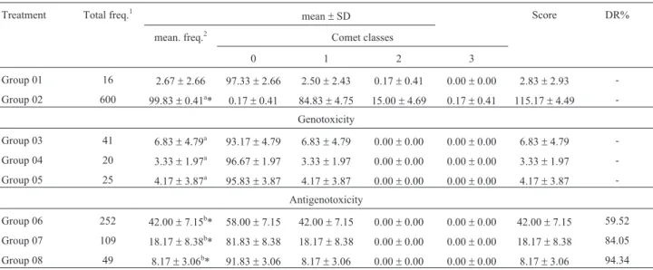
The comet assay results are shown in Table 2 indicat-ing that b-glucan has no genotoxic activity. When com-bined with cyclophosphamide it showed a statistically significant prevention of genotoxic damage caused by the alkylating agent used in the study. The DR%s were 59.52%, 84.05% and 94.34% for the doses of 100, 150 and 200 mg/kg (b.w.), respectively. After taking blood samples, the same animals were kept in the protocol of testing al-ready described and reevaluated in the fifth week of treat-ment.
The data shown in Table 3 correspond to the fifth week. These show that all groups in the time points T0 and T1 had no statistically significant differences with respect to the frequencies of micronuclei, except for Group 7 at T0. The animals in this group, which receivedb-glucan in the concentration of 150 mg/kg associated with cyclophos-phamide, showed a statistically significant reduction rela-tive to its control group. It is necessary to understand that the statistically significant reduction observed does not cor-relate to the treatment of the fifth week, but to the treatment
that had been done before this week, since the administra-tion of cyclophosphamide in the fifth week was done after the analysis. Thus, data on mutagenicity and/or antimu-tagenicity at this initial time points (T0 and T1) from the fifth week of analysis correlate to events accumulated from the past weeks of treatment. However, in Group 2 it appears that, upon assessing the mutagenicity, no damage was found as the averages did not differ statistically, suggesting that this prevention correlates with the baseline micro-nuclei frequency. In the protocol of antimutagenicity, at time point T2, there was a chemoprotective activity for all doses tested and the DR% ranged from 59.77% to 71.63%.
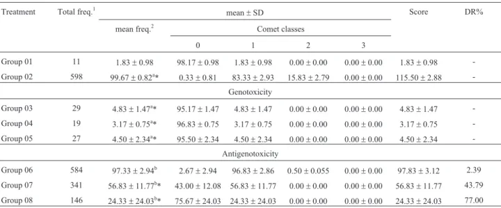
Table 4 shows the data related to the comet assay dur-ing the fifth week of treatment. Here we found that the ad-ministration ofb-glucan lead to a statistically significant increase in the genetic damage frequency. However, the variation found was lower, as the frequency in Group 1 (control group) was 1.83 ± 0.98, while the highest fre-quency was found in Group 3 (which corresponds to the lower dose ofb-glucan), 4.83±1.47. Allied to this increase in the frequency of injured cells in the lower dose of
b-glucan it was possible to see that this same dose showed no chemopreventive activity, as its DR% was only 2.39%. Yet, the other two doses tested showed DR%s of 43.79 and 77.0% for the doses of 150 and 200 mg/kg (b.w.), respec-tively.
During the sixth week of treatment it was possible to see that Group 1 and Groups 3-5, when in T0, T1 and T2, had no statistically significant frequencies of micronuclei.
Table 2- Total and mean frequency of damaged cells, average distribution between the classes of damage, average scoring and damage reduction per-centage related to tests for genotoxicity and antigenotoxicity in peripheral blood of mice during the first week of treatment.
Treatment Total freq.1 mean±SD Score DR%
mean. freq.2 Comet classes
0 1 2 3
Group 01 16 2.67±2.66 97.33±2.66 2.50±2.43 0.17±0.41 0.00±0.00 2.83±2.93 -Group 02 600 99.83±0.41a* 0.17±0.41 84.83±4.75 15.00±4.69 0.17±0.41 115.17±4.49
-Genotoxicity Group 03 41 6.83±4.79a 93.17
±4.79 6.83±4.79 0.00±0.00 0.00±0.00 6.83±4.79 -Group 04 20 3.33±1.97a 96.67±1.97 3.33±1.97 0.00±0.00 0.00±0.00 3.33±1.97
-Group 05 25 4.17±3.87a 95.83±3.87 4.17±3.87 0.00±0.00 0.00±0.00 4.17±3.87 -Antigenotoxicity
Group 06 252 42.00±7.15b* 58.00±7.15 42.00±7.15 0.00±0.00 0.00±0.00 42.00±7.15 59.52
Group 07 109 18.17±8.38b* 81.83±8.38 18.17±8.38 0.00±0.00 0.00±0.00 18.17±8.38 84.05 Group 08 49 8.17±3.06b* 91.83±3.06 8.17±3.06 0.00±0.00 0.00±0.00 8.17±3.06 94.34 Group 01 - control (PBS - 0.1 mL/10.0 g), Group 02 - cyclophosphamide (50 mg/kg), Group 03 -b-glucan (100 mg/kg), Group 04 -b-glucan (150 mg/kg), Group 05 -b-glucan (200 mg/kg), Group 06 -b-glucan (100 mg/kg) + cyclophosphamide (50 mg/kg), Group 07 -b-glucan (150 mg/kg) + cyclophosphamide (50 mg/kg), Group 08 -b-glucan (200 mg/kg) + cyclophosphamide (50 mg/kg).
1Total number of damaged cells by treatment;2Average number of damaged cells by treatment; DR% -Damage reduction percentage. a
However, the time point T0 in Groups 2, 6, 7 and 8 had an increased damage frequency in comparison to Group 1. Due to the administration of cyclophosphamide for five consecutive weeks, the damage frequencies were increased by 3.65, 4.35, 4.64 and 4.38 times the control for Groups 2,
6, 7 and 8, respectively. In the protocol of mutagenicity, once again, we found thatb-glucan did not have any muta-genic activity, and antimutamuta-genicity harm-reduction per-centages ranged from 39.83% to 59.51% (Table 5) at the time point T2. When looking at T1 in this same table, it
ap-Table 3- Frequency, mean, standard deviation and damage reduction percentage related to the micronucleus test in peripheral blood of mice during the fifth week of treatment.
Treatment Micronuclei frequency mean±SD DR%
T0 T1 T2 T0 T1 T2 T2
Group 01 55 52 64 9.17±3.82 8.67±6.19 10.67±7.28 -Group 02 70 55 497 11.7±2.25a 9.17±2.93a 82.33±16.24a*
-Mutagenicity
Group 03 36 52 56 6.00±3.10a 8.67±5.28a 9.33±3.44a -Group 04 55 42 62 9.17±5.23a 7.00±3.46a 10.33±2.42a -Group 05 36 91 46 6.00±3.63a 15.17±8.95a 7.66±5.28a
-Antimutagenicity
Group 06 56 46 237 9.33±5.64b 7.67±2.80b 39.50±6.10b* 59.77
Group 07 39 40 186 6.50±5.64b* 6.67±4.46b 31.00±9.08b* 71.63
Group 08 55 52 218 9.17±4.35b 8.67±4.13b 36.33±13.91b* 64.19 Group 01 - control (PBS - 0.1 mL/10.0 g), Group 02 - cyclophosphamide (50 mg/kg), Group 03 -b-glucan (100 mg/kg), Group 04 -b-glucan (150 mg/kg), Group 05 -b-glucan (200 mg/kg), Group 06 -b-glucan (100 mg/kg) + cyclophosphamide (50 mg/kg), Group 07 -b-glucan (150 mg/kg) + cyclophosphamide (50 mg/kg), Group 08 -b-glucan (200 mg/kg) + cyclophosphamide (50 mg/kg).
Moments T0, T1 and T2: blood samples were taken within an interval of 24 hours, except for T2, which corresponds to 48 hours. DR% - Damage reduc-tion percentage (unpaired Student’s t-test, p < 0.05).
aStatistically compared to the control (Group 01);bstatistically compared to the damage-inducing agent (Group 02); *statistically significant difference.
Table 4- Total and mean frequency of damaged cells, average distribution between the classes of damage, average scoring and damage reduction per-centage related to tests for genotoxicity and antigenotoxicity in peripheral blood of mice during the fifth week of treatment.
Treatment Total freq.1 mean±SD Score DR%
mean freq.2 Comet classes
0 1 2 3
Group 01 11 1.83±0.98 98.17±0.98 1.83±0.98 0.00±0.00 0.00±0.00 1.83±0.98 -Group 02 598 99.67±0.82a* 0.33±0.81 83.33±2.93 15.83±2.79 0.00±0.00 115.50±2.88
-Genotoxicity
Group 03 29 4.83±1.47a* 95.17±1.47 4.83±1.47 0.00±0.00 0.00±0.00 4.83±1.47 -Group 04 19 3.17±0.75a* 96.83±0.75 3.17±0.75 0.00±0.00 0.00±0.00 3.17±0.75
-Group 05 27 4.50±2.34a* 95.50±2.34 4.50±2.34 0.00±0.00 0.00±0.00 4.50±2.34
-Antigenotoxicity
Group 06 584 97.33±2.94b 2.67±2.94 96.83±2.86 0.50±0.055 0.00±0.00 97.83±3.12 2.39
Group 07 341 56.83±11.77b* 43.00±12.08 56.83±11.77 0.00±0.00 0.00±0.00 56.83±11.77 43.79 Group 08 146 24.33±24.03b* 75.67±24.03 24.33±24.03 0.00±0.00 0.00±0.00 24.33±24.03 77.00 Group 01 control (PBS - 0.1 mL/10.0 g), Group 02 - cyclophosphamide (50 mg/kg), Group 03 -b-glucan (100 mg/kg), Group 04 -b-glucan (150 mg/kg), Group 05 -b-glucan (200 mg/kg), Group 06 -b-glucan (100 mg/kg) + cyclophosphamide (50 mg/kg), Group 07 -b-glucan (150 mg/kg) + cyclophosphamide (50 mg/kg), Group 08 -b-glucan (200 mg/kg) + cyclophosphamide (50 mg/kg).
1Total number of damaged cells by treatment;2average number of damaged cells by treatment; DR% - Damage reduction percentage. a
pears that the administration ofb-glucan caused statisti-cally significant a decrease in the frequency of micronuclei when compared to the control. In Table 6 it is possible to see that only the highest dose ofb-glucan showed geno-toxic activity. However, unlike the other time points of
as-sessment, none of the associations showed chemopre-ventive activity, as the DR%s for all were equal to zero. In Figures 2 and 3, the behavior of antimutagenic and antigenotoxic activity is illustrated in terms of the DR%s during the weeks of treatment.
Table 5- Frequency, mean and standard deviation, and damage reduction percentage related to the micronucleus test in peripheral blood of mice during the sixth week of treatment.
Treatment Micronuclei frequency mean±SD DR%
T0 T1 T2 T0 T1 T2 T2
Group 01 63 55 69 10.50±8.14 9.17±5.91 11.50±6.44 -Group 02 230 214 511 38.33±8.14ª* 35.67±2.34ª* 85.17±21.44ª*
-Mutagenicity
Group 03 60 55 62 8.33±4.50ª 9.17±4.79ª 10.33±2.25ª -Group 04 67 44 66 11.17±5.60ª 7.33±3.14ª 11.00±2.19ª -Group 05 68 69 50 11.30±4.08ª 11.50±3.08ª 8.33±4.97ª
-Antimutagenicity
Group 06 274 130 335 45.67±11.20b 21.67±8.62b* 55.83±12.86b* 39.83 Group 07 292 94 264 48.67±11.20b* 15.67±3.72b* 44.00±7.32b* 55.88 Group 08 276 94 248 46.00±5.51b 15.67±2.34b* 41.33±7.84b* 59.51
Group 01 - control (PBS - 0.1 mL/10.0 g), Group 02 - cyclophosphamide (50 mg/kg), Group 03 -b-glucan (100 mg/kg), Group 04 -b-glucan (150 mg/kg), Group 05 -b-glucan (200 mg/kg), Group 06 -b-glucan (100 mg/kg) + cyclophosphamide (50 mg/kg), Group 07 -b-glucan (150 mg/kg) + cyclophosphamide (50 mg/kg), Group 08 -b-glucan (200 mg/kg) + cyclophosphamide (50 mg/kg).
Moments T0, T1 and T2: blood samples were taken within an interval of 24 hours, except for T2, which corresponds to 48 hours; DR% - Damage reduc-tion percentage (unpaired Student’s t-test, p < 0.05).
a
Statistically compared to the control (Group 01);bstatistically compared to damage-inducing agent (Group 02); * statistically significant difference.
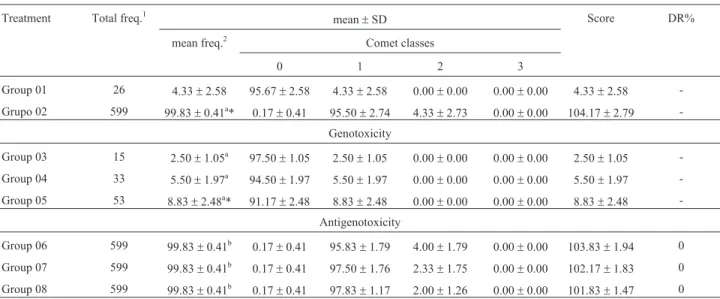
Table 6- Total and mean frequency of damaged cells, mean distribution between the classes of damage, mean scoring and damage reduction percentage related to tests for genotoxicity and antigenotoxicity in peripheral blood of mice during the sixth week of treatment.
Treatment Total freq.1
mean±SD Score DR%
mean freq.2 Comet classes
0 1 2 3
Group 01 26 4.33±2.58 95.67±2.58 4.33±2.58 0.00±0.00 0.00±0.00 4.33±2.58 -Grupo 02 599 99.83±0.41a* 0.17±0.41 95.50±2.74 4.33±2.73 0.00±0.00 104.17±2.79
-Genotoxicity
Group 03 15 2.50±1.05a 97.50±1.05 2.50±1.05 0.00±0.00 0.00±0.00 2.50±1.05 -Group 04 33 5.50±1.97a 94.50±1.97 5.50±1.97 0.00±0.00 0.00±0.00 5.50±1.97 -Group 05 53 8.83±2.48a* 91.17±2.48 8.83±2.48 0.00±0.00 0.00±0.00 8.83±2.48
-Antigenotoxicity
Group 06 599 99.83±0.41b 0.17±0.41 95.83±1.79 4.00±1.79 0.00±0.00 103.83±1.94 0
Group 07 599 99.83±0.41b 0.17±0.41 97.50±1.76 2.33±1.75 0.00±0.00 102.17±1.83 0
Group 08 599 99.83±0.41b 0.17±0.41 97.83±1.17 2.00±1.26 0.00±0.00 101.83±1.47 0 Group 01 - control (PBS - 0.1 mL/10.0 g), Group 02 - cyclophosphamide (50 mg/kg), Group 03 -b-glucan (100 mg/kg), Group 04 -b-glucan (150 mg/kg), Group 05 -b-glucan (200 mg/kg), Group 06 -b-glucan (100 mg/kg) + cyclophosphamide (50 mg/kg), Group 07 -b-glucan (150 mg/kg) + cyclophosphamide (50 mg/kg), Group 08 -b-glucan (200 mg/kg) + cyclophosphamide (50 mg/kg).
1
Total number of damaged cells by treatment;2Average number of damaged cells by treatment; DR% - Damage reduction percentage.
aStatistically compared to the control (Group 01);bstatistically compared to the damage-inducing agent (Group 02); *statistically significant difference
The analysis of data concerning the mutagenicity shows that for the doses of 100 and 150 mg/kg (b.w.) this was gradually reduced over the weeks of treatment. There was a decrease of 40.23 and 15.27 percentage points from the first to the fifth week and 19.94 and 15.75 from the fifth to the sixth week, respectively. Yet, the highest dose tested (200 mg/kg b.w.) showed a different behavior from the oth-ers, as there was first an increase of 2.15 percentage points followed by a decrease of 4.68 from the first to the fifth week and from the fifth to sixth week, respectively.
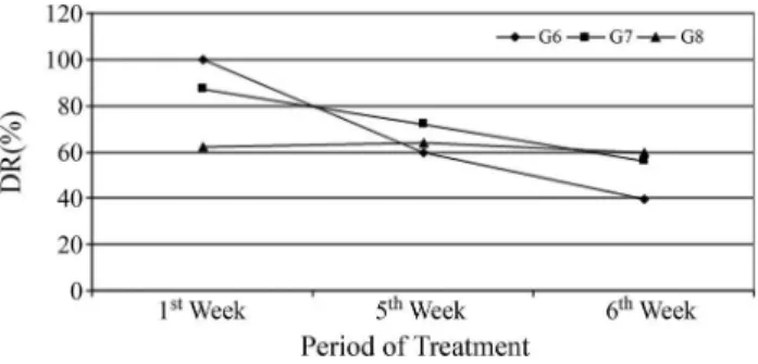
For the DR% observed in the comet assay there was a similar behavior for both doses. The data analysis revealed a reduction of 57.13, 40.26 and 17.34 percentage points for the three different doses (100, 150 and 200 mg/kg b.w.) from the first to the fifth week, respectively. From the fifth to the sixth week there was a reduction of 2.39, 43.79 and 77.0 percentage points for the three different doses, respec-tively. Thus, at the sixth week no antigenotoxic activity was denoted for theb-glucan.
Discussion
In the present study, b-glucan, extracted from
Saccharomyces cerevisiae, was administered
intraperito-neally in mice. The option of using polymer instead of fiber or bran produced fromSaccharomyces cerevisiaewas due to the intention of having a controlled amount administered to the animals, as well as the possibility of finding a more efficient effect of the purified polysaccharide.
In this experimental protocol, an administration of
b-glucan was done before cyclophosphamide; one simulta-neously and another one 24 h after the onset of chemother-apy. The choice of this substance is due to the mechanism of the antimutagenic b-glucan action, which was previ-ously investigated in our laboratory (Oliveiraet al., 2006, 2007). The polysaccharide, regardless of its origin, from cereal or fungus, has its action from both antimutagenesis and bioantimutagenesis. Thus, it is possible to think about a therapy that could combine the protocols for pre-treatment, simultaneous treatment and post-treatment, which would allow greater efficacy of this polysaccharide in the preven-tion of genetic damage and consequent development of a tumor.
The idea proposed previously found support in stud-ies published by other authors. According to these, anti-mutagenic substances are those capable of preventing the action of damage-inducing agents mainly by their adsorp-tion. Therefore these act preferably in the extracellular en-vironment. On the other hand, bioantimutagenic agents are the ones capable of involved in injury prevention or DNA repair, thus acting within the cell (Kadaet al., 1982; Kada and Shimoi, 1987, De Flora, 1998).
The experimental results show that b-glucan pre-sented no mutagenic activity, and in the experimental groups and the groups treated with the lowest and highest dose tested it was capable of reducing the basal frequency of micronuclei. This suggests that in the future this sugar polymer can be an important dietary supplement that can help to prevent the development of cancer because of its chemopreventive ability against spontaneous DNA dam-age.
The study of antimutagenicity showed a high chemo-preventive efficacy against the damage caused. When as-sessing the lowest dose, there was a 100% prevention against these injuries. In this study, there was no correlation between an increased dose of b-glucan and increased chemoprevention. On the contrary, with increasing concen-trations there was actually a reduction in chemopreventive capacity. Although there are no reports yet in the literature to support this, the administration of high doses ofb-glucan could relate to a blockage of cellular metabolism, and thus does not achieve the desired chemoprotective effect but could rather cause cell toxicity. Another possibility would be an inflammatory reaction, and this in turn would lead to an increased frequency of micronuclei.
A possible explanation for the increased frequency of micronuclei in an inflammatory reaction could be the accu-mulation of free radicals that can be generated by the me-tabolism of arginine, as this molecule is the precursor of
Figure 3- Antigenotoxic activity of theb-glucan molecule measured by DR% in the comet assay in peripheral blood. Group 06 - b-glucan (100 mg/kg) + cyclophosphamide (50 mg/kg), Group 07 - b-glucan (150 mg/kg) + cyclophosphamide (50 mg/kg), Group 08 - b-glucan (200 mg/kg) + cyclophosphamide (50 mg/kg).
Figure 2- Antimutagenic behavior of theb-glucan molecule activity measured by DR% in the micronucleus assay in peripheral blood. Group 06
-bglucan (100 mg/kg) + cyclophosphamide (50 mg/kg), Group 07
-bglucan (150 mg/kg) + cyclophosphamide (50 mg/kg), Group 08
nitric oxide. Corroborating this, the study of Oliveiraet al.
(2009b) showed that glutamine can be converted into nitric oxide precursors, and their excess in a body could be related to the increase in DNA damage. A possible explanation for the increased frequency of micronuclei in an inflammatory reaction could be the accumulation of free radicals that can be generated by the metabolism of arginine and/or gluta-mine, as this molecule is the precursor of nitric oxide, which has many functions in the body, including the stimu-lation of the immune response mediated by lymphocytes and macrophages.
In certain situations where the production of nitric ox-ide is increased there may be tissue damage (Dusseet al., 2003; Luikinget al., 2005) and this route is important for this study because it helps in understanding mutagenic data. This would imply a direct proportional relationship be-tween the increase of free radicals and the increase of dam-age to the DNA.
In acute and/or moderate inflammation, such as the possible reaction caused by high doses ofb-glucan admin-istered in the first week of treatment, there could have been a change in arginine metabolism and the use of arginine by the body could be high. To meet this need, there could be a breakdown of muscle protein and the endogenous synthesis of arginine leading to an increase in nitric oxide capable of increasing the level of damage caused by this free radical.
Another subject that needs to be discussed is the po-tential generation of free oxygen radicals due to the oxida-tive burst generated by the activated neutrophils and macrophages. The study made by Demiret al.(2007) dem-onstrated thatb-glucan extracted fromS. cerevisiae, when administered orally during 14 days in women with breast cancer in advanced stage, caused the activation of mono-cytes in peripheral blood, as well as the stimulation of their proliferation. Also, clinical examinations did not show any side effects of oral administration ofb-glucan. Added to this fact, Xiaoet al.(2004) questioned ifb-glucan immu-nomodulatory activity occurs by activating or increasing the host immune response through leukocyte activation and the production of inflammatory cytokines, when treating with anticancer drugs. Thus, to define the exactb-glucan anticancer mechanism, more research is necessary.
Today it is known that cell and/or tissue damage causes the release of a number of cytokines, such as IL-1b, IL-6, IL-8 and TNF-a, that in turn cause neutrophils to be-come activated and produce a host of cytotoxic substances, including reactive oxygen species, such as superoxide an-ions, hypochlorite and hydrogen peroxide (Best et al., 1999; Bricksonet al., 2001). The cytokines IL-1, IL-6 and TNF-aall stimulate pathways that contribute to the activa-tion of the enzyme NADPH-oxidase, which generates a “respiratory burst”. The subsequent release of reactive oxy-gen species (Butterfieldet al., 2006) could then be respon-sible for the increased frequency of micronuclei.
Thus, even thoughb-glucan is described as an antiox-idant agent, and the mechanism of desmutagenic action is its greatest effectiveness (Patchen et al., 1987; Chorva-tovicová, 1991; Slameñová et al., 2003; Oliveira et al., 2006; Oliveiraet al., 2007; Magnaniet al., 2011; Silvaet al., 2012), this could not prevent the damage caused by me-tabolites of arginine generated in the inflammatory process and the metabolism of chemotherapy. On the other hand, when evaluating the comet assay data, a dose-response curve in chemoprevention of genotoxic damage was seen in the first week of treatment.
Notwithstanding, the dose response curve in seen in chemoprevention of genotoxic damage was not observed for mutagenic damage. One possible explanation for this is that the two tests used assessed different types of damage. The micronucleus test quantifies cytogenetic damage, such as mutation events already set in the cell genome (Salvadori
et al., 2003), whereas the comet assay indicates genotoxic damage, which may or may not result in mutations (Oli-veiraet al., 2007).
When assessing the fifth week of treatment we again noticed thatb-glucan showed no mutagenic activity and, again, there was no correlation between an increased dose ofb-glucan and increased chemopreventive activity. How-ever, the intermediate dose was more effective in prevent-ing damage caused by mutagenic alkylatprevent-ing agent. When compared to the first and fifth week of treatment, there was a gradual reduction in chemopreventive ability from the polysaccharide in this study. In the first week, the DR%s showed greater variation (37.96 percentage points between the lowest and highest dose tested) compared to the fifth week (11.86 percentage points between the lowest dose and the intermediate dose).
When analyzing the comet assay, the b-glucan proved to be genotoxic in three of the doses tested in the weeks following the first one. A curve dose response was seen in antigenotoxicity, and it is even possible to infer that the lowest dose tested showed no chemopreventive ability, whereas the higher dose was better at preventing damage to DNA.
b-glucan showed no statistically significant difference over that week and the entire experiment. Thus, once again it can be inferred that the polysaccharide shows no mutagenic ac-tivity.
The analysis of T1 in the sixth week in the protocol of antimutagenicity indicates that the administration ofb -glu-can prior to the new treatment with cyclophosphamide showed a tendency to reduce the incidence of micronuclei observed at the time T0. However, when evaluating T2, 48 hours after the administration of cyclophosphamide combined withb-glucan, we observed a chemopreventive activity of the latter. But this capacity was low compared to the other weeks, with a variation of 19.68 percentage points. Another fact that draws attention is that the in-creased chemopreventive activity was denoted at the high-est dose. But this was still far below the DR% observed in in the first (DR% = 100% - lower dose) and fifth (DR% = 71.63% - intermediate dose) weeks.
In the comet assay, theb-glucan again presented itself as genotoxic, but only for the highest dose. However, be-fore the real genotoxic activity of the polysaccharide can be assessed, further studies are needed to support this finding, since the average values of damage shown in the fifth and sixth weeks were low and close to each other. Therefore, the actual biological significance of these values must be better understood to infer whether or not there is a toxic ef-fect ofb-glucan.
Contrary to what has been observed so far, the comet assay in the sixth week of treatment indicates a total chemo-preventive inefficacy against the genotoxic damage as-sessed. The behavior analysis of the mutagenic and/or genotoxic DR% during the 3 weeks of evaluation in general showed that acute treatment with multiple doses induced a loss in the chemopreventive efficacy ofb-glucan, particu-larly in its antigenotoxic activity, which was canceled dur-ing the last week of the study.
The values found for antimutagenicity and antigeno-toxicity in the acute treatment are very important and en-courage the use of this polysaccharide in the prevention of cancer and/or genetic damage that can lead to the develop-ment of cancer. Another important fact already develop-mentioned by some authors is the possibility of usingb-glucan as an adjunct to chemotherapy, as it is able to prevent some un-wanted side effects (Kanenoet al., 1989; Oliveiraet al., 2006; Oliveiraet al., 2007), which means that the supple-mentation of this polysaccharide could help to decrease mutagenic effects in non-tumor cells.
Facing all these facts, it is still an open question whether and whenb-glucan could has effects that are re-lated to improving the quality of life, both in humans and experimental animal models. Clearly, further work will be necessary to clarify this issue. Nonetheless, some data are of interest. As it is known that the cells are exposed to oxi-dant and antioxioxi-dant sources. The multiple chemical
reac-tions involving oxygen are the most effective mechanisms of energy production and can generate intermediate com-pounds or reactive oxygen species commonly named free radicals. Free radicals are highly reactive compounds with one or more unpaired electrons that are not evenly neutral-ized by enzymatic and non-enzymatic systems (McCord, 1993). Nitric oxide is one of these free radicals and plays an important role in carcinogenesis and tumor progression. Thus, the cellular exposure to high levels of nitric oxide in-duced by nitric oxide synthase during the inflammatory process may induce carcinogenesis due to the mutagenic properties of this compound (Dragstedet al., 1993).
Drawing a parallel with the data obtained herein it is feasible that the exposure to chemotherapy for six consecu-tive weeks, associated with the administration ofb-glucan, might result in releasing free oxygen radicals due to an oxi-dative burst by activating neutrophils and macrophages. These free oxygen radicals could promote a loss in the antigenotoxic activity, mainly during the sixth week, when
b-glucan loses all its antigenotoxic capacity and has its antimutagenic capacity reduced. These effects may occur due to the saturation of repair mechanisms and/or altered cell cycle kinetics. The level of DNA damage that is sus-tained by the cell might be important in this context. At low levels of damage, DNA-repair factors, which are highly specific for damage, could recognize and repair damage be-fore its detection by the checkpoint proteins. However, if damage reaches a higher threshold, checkpoint proteins such as ATR could also find DNA lesions. This repair sys-tem might have been saturated, and it either needs time to work or cannot restore genomic integrity. If ATR signals lead to cell-cycle arrest, ATR might either dissociate from DNA to allow repair enzymes access to the lesion or partic-ipate in as-yet-undiscovered interactions with excision re-pair factors to target the damage for rere-pair (Cline and Hanawalt, 2003).
Based on all these reports, it is suggested thatb -glu-can is a strong -glu-candidate for -glu-cancer prevention and control of genetic damage due to its antioxidant activity. Corrobo-rating this, the work of Patchenet al.(1987) indicates im-provement in the quality of life of animal which had radia-tion exposure, regarding hematopoietic regeneraradia-tion and the ability ofb-glucan in inactivating free radicals. Silvaet al.(2012) showed the anticlastogenic effect ofb-glucan in cells exposed to ultraviolet radiation (UV) suggesting that
b-glucan has more than one mechanism of action, being ca-pable of exerting desmutagenic as well as bioantimutagenic action and, therefore, these results indicate thatb-glucan fromSaccharomyces cerevisiaecan be used in the preven-tion and/or reducpreven-tion of DNA damage. Chorvatovicováet al.(1991, 1996, 1998) reported the prevention of genetic damage caused by cobalt and cyclophosphamide. Oliveira
Sla-menováet al.(2003) and Lazarováet al.(2006) described the sequestrant ability of free radicals in V79 cells and in mice, respectively, when damage is caused by hydrogen peroxide. Adding to these facts, Magnaniet al.(2011) have recently shown a protective effect of Carboxymethyl-Glu-can against DNA damage in patients with advanced pros-tate cancer, which was shown for the first time in humans. These results suggest thatb-glucan is potentially useful in improving the short-term survival.
However, for chronic therapies, there is still doubt about the true activity ofb-glucan and its form of adminis-tration. But the opportunity to use this polymer concur-rently with chemotherapy cannot be dismissed. Certainly, further work is necessary to reproduce other acute models that use multiple doses, as well as protocols where the ad-ministrations of chemotherapeutics are made in at longer intervals, such as chemotherapy protocols in humans.
Acknowledgments
This study was supported by Conselho Nacional de Desenvolvimento Científico e Tecnológico (CNPq), Coor-denadoria de Aperfeiçoamento de Nível Superior (CAPES) and Fundação Araucária.
References
Angeli JP, Ribeiro LR, Gonzaga ML, Soares S de A, Ricardo MP, Tsuboy MS, Stidl R, Knasmueller S, Linhares RS and Man-tovani MS (2006) Protective effects of beta-glucan extracted fromAgaricus blazeiagainst chemically induced DNA dam-age in human lymphocytes. Cell Biol Toxicol 22:285-291. Angeli JP, Ribeiro LR, Angeli JL and Mantovani MS (2009a)
Pro-tective effects of beta-glucan from barley against ben-zo[a]pyrene-induced DNA damage in hepatic cell HepG2. Exp Toxicol Pathol 61:83-89.
Angeli JP, Ribeiro LR, Bellini MF and Mantovani MS (2009b). Beta-glucan extracted from the medicinal mushroom Agaricus blazei prevents the genotoxics effects of ben-zo[a]pyrene in the human hepatoma cel line HepG2. Arch Toxicol 83:81-86.
Best TM, Fiebig R, Corr DT, Brickson S and Ji L (1999) Free radi-cal activity, antioxidant enzyme, and glutathione changes with muscle stretch injury in rabbits. J Appl Physiol 87:74-82.
Brickson S, Hollander J, Corr DT, Ji LL and Best TM (2001) Oxi-dant production and immune response after stretch injury in skeletal muscle. Med Sci Sports Exerc 33:2010-2015. Butterfield TA, Best TM and Merrick MA (2006) The dual roles
of neutrophils and macrophages in inflammation: A critical balance between tissue damage and repair. J Athl Train 41:457-465.
Camargo JLV, Oliveira MLCS, Rocha NS and Ito N (1994) A detecção de substâncias cancerígenas em estudos experi-mentais. Rev Bras Cancerol 40:21-30.
Chorvatovicová D (1991). Suppressing effects of glucan on micromuceli induced by Co60 in mice. Strahlenther Onkol 167:612-614.
Chorvatovicová D, Machová E and Sandula J (1996) Effect of ultrasonicated carboxymethylglucan on cyclophosphamide induced mutagenicity. Mutat Res 371:115-120.
Chorvatovicová D, Machová E and Sandula J (1998) Ultrasoni-cation: The way to achieve antimutagenic effect of carboxy-methyl-chitin-glucan by oral administration. Mutat Res 412:83-89.
Cisneros RL, Gibson FC and Tzianabos AO (1996) Passive trans-fer of poly-(1-6)-b-glucotriosyl-(1-3)-b-glucopyranose glu-cana protection against lethal infection in an animal model of intra-abdominal sepsis. Infect Immun 64:2201-2205. Cline SD and Hanawalt PC (2003) Who’s on first in the cellular
response to DNA damage? Nat Rev Mol Cell Biol 4:361-372.
De Flora S (1998) Mechanisms of inhibitors of mutagenesis and carcinogenesis. Mutat Res 402:151-158.
Demir G, Klein HO, Mandel-Molinas N and Tuzuner N (2007) b-glucan induces proliferation and activation of monocytes in peripheral blood of patients with advanced breast cancer. Int Immunopharmacol 7:113-116.
Di Luzio NR, Williams DL, Mcnamee RB, Edwards BF and Kitahama A (1979) Compartive tumor-innhibitory and anti-bacterial activity of soluble and particulate glucana. Int J Cancer 24:773-779.
Dragsted LO, Strube M and Larsen JC (1993) Cancer-protective factors in fruits and vegetables: Biochemical and biological background. Pharmacol Toxicol 72:116-135.
Dusse LMS, Vieira LM and Carvalho MG (2003) Nitric oxide re-vision. J Bras Patol Med Lab 39:343-350.
Fearon ER and Jones PA (1992) Progressing toward a molecular description of colorectal cancer development. FASEB J 6:2783-2790.
Hayashi M, Morita T, Kodama Y, Sofuni T and Ishidate Jr M (1990) The micronucleus assay with mouse peripheral blood reticulocytes using acridine orange-coated slides. Mutat Res 245:245-249.
Kada T, Inoue T and Namiki N (1982) Environmental desmu-tagens and antidesmudesmu-tagens. In: Klekowski EJ (ed) Environ-mental Mutagenesis and Plant Biology. Praeger, New York, pp 137-151.
Kada T and Shimoi K (1987) Desmutagens and bio-antimutagens: Their modes of action. BioEssays 7:113-115.
Kaneno Y, Chihara G and Taguchi T (1989) Activity of lentinan against cancer and AIDS. Int J Immunother 4:203-213. Kobayashi H, Sugiyama C, Morikawa Y, Hayashi M and Sofuni T
(1995) A comparison between manual microscopic analysis and computerized image analysis in the single cell gel elec-trophoresis assay. MMS Communications 2:103-115. Kumar V, Sinha AK, Makkar HP, de Boeck G and Becker K
(2012) Dietary roles of non-starch polysachharides in hu-man nutrition: A review. Crit Rev Food Sci Nutr 52:899-935.
Lazarová M, Lábaj J, Kogan G and Slamenová D (2006) Carboxy-methyl chitin-glucan enriched diet exhibits protective ef-fects against oxidative DNA damage induced in freshly iso-lated rat cells. Neoplasma 53:434-439.
Luiking YC, Poeze M, Ramsay G and Deutz NE (2005) The role of arginine in infection and sepsis. J Parenter Enteral Nutr 29:70-74.
Magnani M, Castro-Gomez RJH, Mori MP, Kyasne H, Gregório EP, Libos-Jr F and Cólus IMS (2011) Protective effect of carboxymethyl-glucan (CM-G) against DNA damage in pa-tients with advanced prostate cancer. Genet Mol Biol 34:131-135.
Manoharan K and Banerjee MR (1985) beta-Carotene reduces sis-ter chromatid exchanges induced by chemical carcinogens in mouse mammary cells in organ culture. Cell Biol Int Rep 9:783-789.
Mantovani MS, Bellini MF, Angelia JPF, Oliveira RJ, Silva AF and Ribeiro LR (2008)b-Glucans in promoting health: Pre-vention against mutation and cancer. Mutat Res 658:154-161.
Masihi KN (2000) Immunomodulators in infectious diseases: Panoply of possibilities. Int J Immunopharmacol 22:1083-1091.
McCord JM (1993) Human disease, free radicals, and the oxi-dant/antioxidante balance. Clin Biochem 26:351-357. Oliveira RJ, Ribeiro LR, Silva AF, Matuo R and Mantovani MS
(2006) Evaluation of antimutagenic activity and mecha-nisms o action of beta-glucan from barley, in CHO-k1 and HTC cell lines using the micronucleus test. Toxicol in Vitro 20:1225-1233.
Oliveira RJ, Matuo R, Silva AF, Matiazi HJ, Mantovani M and Ribeiro LR (2007) Protective effect of beta-glucan extracted fromSaccharomyces cerevisiae, against DNA damage and cytotoxicity in wild-type (k1) and repair-deficient (xrs5) CHO cells. Toxicol in Vitro 21:41-52.
Oliveira RJ, Salles MJ, da Silva AF, Kanno TYN, Lourenço ACS, Freiria GA, Matiazi HJ, Ribeiro LR and Mantovani MS (2009a) Effects of the polysaccharideb-glucan on clasto-genicity and teratoclasto-genicity caused by acute exposure to cyclophosphamide in mice. Regul Toxicol Pharmacol 53:164-173.
Oliveira RJ, Baise E, Mauro MO, Pesarini JR, Matuo R, Silva AF, Ribeiro LR and Mantovani MS (2009b) Evaluation of che-mopreventive activity of glutamine by the comet and the micronucleus assay in mice’s peripheral blood. Environ Toxicol Pharmacol 28:120-124.
Patchen ML, D’Alesandro MM, Brook I, Blakely WF and MacVittie TJ (1987) Glucan: Mechanisms involved in its radioprotective effect. J Leukoc Biol 42:95-105.
Pinkerton pH and Dubé ID (1991) Chronic myeloid leukemia as a paradigm for oncogenesis. Diagnon Oncol 1:288-297. Pitot HC (1993) The molecular biology of carcinogenesis. Cancer
72:962-970.
Pitot HC and Dragan Y (1991) Facts and theories concerning the mechanisms of carcinogenesis. FASEB J 5:2280-2286.
Salvadori DMF, Ribeiro LR and Fenech M (2003) Teste do micronúcleo em células humanasin vitro. In: Ribeiro, LR, Salvadori DMF and Marques EK (eds) Mutagênese Am-biental. ULBRA, Canoas, pp 201-219.
Silva AF, Oliveira RJ, Niwa AM, D’Epiro GFR, Ribeiro LR and Mantovani MS (2012) Anticlastogenic effect ofb-glucan, extracted fromSaccharomyces cerevisiae, on cultured cells exposed to ultraviolet radiation. Cytotechnology 65:41-48. Singh NP, McCoy MT, Tice RR and Schneider EL (1988) A
sim-ple technique for quantification of low levels of DNA dam-age in individual cells. Exp Cell Res 175:184-191. Slamenová D, Lábaj J, Krizková L, Kogan G, Sandula J, Bresgen
N and Eckl P (2003) Protective effects of fungal (1®3)-b -D-glucan derivatives against oxidative DNA lesions in V79 hamster lung cells. Cancer Lett 198:153-160.
Sugimura T (1992) Multistep carcinogenesis: A 1992 perspective. Science 258:603-607.
Tohamy AA, El-Ghor AA, El-Nahas SM and Noshy MM (2003) b-Glucan inhibits the genotoxicity of cyclophosphamide, adramycin and cisplatin. Mutat Res 541:45-53.
Torrinhas RSMM, Campos LDN and Mazza RPJ (2006) Meto-dologia na pesquisa de dieta, nutrição e câncer: Estudos experimentais em modelos animais e culturas de células. In: Waitzberg DL (ed) Dieta, Nutrição e Câncer. Ateneu, São Paulo, pp 665-674.
Turnbull JL, Patchen ML and Scadden DT (1999) The poly-saccharide, PGG-glucan, enhances human myelopoiesis by direct action independent of and additive to early-acting cytokines. Acta Haematol 102:66-71.
Xiao Z, Trincado CA and Murtaugh MP (2004)b-Glucan en-hancement of T cell IFN-gamma response in swine. Vet Immunol Immunopathol 102:315-320.
Waters MD, Brady AL, Stack HF and Brockman HE (1990) Antimutagenicity profiles for some model compounds. Mutat Res 238:57-85.
Zimmerman JW, Lindermuth J, Fish PA, Palace GP, Stevenson TT and DeMong DE (1998) A novel carbohydrate-glyco-sphingolipid interaction between ab-(1-3)-glucan immuno-modulator, PGG-glucan, and lactosylceramide of human leukocytes. J Biol Chem 273:22014-22020.
Internet Resources
COBEA (2004) Colégio Brasileiro de Experimentação Animal, http://www.cobea.org.br (September 3, 2012).
Associate Editor: Daisy Maria Fávero Salvadori