Joana Lamego
Dissertation presented to obtain the Ph.D. degree in
Engineering and Technology Sciences, Biotechnology
Instituto de Tecnologia Química e Biológica | Universidade Nova de
Lisboa
Oeiras,
December,
Carboxylesterase 2
Unknown sample
Dilute appropriately
Hydrolysis of 4-MUBA?
No CES2 activity detected. N
BNPP inhibition? Y
N
Loperamide inhibition? Y
Quantify other CESs through BNPP inhibition.
Quantify CES2 through loperamide inhibition.
Quantify other CESs through the difference between the two inhibitors. Y
N
Y
Joana Catarina da Rocha Lamego
Dissertation presented to obtain the Ph.D. degree in
Engineering and Technology Sciences, Biotechnology
Instituto de Tecnologia Química e Biológica | Universidade Nova de Lisboa
Oeiras, December 2012
A
A
A
And the feasibility of a more relevant cell model for
nd the feasibility of a more relevant cell model for
nd the feasibility of a more relevant cell model for
nd the feasibility of a more relevant cell model for
intestinal metabolism
by Joana Lamego
Second Edition: December 2012
ITQB-UNL and IBET, Pharmacokinetics and Biopharmaceutical Analysis Laboratory and Cell Line Development and Molecular Biotechnology Laboratory - Animal Cell Technology Unit
Instituto de Tecnologia Química e Biológica, Universidade Nova de Lisboa and Instituto de Biologia Experimental e Tecnológica
Apartado 12, 2781-901 Oeiras, Portugal
Fax: +351 21 442 11 61; Phone: +351 21 446 91 00 http://www.itqb.unl.pt
http://www.ibet.pt http://tca.itqb.unl.pt
Copyright © 2012 by Joana Lamego All Rights Reserved
Dr. Ana Luísa Simplício, Auxiliary Investigator and Head of the Pharmacokinetics and
Biopharmaceutical Analysis Laboratory at ITQB-UNL/IBET and Responsible for the
Quality Assurance Unit of the Analytical Services Unit of IBET (Supervisor)
Dr. Ana Sofia Coroadinha, Auxiliary Investigator and Head of the Cell Line
Development and Molecular Biotechnology Laboratory at ITQB-UNL/IBET
This thesis dissertation is the result of four years of research at the Pharmacokinetics and Biopharmaceutical Analysis Laboratory and at the Cell Line Development and Molecular Biotechnology Laboratory - Animal Cell Technology Unit of Instituto de Tecnologia Química e Biológica – Universidade Nova de Lisboa and Instituto de Biologia Experimental e Tecnológica (Oeiras, Portugal) under the supervision of Dr. Ana Luísa Simplício and co-supervision of Dr. Ana Sofia Coroadinha.
Aos meus pais
O trabalho conducente à elaboração desta tese não teria sido possível sem o apoio financeiro
da Fundação para a Ciência e Tecnologia (FCT) e do FSE no âmbito do Quadro Comunitário de
apoio, BD nº 44025/2008.
Ao longo dos últimos quatro anos foram muitos os que contribuíram para que hoje possa
escrever esta secção onde retribuo, pelo menos em parte, o que me ofereceram: o mérito
desta tese.
À Dra. Ana Luísa Simplício, minha orientadora. Ana, foi uma honra tê-la como mentora ao
longo destes quatro anos repletos de discussão científica onde os nossos pontos de vista nem
sempre coincidentes foram o motor gerador de tantas e boas ideias. O meu agradecimento
sincero por me ter ouvido ao longo deste trabalho, por ter valorizado as minhas ideias e por
me ter concedido a liberdade e autonomia de pensar, experimentar, errar e acertar. A Ana
respeita verdadeiramente os outros, o que são e aquilo que querem ser, ajudando-os a chegar
lá, não só a nível profissional, como também a nível pessoal. Sabe exigir e dar, características
que fazem de si a orientadora que é.
À Dra. Ana Sofia Coroadinha, minha co-orientadora: agradeço todo o apoio e conhecimento
que me transmitiste. Foi um privilégio poder beneficiar desta combinação de conhecimentos
que resultam da orientação por duas cientistas de base científicas diferentes. Agradeço por
me teres também concedido a liberdade de pensar pela minha própria cabeça e me teres
ajudado a crescer cientificamente. O meu sincero obrigado por me teres incentivado a ir para
a bancada testar as minhas mais que muitas hipóteses. As reuniões a par e em grupo que
fomentas são essenciais para o bom desenrolar do trabalho científico bem como para
consolidar o espírito de inter-ajuda entre todos os que delas beneficiam.
Às anteriores e atuais direções das duas instituições, IBET e ITQB, que me receberam e
permitiram o desenvolvimento do meu trabalho de doutoramento, nomeadamente à Prof.
Dra. Paula Alves, Prof. Dr. Manuel Carrondo, e Prof. Dr. Luís Paulo Rebelo. Agradeço em
especial à Dra. Paula Alves: pela oportunidade de trabalhar na Unidade de Tecnologia de
e fazer mais e melhor, contribuindo de uma forma decisiva para o meu crescimento a nível
científico e pessoal.
Aos meus antigos e atuais colegas do Laboratório de Farmacocinética e Análise
Biofarmacêutica e da Unidade de Tecnologia de Células Animais, por toda a ajuda e pelo
saudável ambiente que aí se respira.
À Cristina Peixoto e ao Marcos Sousa, co-autores do trabalho da produção e purificação da
CES2, descrito na Secção 3 desta tese.
Um obrigada muito especial à Bárbara Cunha, co-autora desse mesmo trabalho e companheira
de experiências “malucas” (e não tão “malucas” assim) que nunca vira a cara a um bom
desafio, a uma boa aventura, mesmo que isso implique trabalhar desenfreadamente até à
exaustão. Obrigada pela tua fundamental ajuda, foi um verdadeiro privilégio trabalhar ao teu
lado. Serás uma brilhante cientista e podes contar com esta tua amiga para o que precisares.
À “equipa” do almoço e da boa disposição, um obrigada especial à Carina Brilha, à Vanessa
Bandeira, ao Luís Marques e ao Hugo Soares pelas tertúlias “intelectuais”, “sérias” e
“extremamente silenciosas”, fundamentais para manter a sanidade mental, e que serão
continuadas e upgraded a jantaradas, passeatas e outros que tais do mesmo nível!
À Patrícia Alves, parte fundamental do trio (duo e três quartos, na recta final) dinâmico dos
pequenos-almoços bem dispostos e energizantes. Giraça, obrigada pelo teu apoio!
Aos meus colegas das aulas de doutoramento, um agradecimento especial ao Fábio Silva pelas
aventuras das aulas, do Conselho Pedagógico do ITQB, da Escola Doutoral da Nova, do
COHiTEC. Sei que a palavra “amarelo” ganhou toda uma nova cor para ti.
Aos amigos, os de sempre e os mais recentes, Madalena Carido, Cláudia Vicente, Pedro
Aos que que já partiram mas que continuam.
A um ser que apesar de não humano foi mais pessoa que muitos Homens, membro inegável
da família, companheira de uma boa parte da minha vida, fonte incansável de alegria,
brincadeira, companhia, ternura e de muitas saudades.
Um agradecimento especial aos meus avôs. O vosso orgulho está sempre comigo.
Aos que sempre estão.
Às minhas avós, matriarcas da família. À minha irmã e recentes adições à família, a Francisca e
a Constança, pequeninas ainda para perceberem a influência boa que são já na vida da tia:
terapeutas milagrosas dos momentos de maior neura.
Aos meus pais. Faltam as palavras para vos agradecer a forma como me educaram, como me
ensinam e como me apoiam todos os dias. À minha mãe: pilar inabalável, companheira de
todos os dias, fonte inesgotável de apoio, amor e compreensão, por estares sempre lá, por
partilhares toda a emoção em primeira mão, da alegria das vitórias à tristeza das pequenas
derrotas. Ao meu pai: apesar da distância soubeste e sabes sempre estar presente. És o meu
exemplo de integridade, dedicação, trabalho, exigência, rigor, orgulho de fazer bem feito,
vontade de mudar o que não está bem e de chegar mais longe. De ti absorvi características
únicas que definem a minha forma de estar, de agir e de pensar. Com um orgulho
inconfessável por ser vossa filha, posso apenas desejar um dia ser para alguém aquilo que
vocês foram e são para mim.
Ao Miguel. O meu outro pilar. Companheiro de aventuras, de sonhos e de realidades. Sempre
disponível, fisicamente ou à distância de um “trim trim”, para viver as nossas peripécias
quotidianas. Fonte inesgotável de criatividade, carinho, alegria, boa disposição, paciência e
compreensão. Motor gerador de motivação, nos momentos onde esta se perdia (o cliché neste caso bem verdadeiro): acreditaste em mim e deste-me força para continuar quando eu própria
não acreditava ser possível. Agradeço-te tudo isto mas principalmente a dose certa de loucura,
infantilidade e irreverência que nos permite olhar para as nuvens, dar gargalhadas e ser feliz.
The first barrier oral drugs and prodrugs encounter prior to reaching an organism’s systemic circulation is the gastrointestinal (GI) tract, specifically the intestine, which is the primary section for absorption. Therefore, it is fundamental to understand the permeability of the therapeutic agent as well as its potential metabolism by human enterocytes, since biotransformation may result in the inactivation of the therapeutic agent or, to the contrary, in the formation of more therapeutically active metabolites. Carboxylesterases (CESs), phase I metabolising enzymes, are important in the metabolism of several drugs and prodrugs with amide, ester, or thioester functional groups. After cytochrome (CYP) P450s and UDP-glucuronosyltransferases (UGTs), CESs are the most relevant enzymes for the metabolism of therapeutic agents.
Carboxylesterase 2 (hCES2) is the main CES expressed in the human intestine and is an increasingly important enzyme in anti-cancer combined therapies for the treatment of different pathologies such as colon adenocarcinoma and malignant glioma, due to its potential to improve the anti-cancer effect of certain therapeutic compounds. Notwithstanding, it is down regulated in the most used intestinal model for permeability, the human colon adenocarcinoma derived Caco-2 cell line. On the contrary, carboxylesterase 1 (hCES1), the main CES expressed in the human liver, is highly expressed in Caco-2 cells, unlike what is known to occur in the intestine. The main goal of the work presented in this thesis was to increase the fundamental knowledge of hCES2 and to improve the in vitro to in vivo relevance of Caco-2 cells, by increasing the expressed hCES2 levels.
The developed methodology was applied to a variety of samples and proved to be suitable for the quantitation of hCES2-specific activity in mixtures of expressed and purified CES1 and hCES2 enzymes. Moreover, the developed method showed that transiently transfected HEK-293T cells had 15-fold higher hCES2-specific activity than non-transfected cells. This method also showed effectiveness in distinguishing CES2-specific activity in different mammalian sera containing a variety of esterases. The developed methodology should be applicable to a wider variety of samples with esterase activity, as well as for the analysis of the activities of other esterases, once appropriate substrates and inhibitors are used.
Using the appropriate tools to study hCES2, a method enabling the production and purification of this enzyme was developed. This successfully strategy, described in Section 3, resulted in the first recombinant human hCES2 enzyme produced using human HEK-293T cells in suspension. The utilized strategy demonstrated that the addition of an in-frame, C-terminally localized 10x histidine tag, was sufficient to promote hCES2 secretion. This avoided the need of additional N-terminal signal sequences or the modification or deletion of the Endoplasmic Reticulum (ER) retention sequence, which are the traditional strategies. Moreover, using both standard and in-house developed biochemical and analytical techniques, new fundamental features were unravelled, such as the presence of oligomeric active and inactive forms of hCES2, which was previously reported as existing only in a monomeric 60 kDa form, leading the way to possible new discoveries concerning hCES2 properties. Different oligomeric forms had previously been reported for hCES1; the present work suggests this might be a common feature for CESs.
cell line resulting in a population with increased hCES2 mRNA, protein expression, and activity levels. No changes in cell differentiation and polarization ability were detected in comparison with the parental cell line, as the overexpressing population retained the capacity to express similar levels of alkaline phosphatase. A hCES2-expressing Caco-2 cell line was generated through clonal selection and its stability during passaging and differentiation, was evaluated. An obvious decay of enzyme expression was observed at higher passage numbers, confirming a previously reported hurdle to Caco-2 manipulation. New clues to understand and overcome this limitation of the Caco-2 model are provided.
Upon complete validation with reference compounds,
the newly developed cell line has the potential to become a useful tool for coupling the study of intestinal absorption with intestinal metabolism, especially when it involves the hydrolysis of ester-containing drugs and prodrugs.Fármacos e pró-fármacos administrados por via oral encontram no trato gastrointestinal, em especial no intestino delgado, região primordial de absorção, a primeira barreira antes de alcançarem o sistema circulatório. Torna-se, desta forma, fundamental, compreender as propriedades dos agentes terapêuticos em termos de permeabilidade, bem como a possível susceptibilidade a serem metabolizados nos enterócitos humanos, uma vez que a sua bio-transformação pode conduzir à inativação, ou, pelo contrário, à formação de metabolitos de maior ação terapêutica. Carboxilesterases (CESs) são enzimas metabólicas de fase I importantes no metabolismo de fármacos e pró-fármacos contendo grupos químicos funcionais específicos tais como amidas, ésteres ou tio-ésteres. Carboxilesterases são, depois das citocromo (CYP) P450 e das UDP-glucoronosil-transferases (UGTs), as enzimas de maior relevância para o metabolismo de agentes terapêuticos.
A carboxilesterase 2 (hCES2) é a principal CES expressa no intestino humano, sendo uma enzima de importância crescente em terapias anti-cancerígenas combinadas para o tratamento de diversas patologias tais como o adenocarcinoma do colon ou o glioma maligno, devido ao potencial que tem de aumentar o efeito anti-cancerígeno de certos compostos terapêuticos. Esta enzima encontra-se, contudo, regulada negativamente no modelo intestinal mais utilizado para a avaliação da permeabilidade intestinal, a linha celular Caco-2, derivada de um adenocarcinoma de colon humano. Pelo contrário, a carboxilesterase 1 (hCES1), a CES de maior expressão no fígado humano, é altamente expressa nas células Caco-2, de modo oposto ao que se sabe ocorrer no intestino humano.
amostras biológicas complexas, tiveram de ser desenvolvidas, tal como descrito na Secção 2. A utilização de um inibidor específico da CES2, a loperamida, e de um inibidor geral das CESs, o BNPP, possibilitou o desenvolvimento com sucesso de um método rápido, simples, repetível e necessitando de pequenas quantidades de amostra, para análise em eletroforese capilar, com aplicabilidade mesmo em situações nas quais substratos e produtos de reação absorvem no mesmo comprimento de onda. A metodologia assim desenvolvida foi aplicada em diversos tipos de amostras e provou ser adequada para a quantificação da atividade específica da hCES2, mesmo quando presente em misturas de enzimas expressas e purificadas, contendo CES1. Adicionalmente, o método desenvolvido possibilitou demonstrar que a atividade específica de hCES2 aumenta 15 vezes em células HEK-293T transfectadas de forma transiente em comparação com as mesmas células não transfectadas. Este método provou ainda ser eficaz para distinguir a atividade específica de CES2 presente em diferentes soros de mamíferos, contendo diversas esterases. A metodologia desenvolvida poderá ser adicionalmente aplicada não só a uma maior variedade de amostras contendo atividade de esterases, bem como para a avaliação de atividades de outras esterases desde que sejam utilizados substratos e inibidores apropriados.
técnicas analíticas e bioquímicas quer tradicionais quer desenvolvidas no laboratório, foi possível revelar novas propriedades fundamentais da hCES2 tais como a presença de formas oligoméricas ativas e inativas, desta proteína anteriormente descrita como existindo apenas na forma de monómero com 60 kDa, abrindo caminho a eventuais novas descobertas relativamente às suas propriedades. De facto, diferentes formas oligoméricas foram já descritas para a hCES1; o trabalho aqui apresentado sugere que esta possa ser uma característica comum a todas as CESs.
caminhos na descoberta de mistérios adicionais da hCES2 e das células Caco-2.
Peer-reviewed Articles
Lamego J, Coroadinha AS, Simplício AL. Detection and quantification of carboxylesterase 2 activity by capillary electrophoresis. Analytical Chemistry 2011; 83: 881-887.
Lamego J, Cunha B, Peixoto C, Sousa MF, Alves PM, Simplício AL, Coroadinha AS. Carboxylesterase 2 production and characterization in human cells: new insights into enzyme oligomerization and activity. Applied Microbiology and Biotechnology 2012; doi 10.1007/s00253-012-3994-3.
Lamego J, Simplício AL, Coroadinha AS. Development of a Caco-2 cell line expressing human Carboxylesterase 2. Toxicology in vitro 2012; submitted.
Book Chapters
Simplício AL, Coroadinha AS, Gilmer JF, Lamego J. A methodology for detection and quantification of esterase activity. Methods in Molecular Biology: Electrophoresis of Biomolecules 2013; in press.
Published Abstracts and Proceedings
Lamego J, Coroadinha AS, Simplício AL. Evaluation of carboxylesterases activity in biological samples. Revista Portuguesa de Farmácia 2010; Volume LII (n. º4) ISSN 0484-811X
Lamego J, Coroadinha A, Simplício A. Bridging the gap between Caco-2 cells and human carboxylesterases. Toxicology Letters 2011; 205: S165-S165 doi 10.1016/j.toxlet.2011.05.576 Lamego J, Ferreira P, Cunha B, Coroadinha AS and Simplício AL. Unraveling human carboxylesterase 2 activity-expression mismatch. Revista Portuguesa de Farmácia 2011; Volume LII (n. º6) ISSN 0484-811X
Additional Publications
Peer-reviewed Articles
4-MUB 4-Methylumbelliferone
4-MUBA 4-Methylumbelliferyl acetate
4-MUP 4-Methylumbelliferyl phosphate
ABCC1 ATP-binding cassette sub-family C (CTFR/MRP), member 1 (also known as multidrug resistance-associated protein 2 – MRP1)
ABCC2 ATP-binding cassette sub-family C (CTFR/MRP), member 2 (also known as multidrug resistance-associated protein 2 – MRP2)
ABCG2 ATP-binding cassette sub-family G (WHITE), member 2 (also known as brest cancer resistance protein – BCRP)
AcChE Acetylcholinesterase
AcTCh Acetylthiocholine
ADME Absorption, distribution, metabolism and excretion
AMEM Minimum Essential Medium Alpha
ALP Alkaline phosphatase
APC CPT-11 aminopentane carboxylic acid metabolite
ATCC American Type Culture Collection
AU Arbitrary Units
BAP Bovine alkaline phosphatase
BCA Bicinchoninic acid
BES Background electrolyte solution
BNPP Bis-p-nitrophenyl phosphate BuChE Butyrylcholinesterase
BuTCh Butyrylthiocholine
CE Capillary electrophoresis
CES Carboxylesterase
CES1 Carboxylesterase 1
CES2 Carboxylesterase 2
CES2-10xHis Recombinant CES2 with an in-frame C-terminal 10× histidine tag
CES3 Carboxylesterase 3
CHO Chinese hamster ovary cells
CO2 Carbon dioxide
COS-7 African green monkey kidney fibroblast-like cell line
CPT-11 (Irinotecan) (4S)-4,11-Diethyl-3,4,12,14-tetrahydro-4-hydroxy-3,14-dioxo-1H -pyrano[3’,4’:6,7]indolizino[1,2-b]quinolin-9-yl-[1,4’-bipiperidine]-1’-carboxylic acid ester hydrochloride
CVrCoefficient of variation of repeatability
CVi Coefficient of variation of intermediate precision
CYP Cytochrome P450
DMEM Dulbecco’s Modified Eagle Medium
DMSO Dimethyl sulfoxide
DMSZ Leibniz Institute DSMZ-German Collection of Microorganisms and Cell Cultures
ECACC European Collection of Cell Cultures
ECVAM European Centre for the Validation of Alternative Methods
EDTA Ethylenediaminetetraacetic acid
Endo H Endoglycosydase H
FBS Foetal bovine serum
GAPDH Glyceraldehyde-3-phosphate dehydrogenase
GPx4 Glutathione peroxidase 4
hCES Human carboxylesterase
HEK-293 Human embryonic kidney 293 cells
HEK-293T HEK-293 cells constitutively expressing SV40 large T antigen
HEPES 4-(2-Hydroxyethyl) piperazine-1-ethanesulfonic acid N-(2-Hydroxyethyl) piperazine-N’-(2-ethanesulfonic acid)
HIEL CES1 Histidine-isoleucine-glutamic acid-leucine CES1 motif
HPLC High-performance liquid chromatography
Hpt Hours post-transfection
HRP Horseradish peroxidase
HTEL CES2 Histidine-threonine-glutamic acid-leucine CES2 motif
i.d. Internal diameter
IL2RG Common gamma chain or Interleukin-2 receptor subunit gamma
LLC-PK1 Lewis lung carcinoma-porcine kidney 1 cells
IMAC Immobilized metal affinity chromatography
IV Intravenous
IVIVC In vitro-in vivo correlation kDa Kilodalton
KO Knockout
MDCK Mardin-Darby canine kidney cells
NBT/BCIP Nitro-blue tetrazolium/ 5-bromo-4-chloro-3’-indolyphosphate
NIH National Institutes of Health
NME New molecular entity
NPC CPT-11 primary amine metabolite
ON Overnight
p p-value
p.a. pro analysis
p-ABA p-aminobenzoic acid
PALP Human placental alkaline phosphatase
PAMPA Parallel artificial membrane permeability assay
PBPK Physiologically based pharmacokinetic
PBS Phosphate-buffered saline
PCR Polymerase chain reaction
PEI Polycation polyethylenimine
P-gp P-glycoprotein (also known as multidrug resistance protein 1 – MDR1 and ATP-binding cassette sub-family B member 1 – ABCB1)
PNGase F Peptide: N-glycosidase F
p-NP p-Nitrophenol
p-NPA p-Nitrophenyl acetate
pO2 partial pressure of oxygen
PVDF Polyvinylidene difluoride
QEDL CES3 Glutamine-glutamic acid-aspartic acid-leucine CES3 motif
R2R-squared coefficient
RAG2 Recombinant Activating Gene 2
RAJI Burkitt’s lymphoma-derived cell line
RNase B Ribonuclease B
RPMI Roswell Park Memorial Institute medium
SDS-PAGE Sodium dodecyl sulfate polyacrylamide gel electrophoresis
SN-38 (7-Ethyl-10-hydroxycamptothecin) (4S)-4,11-Diethyl-4,9-dihydroxy-1H- pyrano[3’,4’:6,7]indolizino[1,2-b]quinolone-3,14(4H,12H)dione
SNP Single nucleotide polymorphism
TCh Thiocoline
TEER Trans-epithelial resistance
UGT Uridine 5’-diphospho(UDP)-glucuronosyltransferase
UV Ultraviolet
V Volume
V79 Chinese Hamster Fibroblast cell line
Vvm Gas volume flow per unit of liquid volume per minute
cm Centimetre
g Times gravity
h Hour
M Molar
mg Milligram
min Minute
mL Millilitre
mM Millimolar
mU Milliunit
ng Nanogram
nm Nanometre
nmol Nanomole
rpm Revolutions per minute
s Second
vvm vessel volumes per minute
μA Microampere
μg Micrograms
μL Microliter
µm Micrometre
µM Micromolar
µmol Micromole
Section 1 – Introduction . . . 1
Section 2 – Analytical methodology development for CES2 activity evaluation . . . . 49
Section 3 – Production and characterisation of human recombinant CES2 . . . 75
Section 4 – Development of a Caco-2 cell line expressing human carboxylesterase 2 . .
. . . 109
Section 5 – Discussion and Future Work . . . 139
S
ECTION
1
Contents
1.1 The fundamentals of carboxylesterases ... 3
1.1.1 Carboxylesterases across species ... 4
1.1.2 Looking in more detail to carboxylesterases in mammals ... 5
1.1.3 Human carboxylesterase 2 ... 6
1.2 The tools to study carboxylesterases ... 12 1.3 From drug metabolising enzymes to intestinal permeability models ... 14
1.3.1 The importance of seeing both sides - The irinotecan pathway in a human organism as a case study ... 14
1.3.2 Intestinal permeability and metabolism overview ... 16
1.3.2.1 Architecture and function of intestinal epithelium ... 17
1.3.2.2 Enterocytes – permeability and metabolism ... 19
1.4 Tools to study intestinal permeability and metabolism... 22
1.4.1 In vivo ... 22 1.4.2 In situ ... 23 1.4.3 Ex vivo ... 23 1.4.4 In silico ... 24 1.4.5 In vitro ... 25
1.4.5.1 Non-cellular models ... 25
1.4.5.2 Cellular models ... 25
1.4.6 The need for improved in vitro models ... 29
1.1 The fundamentals of carboxylesterases
Carboxylesterases (CESs), generally regarded as hydrolytic enzymes, are involved in
the conversion of ester containing compounds into their carboxylic acid and alcohol
metabolites. The enzymatic mechanism, through which these enzymes act upon their
substrates, has been previously reviewed (Satoh and Hosokawa 2006) and new
insights continuously arise through classical enzymatic activity assays as well as more
recent techniques such as X-ray crystallography (Redinbo and Potter 2005).
Carboxylesterases have been traditionally classified with acetylcholinesterases
(AcChE) and butirylcholinesterases (BuChE) as type-B esterases. These enzymes are
inhibited by organophosphates, unlike type-C (such as acetylesterases) and type-A
hydrolases (such as paraoxonase; Table 1.1). Type-A esterases hydrolyse these
compounds, such as paraoxon, but type-C esterases do not interact with them
(Aldridge 1993; Liederer and Borchardt 2006).
Table 1.1 Esterases differentiation according with organophosphates hydrolisys/inhibition
Esterase type
Hydrolyse organophosphates?
Are inhibited by organophosphates?
A Yes No
B No Yes
C No No
Aldridge 1993;Liederer and Borchardt 2006.
After oxidative enzymes such as cytochrome (CYP) P450 enzymes and
UDP-glucuronosyltransferases (UGTs), esterases are the third major class of enzymes
involved in the metabolic clearence of currently administered therapeutic drugs,
(Williams et al. 2004; Liederer and Borchardt 2006). Esterases B are additionally
classified as belonging to the α/β-hydrolase fold superfamily. α/β-Hydrolases have
their secondary structure characterised by an α/β/α sandwich in which five to eight
β-sheets form a core connected by α helices (Hotelier et al. 2004). Type-B esterases are
histidine aminoacids connected by hydrogen bonds is essential for their enzymatic
activity (Liederer and Borchardt 2006).
1.1.1 Carboxylesterases across species
Carboxylesterases are ubiquitously present throughout all forms of life, from bacteria
to man. Their enzymatic capability has been explored through diverse industrial
applications such as organic chemical synthesis and agrochemical industry, being this
a very active research field (Jeon et al. 2011). Special attention has been devoted to
thermostable carboxylesterases isolated from different archea, due to their possible application as industrial biocatalysts (Angkawidjaja et al. 2012). Carboxylesterases
role in the metabolism of xenobiotic compounds is well known. By acting as
detoxifying agents, they have, for instance, been applied in environmental monitoring
(Wheelock et al. 2006). Other functions, however, have been attributed to
carboxylesterases. In fungi, best known as feruloyl esterases, they have been
implicated in hemicellulose solubilisation (Tartar et al. 2009). In insects, their
functions range from insecticide resistance and other detoxifying functions, to
pheromone-degrading enzymes, the first being generally intracellular and the second,
extracellular (Durand et al. 2012; Claudianos et al. 2006). In plants, they have been
described as having important defensive functions without intrinsic catalytic activities
(Akashi et al. 2005). In olive (Olea europaea) pollen, for instance, they have been identified and implicated in its germination (Rejón et al. 2012).
Being so diverse and widely represented, carboxylesterase family
classification is not a trivial task. It has been proposed to divide it into thirteen clades:
eight exclusive plant carboxylesterases clades (I to VIII); clade C containing the fungi
Aspergillus nidulans genes and clades A, B, D and E all containing representatives of microorganisms, other fungi and mammals (Akashi et al. 2005). Although a good
effort, this classification has been based in only one hundred and two sequences.
usually classified on the basis of conserved sequence motifs and biological properties,
comprising eight families (I to VIII; Jeon et al. 2011). Insect carboxylesterases have
been classified into three major classes, subdivided into thirteen clades (Claudianos et
al. 2006). The ESTHER database has a comprehensive amount of available
α/β-hydrolases gene and protein information from all species. Useful links to other
databases as well as other relevant information are also available (Renault et al.
2005).
1.1.2 Looking in more detail to carboxylesterases in mammals
Mammalian carboxylesterases, the most studied, also have their own
classification, into five groups (CES1 to CES5), mainly attending to sequence identity
(Satoh and Hosokawa 2006). Due to recent advancements in whole genome
sequencing, several new CESs genes have been unravelled in different species, such as opossum, a marsupial, and primates . Their comparison with the already known CESs has been performed (Holmes et al. 2008; Holmes et al. 2009; Williams et al. 2010).
This diversity has converged to the proposal of a new nomenclature for mammalian
carboxylesterases (Holmes et al. 2010). The newly proposed nomenclature was not
followed in this thesis as it is restricted to few mammalian species, posing difficulties
in its usage.
Mammalian CESs are intra or extracellularly localised in diverse tissues.
Intracellular CESs are usually found inside the endoplasmic reticulum (ER) having a
specific retention sequence that interacts with the KDEL receptor (Satoh and
Hosokawa 2006) as it happens with human carboxylesterases (hCESs; Satoh and
Hosokawa 2006). Nonetheless, carboxylesterase 1 isoform 1 (CES1A1 or
CES1_AB119997) has been reported to exist in hepatic cytosol (Tabata et al. 2004).
Differences in the number, tissue distribution, substrate selectivity as well as
sensitivity towards different inhibitors have been previously reported for different
than humans, they also present several secreted forms in the plasma and different
substrate specificity: the pranlukast drug was found to be hydrolysed in rats but not in
humans (Fukami and Yokoi 2012).
In terms of regulation, mammalian carboxylesterases share similarities with
other xenobiotic-metabolising enzymes since they may be induced by similar agents.
An example is their induction by chemicals such as phenobarbital, well known to
induce CYP enzymes (Satoh and Hosokawa 1998). In the case of down-regulation,
hCES1 and carboxylesterase 2 (hCES2) as well as CYP enzymes are suppressed by
Interleukin-6 (Yang et al. 2007). Nonetheless, specific inhibitors for CESs have been
reported, such as bis-p-nitrophenyl phosphate (BNPP) and benzyl (Yoon et al. 2004; Tsurkan et al. 2012). Due to their potential pharmacological application, the search
for CESs specific inhibitors is, in fact, an active research field.
Human CES1 has traditionally been the most studied hCES, being the only
human form that has, so far, a fully known structure (Bencharit et al. 2003). However,
increased attention has been devoted to hCES2 due, for example, to its role in the
activation of the anti-cancer prodrug irinotecan (CPT-11) and its potential application
in prodrug-activating gene therapies (Yano et al. 2008; Uchino et al. 2008).
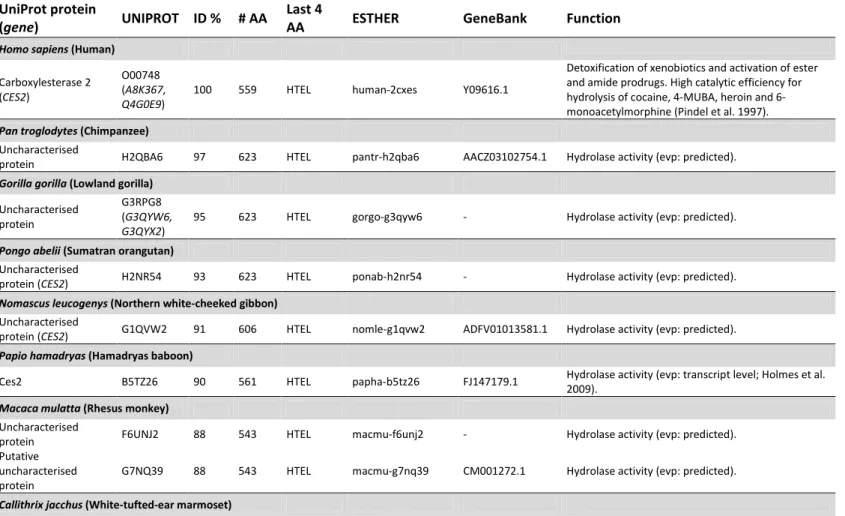
1.1.3 Human carboxylesterase 2
Human CES2 is found in different tissues, especially in the liver, intestine,
and kidney (Fukami and Yokoi 2012). It constitutes a good example of the
aforementioned species variation of CESs. Through a BLAST-P (Altschul et al. 1997)
search, the first fifty hits correspond exclusively to eighteen mammalian species
(Table 1.2). The search was performed in ESTHER database against all protein
sequences available (http://bioweb.ensam.inra.fr/ESTHER/general?what=index),
using as query the amino acid sequence of hCES2, which is shown as the first entry of
sequence identities towards the used query, as well as additional information are
provided. These include: protein accession numbers in ESTHER and UniProt
databases; alternative protein entries (italicized); protein name and function
information available at UniProt database; gene designation (italicized) when existing;
GeneBank accession number; level of experimental evidence of protein existence;
length of the full protein as well as the last four amino acids. The first non-mammalian
hydrolase arising in the search is an uncharacterised protein from Anolis carolinensis, an American chameleon (class Reptilia), showing 50% amino acid sequence identity to hCES2, whereas hCES1 only appears later, with 46% identity towards the query (See
Appendice Table).
Genetic expression of hCES2 has been well documented, ranging from gene structure to the potential promoters involved in the transcription initiation in
different human tissues (Wu et al. 2003). Moreover, 3 trancript variants of the gene
(Schiel et al. 2007) as well as several single nucleotide polymorphisms (SNPs) have
been documented (Wu et al. 2004; Kubo et al. 2005). Some of these, such as the
Table 1.2 CES2 protein across different species
UniProt protein
(gene) UNIPROT ID % # AA
Last 4
AA ESTHER GeneBank Function
Homo sapiens (Human)
Carboxylesterase 2 (CES2)
O00748 (A8K367, Q4G0E9)
100 559 HTEL human-2cxes Y09616.1
Detoxification of xenobiotics and activation of ester and amide prodrugs. High catalytic efficiency for hydrolysis of cocaine, 4-MUBA, heroin and 6-monoacetylmorphine (Pindel et al. 1997).
Pan troglodytes (Chimpanzee)
Uncharacterised
protein H2QBA6 97 623 HTEL pantr-h2qba6 AACZ03102754.1 Hydrolase activity (evp: predicted).
Gorilla gorilla (Lowland gorilla)
Uncharacterised protein
G3RPG8 (G3QYW6, G3QYX2)
95 623 HTEL gorgo-g3qyw6 - Hydrolase activity (evp: predicted).
Pongo abelii (Sumatran orangutan)
Uncharacterised
protein (CES2) H2NR54 93 623 HTEL ponab-h2nr54 - Hydrolase activity (evp: predicted). Nomascus leucogenys (Northern white-cheeked gibbon)
Uncharacterised
protein (CES2) G1QVW2 91 606 HTEL nomle-g1qvw2 ADFV01013581.1 Hydrolase activity (evp: predicted).
Papio hamadryas (Hamadryas baboon)
Ces2 B5TZ26 90 561 HTEL papha-b5tz26 FJ147179.1 Hydrolase activity (evp: transcript level; Holmes et al. 2009).
Macaca mulatta (Rhesus monkey)
Uncharacterised
protein F6UNJ2 88 543 HTEL macmu-f6unj2 - Hydrolase activity (evp: predicted).
Putative uncharacterised protein
Uncharacterised protein
F6ZPL6
(F6Z7R7) 83 620 HTEL calja-f6zpl6
ACFV01013743.1, ACFV01013744.1, ACFV01013745.1
Hydrolase activity (evp: predicted).
Ailuropoda melanoleuca (Giant panda)
Uncharacterised protein (CES2)
D2H9C9
(G1MFN8) 77 534 HTEL ailme-d2h9c9
GL192596.1,
ACTA01043044.1 Hydrolase activity (evp: predicted; Li et al. 2010).
Equus caballus (Horse)
Uncharacterised
protein (CES2) F7BJ10 77 579 HTEL horse-f7bj10 - Hydrolase activity (evp: predicted; Wade et al. 2009). Loxodonta africana (African bush elephant)
Uncharacterised
protein (CES2) G3TN98 76 554 HTEL loxaf-g3tn98 - Hydrolase activity (evp: predicted).
Canis lupus familiaris (Dog)
Uncharacterised
protein (Ces2) F1P6W8 74 585 HTEL canfa-f1p6w8 -
Hydrolase activity (evp: predicted; Lindblad-Toh et al. 2005).
Oryctolagus cuniculus (European rabbit)
Uncharacterised
protein G1TZV1 74 558 HTEL rabit-g1tzv1 AAGW02053044.1 Hydrolase activity (evp: predicted). Uncharacterised
protein G1SJQ8 74 621 HTEL rabit-g1sjq8 AAGW02067905.1 Hydrolase activity (evp: predicted). Uncharacterised
protein G1T6X7 74 558 HTEL rabit-g1t6x7 AAGW02053044.1 Hydrolase activity (evp: predicted). Uncharacterised
protein G1TDR0 74 534 HTEL rabit-g1tdr0 AAGW02067906.1 Hydrolase activity (evp: predicted). Uncharacterised
protein G1T7P3 73 532 HTEL rabit-g1t7p3 AAGW02053044.1 Hydrolase activity (evp: predicted). Uncharacterised
protein G1T7Q5 73 561 HTEL rabit-g1t7q5 AAGW02053044.1 Hydrolase activity (evp: predicted). Uncharacterised
protein G1SN51 72 561 HTEL rabit-g1sn51
AAGW02067906.1
AGW02067907.1 Hydrolase activity (evp: predicted). Uncharacterised
Liver carboxylesterase
2 (CES2) P14943 72 532 HTEL rabit-2cxes -
Detoxification of xenobiotics and activation of ester and amide prodrugs (evp: protein level; Ozols 1989). Uncharacterised
protein G1T6L1 71 528 HTEL rabit-g1t6l1 AAGW02053044.1 Hydrolase activity (evp: predicted).
Rattus norvegicus (Brown rat)
Ces2h (Ces2h) Q32Q55 73 558 HTEL ratno-q32q55 BC107806.1 Carboxylesterase activity (evp: transcript level; Gibbs et al. 2004).
LOC679149 protein Q4QR68 70 561 HTEL ratno-q4qr68 BC097486.1 Carboxylesterase activity (evp: transcript level). Carboxylesterase 5,
isoform CRA_a (Ces2e) G3V7J5 70 557 HTEL ratno-phebest
D50580.1,
CH474006.1 Hydrolase activity (evp: predicted; Gibbs et al. 2004). Ces2c (Ces2c) G3V9D8 70 561 HAEL ratno-pbcxe AB010635.1,
CH473986.1 Hydrolase activity (evp: predicted; Gibbs et al. 2004). Carboxylesterase
(Ces2) O70177 69 561 HAEL ratno-sicxe AB010632 Carboxylesterase activity (evp: predicted).
Ces2g (Ces2g) D3ZXQ0 67 560 HKEL ratno-d3zxq0 CH473972.1 Carboxylesterase activity (evp: predicted; Gibbs et al. 2004).
Carboxylesterase
(Protein Ces2a) Q8K3R0 66 558 HAEL ratno-LOC246252
NM_144743, AY034877
Carboxylesterase activity (evp: transcript level; Gibbs et al. 2004).
Ces2i (Ces2i) D3ZE31 66 559 HAEL ratno-d3ze31 - Carboxylesterase activity (evp: predicted; Gibbs et al. 2004).
Cavia porcellus (Guinea pig)
Uncharacterised
protein (CES2) H0V5V8 73 568 HTEL cavpo-h0v5v8 - Carboxylesterase activity (evp: predicted).
Bos taurus (Aurochs)
Carboxylesterase 2 (intestine, liver; CES2)
Q3T0R6
(F1MU22) 72 553 HTEL bovin-q3t0r6
BC102288.1,
AAFC03046191.1 Carboxylesterase activity (evp: transcript level).
Heterocephalus glaber (Naked mole rat)
Carboxylesterase 2 G5BZE3 72 553 HAEL hetga-g5bze3 JH172552.1 Hydrolase activity (evp: predicted; Kim et al. 2011). Carboxylesterase 2 G5BP68 70 570 AAQE hetga-g5bp68 JH171227.1 Hydrolase activity (evp: predicted; Kim et al. 2011). Carboxylesterase 2 G5BP66 69 562 HAEL hetga-g5bp66 JH171227.1 Hydrolase activity (evp: predicted; Kim et al. 2011).
Ces2h (Ces2h) F6Z9B9 72 558 HKEL mouse-Ces2h AC166833.4, XM_488149.1
Carboxylesterase activity (evp: predicted; Church et al. 2009).
Ces2c (Ces2c) Q91WG0 71 561 HREL mouse-Ces2c BC015290.1, AC166833.4
Carboxylesterase activity (evp: transcript level; Furihata et al. 2003).
Uncharacterised
protein (Ces2d-ps) D3YWM6 71 558 HREL mouse-Ces2d-ps -
Hydrolase activity (evp: predicted; Church et al. 2009).
Ces2b (Ces2b) Q6PDB7 71 556 HTEL mouse-Ces2b BC058815.1 Carboxylesterase activity (evp: transcript level; Mural et al. 2002).
Carboxylesterase 5
(Protein Ces2e) Q8BK48 70 559 HkEL mouse-Ces2e
XM_134366,
BC022148 Carboxylesterase activity (evp: transcript level). Ces2g (Ces2g) E9PV38 68 560 HKEL mouse-Ces2g
BC027185.1, BC024548.1, BC026641.1
Carboxylesterase activity (evp: predicted; Church et al. 2009).
Putative uncharacterised protein (Ces2a; Ces6)
Q3TMR2
(E9Q3D0) 67 525 HAEL mouse-Ces2a
BC024491.1, BC024517.1, BC025537.1
Carboxylesterase activity (evp: transcript level).
Cricetulus griseus (Chinese hamster)
Carboxylesterase 2 G3IIG3 71 511 HGEL crigr-g3iig3 JH003006.1 Hydrolase activity (evp: predicted; Xu et al. 2011). Liver carboxylesterase G3IIG1 70 561 HKEL crigr-g3iig1 JH003006.1 Hydrolase activity (evp: predicted; Xu et al. 2011). Liver carboxylesterase G3I767 70 535 HKEL crigr-g3i767.2 JH001411.1 Hydrolase activity (evp: predicted; Xu et al. 2011). Liver carboxylesterase G3I766 69 561 HQEL crigr-g3i766 JH001411.1 Hydrolase activity (evp: predicted; Xu et al. 2011). Liver carboxylesterase G3I769 67 545 HAEL crigr-g3i769: JH001411.1 Hydrolase activity (evp: predicted; Xu et al. 2011).
Mesocricetus auratus (Golden hamster)
Carboxylesterase O35533 70 559 HQEL mesau-cxest2 D50577 Carboxylesterase activity (evp: trancript level; Sone et al. 1994)
Liver carboxylesterase Q64419 66 561 HSEL mesau-cxest D28566.1
Detoxification of xenobiotics and activation of ester and amide prodrugs. (evp: transcript level; Sone et al. 1994).
1.2 The tools to study carboxylesterases
The study of CESs, such as their quantification and differentiation from other
enzymes, may be performed through different techniques targeting expression levels,
from gene to protein expression, by generating specific primers and antibodies
(Sanghani et al. 2003; Morgan et al. 1994). Recently, simultaneous detection and
quantification of hCES1 and hCES2 proteins by liquid chromatography coupled with
tandem mass spectrometry (LC-MS/MS) was also shown to be possible (Sato et al.
2012). A direct correlation between protein levels and enzyme activity is not always
possible for these enzymes. Reports of both inability (Ross et al. 2012) and ability of
performing such correlations (Sato et al. 2012) may be found in the literature.
Different tools to evaluate CESs activity exist and have been reviewed, such as
spectrophotometry, high-performance liquid chromatography (HPLC), and in-gel
activity assays (Ross and Crow 2007).
Carboxylesterase-mediated hydrolysis has been thoroughly studied and it is
clear that many substrates have recognittion among different CESs as well as with
other esterases. For example, p-nitrophenyl acetate (p-NPA), a classical CESs substrate, is hydrolysed not only by the different CESs but also by cholinesterases
(Satoh and Hosokawa 2006). Even pharmacologically relevant compounds known for
their higher specificities towards CES2, such as aspirin (Tang et al. 2006), irinotecan
(Humerickhouse et al. 2000), and cocaine (Hatfield et al. 2010) are also hydrolysed by
BuChE (Li et al. 2005).
Due to this promiscuity in substrate hydrolysis, it is very difficult to
differentiate a single carboxylesterase activity in samples containing multiple CESs or
other esterases by simple spectrophotometric assays. A classic example is the late
demonstration of the absence of CES activity in healthy human plasma through an
in-gel activity assay with different substrates and inhibitors for CESs, cholinesterases,
detection in the presence of certain substrates and inhibitors with protein detection
through Western blot, it was recently proposed to be possible to characterise an
individual’s liver in terms of CES profile (Ross et al. 2012).
A different perspective in CES analysis may arise upon looking to more
physiological relevant conditions such as protein localisation and/or activity in
whole-living cells, instead of analysing purified enzymes, cell lysates, or tissue homogenates.
The importance of addressing how enzymes behave in living cells, where complex
networks of protein interactions occur, is detailed in Section 1.3.1. Recent advances in
whole-living cell analysis have been made for cytoplasmic membrane anchored
enzymes with the use of fluorescent probes (Ferruzza et al. 2012). Evaluating the
activity of these types of enzymes is by far an easier task than to quantify the activity
of ER localised enzymes, as it is the case of the majority of CESs. Nonetheless, recent
advances have been made, such as the development of specific CES fluorescent
probes (Wang et al. 2011) that have been applied to live imaging of whole-living cells
(Hakamata et al. 2011).
The development of simple, fast, and reliable tools enabling the
differentiation and quantification of different CESs expression and activity is thus a
very active research field, where some controversial questions still remain. To
develop and improve the methodologies, as well as to test new substrates and
inhibitors that will also improve already existing methodologies, purified CES enzymes
are needed, as they constitute an easier step towards the analysis of more complex
samples, such as cell extracts and tissue homogenates. Insights into the latest
1.3 From drug metabolising enzymes to intestinal permeability models
1.3.1 The importance of seeing both sides - The irinotecan pathway in a human organism as a case study
Notwithstanding the importance of studying purified or recombinant enzymes, it is
crucial to understand their behaviour in the most physiologically relevant level,
meaning the environment where native enzymes exist. This may mean that studies
involving cells and/or organisms may have to be performed. An example involves the
metabolism of the chemotherapeutic drug irinotecan. As mentioned above, hCES2
hydrolyses irinotecan into its active metabolite, SN-38. The enzymatic reaction has
been characterised with purified enzyme (Humerickhouse et al. 2000), resulting in the
determination of important kinetic parameters. Nonetheless, the pathway of
intravenous-delivered (IV) irinotecan transport and metabolism in the organism is
complex involving the interplay of a complex network of proteins (Figure 1.1) in
different cellular compartments. Phase I metabolising enzymes, including CES and
CYP3A family members, as well as Phase II enzymes, such as UGTs and the interplay
with drug efflux pumps from the ABC family. Both CESs and BuChEs are able to
hydrolyse irinotecan, with CESs being more efficient to convert to SN-38. Human CES2
was shown to be one hundred-fold more efficient than hCES1, and hCES3 the least
efficient (Humerickhouse et al. 2000). Due to the differences in the hCES1 and hCES2
expression in the liver and intestine, hCES2 accounts for the majority of irinotecan
activation in the intestine and kidney while hCES1 has an important function in the
activation of this pro-drug in the liver (Hatfield et al. 2011).
Irinotecan is inactivated through oxidation by CYP3A4/5 into aminopentane
carboxylic acid (APC), M4, and primary amine metabolite (NPC; Santos et al. 2000;
Innocenti et al. 2009). SN-38 is inactivated by UGT Phase II enzymes, namely
Active transport of irinotecan, SN-38, and SN-38G to the extracellular space is
facilitated by a few members from the ATP-binding cassette (ABC) family. Irinotecan is
transported by ABCC1, ABCB1, and ABCC2; SN-38G is transported by ABCC2, and
ABCG2; all of these may be involved in pumping SN-38 (Kweekel et al. 2008).
The chemotherapeutic action of irinotecan is achieved through the binding of
SN-38 to topoisomerase I (TOPO-1) during cell division, more specifically, during DNA
replication. This complex, formed by SN-38 and TOPO-1, becomes blocked leading to
the destruction of the DNA when the replication machinery clashes with it (Marsh and
Hoskins 2010). Delayed-type diarrhoea and neutropenia, a decrease in neutrophils
increasing the risk of infection (Ammann et al. 2012), are the most common toxic
effects of irinotecan and their severity, reaching life-threatening cases, has been
linked to some polymorphisms in certain genes, such as UGT1A1*28 (Marsh and Hoskins 2010; Innocenti et al. 2009; de Jong et al. 2007).
Other examples of the interaction between different metabolic enzymes and
efflux transporters have also been studied, such as the case of the interplay between
P-glycoprotein (P-gp), multidrug resistance-associated protein 2 (Mrp2), CYP3A, and
CES2 in the oral availability of vinorelbine, a pharmaceutical indicated for the
treatment of some types of lung and breast cancer. Looking to these interactions in
living cells or organisms may be crucial to better understand the reasons behind
inter-individual variability in oral dosing (Lagas et al. 2012).
1.3.2 Intestinal permeability and metabolism overview
Oral delivery is still the most common way of dosing drugs to the patients (Buckley et
al. 2012). Every drug undergoes a series of transformations inside the organism that
depends not only on the type of drug but also on the individual, due to the genetic
variability found in humans. Overall, this path is refered to as the ADME (absorption,
such as intestine, liver, and kidney, where different transformations may occur. It may
be categorised in different stages, from Phase I-III. Phase I involves the modification
of the compounds which may occur through oxidation or hydrolysis, for example. In
Phase II, conjugation enzymes usually increase the hydrophilicity of the metabolised
compound through the addition of a hydrophilic molecule, such as glucoronic acid,
sulphate, or glutathione, to facilitate its elimination. Phase III involves the elimination
of the compounds through active transporters (Huynh et al. 2009). Tissue distribution
of these proteins has been previously reported (Nishimura and Naito 2006). Being the
primary site for xenobiotic absorption and with an important function in drug
metabolism (Shen et al. 1997), a special focus is devoted to the small intestine in the
following sections.
1.3.2.1 Architecture and function of intestinal epithelium
The intestinal epithelium is found in two, the small and large intestine, of the four
segments composing the gastrointestinal tract (GI). In the large intestine, the
intestinal epithelium has a flat shape, punctuated with invaginations. In the small
intestine, it is composed of projections, the villi, and invaginations, the crypts of
Lieberkühn (Figure 1.2). This different morphology is related with different functions
of both segments, where the small intestine is mainly responsible for absorption and
re-absorption of water is mainly performed by the large intestine. (Rizk and Barker
2012; Dubreuil 2012; Vereecke et al. 2011). The intestinal epithelium protects the
internal environment of the organ acting as a selective barrier, due to the tight
junctions established between adjacent cells that also contribute to the integrity of
the epithelial layer.
There are different types of junctions characterised by their localisation and
by the types of proteins involved. From apical to basolateral, one finds tight junctions
regulating the paracellular permeability, a type of transport occurring between cells.
Gap junctions and desmosomes are the more basolateral types of junctions (Elamin et
al. 2012; Ashida et al. 2011; Gumbiner 1996).
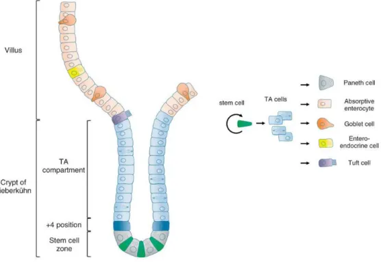
Figure 1.2 Intestinal epithelium architecture and renewal.Intestinal epithelial cells differentiate as they migrate along the crypt-villus axis. Stem cells, housed at the crypt, give rise to the transient amplifying (TA) cells, which move forward originating the different intestinal epithelial cells. Fully differentiated cells reaching the tip of the villi enter programed cell death (apoptosis) being liberated into the lumen. Paneth cells are the exception cells migrating downwards. Adapted and reprinted with permission from Wiley Periodicals, Inc.: Wiley Interdisciplinary Reviews: Systems Biology and Medicine (Rizk and Barker 2012), copyright (2012).
Mammalian intestinal epithelia are composed of different differentiated cell
types: enterocytes (or absorptive cells), enteroendocrine, goblet, Paneth, Tuft, and M
cells. All of these originate from transient amplifying cells (TA) which in turn derive
from the stem cells housed at the base of the crypt. These cells differentiate along
their migration path across the crypt-villus axis with a rapid renewal of 2-5 days. The
(Dubreuil 2012; Vereecke et al. 2011; Nicoletti 2000). The majority of intestinal
epithelium is composed of enterocytes, polarised cells specialised in absorption,
bearing microvilli at their apical surface, also called the brush border membrane.
Goblet cells are responsible for mucus production and are found both in small and
large intestinal epithelia. Paneth cells are specialised in the production of
antimicrobial factors being exclusive to the small intestine (Vereecke et al. 2011;
Simon-Assmann et al. 2007). Enteroendocrine, Tuft, and M cells are less represented
cell populations. The first are responsible for the secretion of diverse hormones thus
contributing to the overall homeostasis of the epithelium. The functional significance
of Tuft cells remains uncertain (Rizk and Barker 2012). M cells, present in both small
and large intestinal epithelia, have defensive roles, being a route for the entry and
contact between antigens (such as those from bacteria and virus) and the intestinal
immune system (Nicoletti 2000). M cells have also been shown to be able to derive
from differentiated absorptive enterocytes. In a curious experimental approach, fully
differentiated enterocyte-like Caco-2 cells cultured in the presence of primary
lymphocytes were converted into cells having M cell characteristics (Kernéis et al.
1997).
1.3.2.2 Enterocytes – permeability and metabolism
Until reaching the systemic circulation, orally delivered therapeutics must cross the
intestinal epithelium followed by the liver, through the hepatic portal vein. The
exceptions, those not reaching the liver, are absorbed but directly enter the lymphatic
system or are absorbed in the distal rectum (Shen et al. 1997). Oral bioavailability
(Foral) corresponds to the fraction of dose that is absorbed (Fa) and not metabolised,
crosses the intestinal epithelia to the hepatic portal vein (Fg) and not metabolised in
the liver (Fh). Thus, the fraction of the administered drug that effectively reaches its
known as first-pass biotransformation, where several metabolic enzymes and
transporters are invovled (Thelen and Dressman 2009).
The players involved in transport and metabolism in enterocytes have been
extensively studied through several techniques. Different layers of expression and
function, from genomics to metabolomics and transportomics have been thoroughly
evaluated under various physiological conditions including inflammation (Stegmann
et al. 2006; Fleet 2007; Béaslas et al. 2008; Romero-Calvo et al. 2011). Orally delivered
xenobiotics, such as nutrients and drugs, have various routes to cross the intestinal
epithelium (Figure 1.3).
Figure 1.3 Intestinal absorption routes. Xenobiotic compounds may cross the intestinal epithelium through (1, 2, 3, 5, 6 and 7) or in between enterocytes (4). 1. Transcellular passive transport. 2. Active transport. 3. Facilitated passive diffusion. 4. Paracellular passive transport. 5. Efflux active transport. 6. Metabolic reaction. 7. Endocytosis. Reprinted with permission from Springer (Springer, Part of Springer Science+Business Media): The AAPS Journal (Balimane et al. 2006), copyright (2012).
Intestinal absorption may occur through the intestinal cells or between cells
and may involve the interplay of both transport proteins as well as metabolic
enzymes. Passive absorption most commonly occurs through the cells, known as
transcellular transport, where compounds are able to cross the cell membrane due to
an adequate lipophylicity. Transport across channel proteins without energy
(uniport), is another possibility. Absorption can also occur between cells, known as
paracellular transport, where compounds transverse the junctions between cells.
Drugs and other compounds may also be transported through the cells by
endocytosis and active primary transport, utilising ATP, or active secondary transport,
such as coupled carrier-mediated transport (symport or antiport). Efflux transporters
are examples of active primary transporters and counter the apical to basolateral
transport by the active transport of compounds towards the lumen of the intestine.
They are members of the ABC family such as the breast cancer resistance protein
(BCRP), P-gp and multidrug resistance-associated protein (MRP). Some Phase I
metabolic enzymes, such as those from CYP and CES families, may metabolise the
transported compound thus interfering with its absorption rate (Balimane et al. 2006;
Buckley et al. 2012; Alberts et al. 2002). An example is the interplay between CYP3A
metabolic enzymes and P-gp transport, where several drugs are substrates of both
proteins (Christians et al. 2005; van Waterschoot and Schinkel 2011).
The liver is the organ traditionally regarded as the primary site of drug
metabolism. Nonetheless, the importance of intestinal metabolism should not be
overlooked. It has been demonstrated that the majority of the metabolic enzymes
found in liver cells are also present in the enterocytes. The enzymes found in small
intestine have been extensively reviewed and include Phase I (CYP enzymes, with
CYP3A4 being the most abundant; esterases; epoxide hydrolase; alcohol
dehydrogenase) and Phase II (UGTs, sulfonotransferases, acetyl transferases, and
glutathione S-transferases) metabolic enzymes (Bonnefille et al. 2011; Thelen and
1.4 Tools to study intestinal permeability and metabolism
Several tools are available to evaluate intestinal permeability of drugs. Ranging from
in vivo, in situ, ex vivo, in silico, and in vitro assays, they have been extensively reviewed in the literature (Cheng et al. 2008; Buckley et al. 2012; Volpe 2010; Geerts
et al. 2011). The suitability of any given experimental model may be evaluated
according to the accuracy of the in vitro-in vivo correlation (IVIVC) (Volpe 2010). No perfect model exists and the choice of which model(s) to use must rely on the
knowledge of their main advantages and limitations. Several authors have proposed
different strategies and outcomes on how to perform this choice (Fagerholm 2007;
Christensen et al. 2012). Under the scope of this thesis, a brief overview of the
possible approaches is performed with a special emphasis on in vitro models and Caco-2 cells.
1.4.1 In vivo
The usage of experimental or laboratory animal models constitutes the only whole
living organism approach possible, besides the clinical studies performed with
humans. Rat, dog, monkey, sheep, mouse, and pig are some of the most commonly
used animals (Harrison et al. 2004; Fagerholm 2007; Cheng et al. 2008).
In addition to the regulatory, ethical and economic constraints inherent to
the use of animals (please refer to Section 1.4.6) another severe limitation arises from
the demonstrated species differences that may affect the accuracy of the IVIVC (Crow
et al. 2007; Williams et al. 2011). In fact, species differences may impact more than
the extrapolation of absorption data to humans. An unfortunately notorious example
is thalidomide, a mild sleeping pill that reduced morning sickness and was
commercialized in the mid 1960’s. It caused the birth of several thousand impaired
discovered to impair the correct development of rabbit foetuses (Harrison et al.
2004).
1.4.2 In situ
In the perfusion in situ technique, usually, a segment of the intestine of a numbed animal is perfused with a drug solution containing a predetermined concentration. By
measuring the amount of drug that leaves the segment in the original or metabolised
form, one may indirectly determine absorption. Different techniques were developed
such as open, semi open, or closed perfusion (Lennernäs 1998; Volpe 2010).
Perfusion models constitute the best approximation to the anatomy found
in the living organ but require the usage of animals, their manipulation through
anaesthesia and surgery, thus being an invasive, time consuming, and low throughput
technique (Volpe 2010; Harrison et al. 2004). Resorting to animals, it shares the
limitations of in vivo assays, mentioned above, using animal experimental models.
1.4.3 Ex vivo
In explant cultures, portions of the GI tissues are removed and cultured according to
different methods and culture techniques (Randall et al. 2011). Two examples are the
everted gut sac model and the diffusion chamber. In the first, a portion of everted
intestine is filled, tied up and placed in a chamber containing the drug solution. The
permeation of the drug is determined by measuring how much appears inside the sac.
Different animals have been used for the application of this technique, with rat being
the most common (Volpe 2010; Alam et al. 2012). In the diffusion chamber method, a
portion of excised tissue is opened and cultured as a single layer in the interface of
two chambers. The absorption rate is determined by measuring the amount of drug,