89 89 89 89 89 Mem Inst Oswaldo Cruz, Rio de Janeiro, Vol. 96(1): 89-98, January, 2001
Immunochemotherapy in American Cutaneous
Leishmaniasis: Immunological Aspects before and after
Treatment
VPCP Toledo, W Mayrink*, KJ Gollob**, MAP Oliveira**, CA da Costa,
O Genaro**, JA Pinto***, LCC Afonso****/
+Departamento de Análises Clínicas e Toxicológicas, Faculdade de Farmácia *Departamento de Parasitologia **Departamento de Bioquímica e Imunologia, Instituto de Ciências Biológicas ***Departamento de Pediatria, Faculdade de Medicina, Universidade Federal de Minas Gerais, Belo Horizonte, MG, Brasil ****Departamento
de Ciências Biológicas/Nupeb, Instituto de Ciências Exatas e Biológicas, Universidade Federal de Ouro Preto, Campus Universitário, Morro do Cruzeiro, 35400-000 Ouro Preto, MG, Brasil
In this study, we evaluated the immune response of patients suffering from cutaneous leishmaniasis treated with two distinct protocols. One group was treated with conventional chemotherapy using pen-tavalent antimonium salts and the other with immunochemotherapy where a vaccine against cutaneous leishmaniasis was combined with the antimonium salt. Our results show that, although no differences were observed in the necessary time for complete healing of the lesions between the two treatments, peripheral blood mononuclear cells from patients treated by chemotherapy showed smaller lymphoproliferative responses at the end of the treatment than those from patients in the immunochemotherapy group. Furthermore, IFN-γ production was also different between the two groups. While cells from patients in the chemotherapy group produced more IFN-γ at the end of treatment, a significant decrease in this cytokine production was associated with healing in the immunochemotherapy group. In addition, IL-10 production was also less intense in this latter group. Finally, an increase in CD8+ -IFN-γ producing cells was detected in the chemotherapy group. Together these results point to an alternative treatment protocol where healing can be induced with a decreased production of a poten-tially toxic cytokine.
Key words: leishmaniasis - immunochemotherapy - human - interferon-γ
Cutaneous leishmaniasis is caused by a proto-zoa from the genus Leishmania that infect mac-rophages from many mammals including humans. The disease, depending on the species of the para-site, is characterized by occurrence of ulcerative lesions in the skin and/or mucosae that frequently result in permanent disfiguration of the patient. Ac-cording to the World Health Organization, 12 mil-lion people in 88 countries are currently affected by the various forms of the disease, with an inci-dence of 1-1.5 million new cases per year. Treat-ment of leishmaniasis is based, primarily, on the administration of antimonium salts which, due to
their toxicity (WHO 1990), may not be used in several cases. This fact, associated with the oc-currence of resistance to treatment (Marsden 1985, Grogl et al. 1992), has increased the search for new alternative therapies.
A vaccine against cutaneous leishmaniasis de-veloped by Mayrink et al. (1979) has been used not only in the prevention of the disease (Mayrink et al. 1985, 1986, Antunes et al. 1986, Nascimento et al. 1990) but also as an immunotherapeutic agent in cases where the antimonium salts can not be used (Mayrink et al. 1992, Hermeto et al. 1994), con-firming the results of Convit et al. (1987) who used a different promastigote antigen preparation in association with BCG. This protocol, although highly successful, had the caveat of being substan-tially longer than conventional chemotherapy with antimonium salts. An attempt to reduce the treat-ment period was made by combining antimonium and vaccine administration (Genaro et al. 1996). This study showed that the combined therapy was able to reduce the time necessary for complete heal-ing of the lesions when compared to conventional chemotherapy.
Financial support: UNDP/World Bank/WHO Special Programme for Research and Training in Tropical Dis-eases (TDR), CNPq, Finep/CNPq/Pronex, Biobrás SA and Universidade Federal de Ouro Preto.
+Corresponding author. Fax: +55-31-559.1680. E-mail:
90 90 90 90
90 Immunochemotherapy in Cutaneous Leishmaniasis VPCP Toledo
Several studies have demonstrated that there is an important immunological component in the re-sponse to treatment with antimonials (Berger & Fairlamb 1992, Coutinho et al. 1996, 1998). Murray et al. (1989) demonstrated that T cell-de-ficient mice were unable to control L. donovani proliferation when treated with Pentostam®, sug-gesting that these cells (probably through cytokine production) are involved in the healing process following treatment. In humans, CD8+ T cells are, apparently, involved in the healing process and elevated IFN-γ production at the end of the treat-ment has also been shown (da Cruz et al. 1994a). Furthermore, immunocompromised patients have been shown to be refractory to conventional che-motherapy, at least in some cases of visceral leish-maniasis (Peters et al. 1990).
Our main objective in this study was to under-stand the immunological aspects involved in the healing process associated with an alternative pro-tocol based on the combined action of Glu-cantime® and a vaccine against cutaneous leish-maniasis (Leishvacin®). The results described here, although not showing the same efficacy as previous trials, present an intriguing aspect regard-ing the immune mechanism underlyregard-ing the heal-ing process. The immune mechanism involved in the healing process was probably different in each treatment, since cells from patients treated by the combined therapy produced significantly less IFN-γ in vitro than those from patients treated by the conventional protocol.
MATERIALS AND METHODS
Antimonial and vaccine - Glucantime® – Rhodia Farma Ltda. (SP, Brazil) – was used as pro-vided, in 5 ml ampoules containing 425 mg of meglumine antimoniate. Leishvacin® – Biobrás S.A. (Montes Claros, MG, Brazil) – was prepared, under Good Manufacturing Procedures (GMP), as a concentrate containing 240 µg/ml total nitrogen of killed promastigotes from five different Leish-mania strains diluted in phosphate buffer 0.1 M pH 7.4, with timerosal phosphate 0.01% as a pre-servative (Mayrink et al. 1979). Staphylococcus aureus Cowan strain – SAC – (Calbiochem, La Jolla, CA, USA) was used at a concentration of 0.01% to stimulate IL-10 production by periph-eral blood mononuclear cells (PBMC).
Antigen for immunological assays - L. V. braziliensis (MHOM/BR/2903) stationary phase promastigotes (seven days) were harvested from LIT medium (Camargo 1964), washed three times in sterile PBS by centrifugation at 1,000 x g for 10 min at 4ºC. Cells were disrupted by three cycles of sonication 1 min, 20 Hz (Ultrasonic Disrupter, Tekmar Co., Cincinnati, OH). The suspension was
sedimented at 4,000 x g for 10 min and the super-natant collected for use as antigenic stimulus after protein quantification (Lowry et al. 1951). Aliquots (1 mg/ml) were stored at -70ºC until needed.
Study subjects - A total of 117 patients suffer-ing from cutaneous leishmaniasis volunteered to take part in the trial. Written consent was obtained from the volunteers or from parents/guardians of those under 18 years of age. Patients were diag-nosed on basis of clinical and parasitological cri-teria. Patients included in the trial patients were older than five years and had the cutaneous form of the disease with confirmed diagnostic. Preg-nant women or those with debilitating diseases were not included in the trial as well as those who refused to sign the informed consent. The study protocols were in accordance with The Helsinki Declaration and has been approved by the Ethical Committee from the Escola de Medicina, Universidade Federal de Minas Gerais.
Treatment protocols - The study was designed as a double blinded randomized trial. Based on previous non-randomized trials (Genaro et al. 1996) and estimating an α value of 0.01 (type I error) and a β value of 0.05 (type II error) a sample size of 50 volunteers per group was calculated for the trial, including a 10% chance of withdrawal. Patients were allocated at a 1:1 ratio into two groups. Both groups received i.m. injections of Glucantime® at daily doses of 1 ml/5 kg body weight, until a maximum dose of 10 ml/day. Pa-tients treated by immunochemotherapy (IC) re-ceived simultaneous subcutaneous injections of vaccine in the forearm, according to the following protocol (Mayrink et al. 1991): day 1: 100 µl; day 2: 200 µl; day 3: 300 µl; day 4: 400 µl and days 5 to 10: 500 µl. After a resting period of 10 days another series of antimonial and vaccine injections (day 1 to 10: 500 µl) was administered. This treat-ment/rest/treatment protocol was maintained until complete healing of lesions. The control group (CH) received, besides Glucantime®, placebo in-jections (sterile PBS with 0.01% timerosal phos-phate) in the forearm according to the same proto-col described for the previous group. The occur-rence of adverse reactions was monitored along the treatment period. No report of adverse reac-tion was filed in both treatment groups during the whole trial.
91 91 91 91 91 Mem Inst Oswaldo Cruz, Rio de Janeiro, Vol. 96(1), January, 2001
Lymphocyte proliferation assays - This assay was performed as previously described (Mendonça et al. 1986). Briefly, PBMC were isolated from blood samples by centrifugation over a Ficoll-Hypaque gradient. 3.5 x 10⁶ cells were stimulated in RPMI medium supplemented with 5% human AB serum for 24 h with 0.1 µg Phytohemaglutinin A (PHA) or with 10 µg (protein content) of L. braziliensis antigen for 72 h. Proliferation was assessed by incorporation of [3H]-Thymidine (SIGMA, St. Louis, MO, USA, 5.3 Ci/mmol) for the last 18 h of culture. Results were expressed as
∆kcpm (kcpm of stimulated - kcpm of control cul-tures).
Cytokine production - Cytokine production was assayed on supernatants from PBMC (5 x 10⁶ cells/ ml) stimulated with 50 µg/ml of L. braziliensis an-tigen. IL-2, was assayed on 24 h supernatants (Intertest 2, Genzyme Immunobiologicals, USA). IL-10 was assayed on 72 h supernatants (Quantikine Human IL-10 Immunoassay, R & D systems, USA). All tests were performed accord-ing to manufacturer’s instructions. IL-12 and IFN-γ assays were performed on 24 and 72 h su-pernatants, respectively, according to the protocols described below.
IFN-γ ELISA assay - IFN-γ production was measured in three day culture supernatants by a sandwich ELISA. Plates were coated with a mouse monoclonal antibody (B133.1 – a kind gift from Dr Giorgio Trinchieri, The Wistar Institute, Phila-delphia, PA, USA) at 5 µg/ml in 0.1 M NaHCO3/ NaCO3 buffer, pH 8.6, overnight at 4oC. After blocking with phosphate buffered saline (PBS)/5% bovine fetal serum (BFS) for 1 h, plates were washed three times with PBS/Tween 20 0.05%. Samples were added directly to the plate and incu-bated for 2 h at 37oC. Standard curve was per-formed using recombinant human IFN-γ diluted in PBS/5% BFS at concentrations which varied from 12.5 to 0.02 ng/ml. After washing, plates were incubated with biotinylated-B133.5 mono-clonal antibody, at a concentration of 0.5 µg/ml, for 1 h. This incubation was followed by washing and incubation with streptoavidin-peroxidase for another hour. ABTS (SIGMA - 0.5 mg/ml) and hydrogen peroxide (0.01%) were used as sub-strates. Readings were performed at 415 nm.
IL-12 ELISA assay - IL-12 p40 detection was carried out by a ELISA sandwich using mouse anti human IL-12 p40 monoclonal antibodies (C11.15 and biotinylated-C8.6). The same procedure and concentrations described above were used in this assay.
Flow cytometry analysis - PBMC were col-lected before and after treatment and stimulated in vitro with L. braziliensis. After 72 h, 2 x 10⁶ cells
were harvested, washed in sterile PBS and stained with CD3-FITC, and rodamine labeled anti-CD4 or anti-CD8 monoclonal antibodies (Beckton-Dickson, San Jose, CA, USA). Cytometric analy-sis was performed on a Coulter – EPICS 751 cy-tometer and data analyzed by the LYSIS II soft-ware. A minimum of 30,000 events were analyzed per sample.
IFN-γ intracellular staining - PBMC were stimulated in vitro with L. braziliensis for 72 h. Brefeldin A (1 mg/ml) was added during the last 6 h of culture. Cells were harvested and stained for CD4 or CD8 markers as described above and then washed with PBS/0.02% sodium azide/0.2% bo-vine soroalbumin. Cells were then fixed with 2% paraformaldehyde and washed again. Aliquots of 2 x 10⁶ permeabilized cells (50 µl) were incu-bated with FITC labeled anti-human IFN-γ mono-clonal antibody (Pharmingen, San Diego, CA, USA), washed and analyzed by flow cytometry immediately after (Prussin & Metcalfe 1995).
Statistical analysis - Analysis of patient distri-bution between groups was made by univariated analysis. Laboratorial data were analyzed by paired two tail Student’s t test, regression analysis and correlation. p values ≤ 0.05 were considered sig-nificant.
RESULTS
Demographic and clinical data - A total of 117 patients involved in this study were allocated to the two treatment groups on a 1:1 ratio. Demo-graphic data obtained from the two groups showed that they were homogenous in regard to age [30.1 ± 13.4 and 26.2 ± 17.6 years for the chemotherapy (CH) and immunochemotherapy (IC) groups, re-spectively] and sex (53.1% male – CH and 54.7% male – IC). Clinical data regarding the leishmanial infection were also similar between the two groups (Table). Patients from the two groups had lesions that were similar in evolution time, size and num-ber. It is important to mention that the majority of the patients (76%) presented only one well defined lesion, usually in an uncovered part of the limbs. Both groups also presented similar rates of posi-tive Montenegro’s skin test prior to treatment. Although patients in the IC group presented a ten-dency to heal faster, no statistical differences were observed between the two groups when healing rate was measured by the number of treatment series necessary to induce complete healing of the lesions. It should be emphasized, however, that regardless of the treatment adopted, complete healing of the lesions was always obtained at the end of treat-ment.
92 92 92 92
92 Immunochemotherapy in Cutaneous Leishmaniasis VPCP Toledo
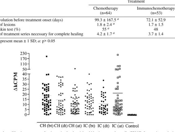
associated to each treatment, PBMC obtained from blood samples taken before treatment, after two series and one month after the end of treatment were stimulated in vitro with Leishmania antigen. While cells from uninfected volunteers did not liferate in response to the antigenic stimulus, pro-liferative responses, measured five days after stimu-lation, varied significantly among the patients in both treatment protocols (Fig. 1). Nonetheless, it was possible to observe that in the CH group there was a decrease in the mean proliferative response after the end of treatment (∆kcpm = 10.4 ± 10.0 – mean ± SD) as compared to results observed be-fore treatment (∆kcpm = 18.2 ± 26.2, p < 0.05). On the hand, no alteration in lymphoproliferation was observed within the IC group, with mean
∆kcpm values remaining practically constant dur-ing the whole treatment period (before treatment 13.3 ± 10.7; after treatment 21.3 ± 35.2, p > 0.05). Cytokine production by stimulated PBMC - The role of cytokines in the control of leishmaniasis has been clearly demonstrated in many animal
Fig. 1: lymphoproliferative responses of antigen stimulated peripheral blood mononuclear cells (PBMC) from patients submit-ted to chemotherapy or immunochemotherapy. Symbols represent ∆kcpm (cpm stimulated cultures - cpm control cultures) for each individual before treatment onset (bt), after the second series of treatment (dt), and after complete healing of lesions (at). Horizontal bars represent the mean value for each group. Treatments – CH: chemotherapy; IC: immunochemotherapy; n = 56 (CH) and 49 (IC); control cultures were performed using PBMC from healthy individuals living outside the endemic area.
TABLE I
Clinical characteristics of patients submitted to chemotherapy or immunochemotherapy
Treatment
Chemotherapy Immunochemotherapy
(n=64) (n=53)
Lesion evolution before treatment onset (days) 99.3 ± 167.5 a 72.1 ± 52.9
Number of lesions 1.8 ± 2.4 a 1.7 ± 1.5
Positive skin test (%) 55 a 48
Number of treatment series necessary for complete healing 4.2 ± 1.7 a 3.7 ± 1.4
Values represent mean ± 1 SD; a: p> 0.05
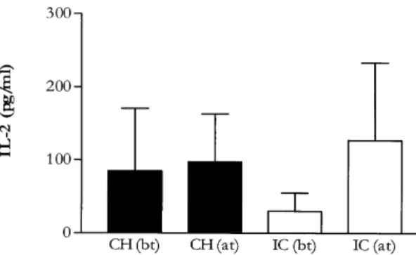
models as well as in the human disease (for review see Scott 1989, Kemp 1997, Etges & Müller 1998). We initially investigated the role of IL-2 during both treatment protocols by measuring its levels in culture supernatants from antigen stimulated PBMC. Fig. 2 shows the results obtained before and after treatment in both groups. While no dif-ferences were observed in the CH group, cells from patients from the IC group produced significantly more IL-2 after treatment than they did before treat-ment (p<0.05). As expected, no significative dif-ferences were observed between the treatment groups before treatment onset (p>0.05).
93 93 93 93 93 Mem Inst Oswaldo Cruz, Rio de Janeiro, Vol. 96(1), January, 2001
IL-12 stimulates IFN-γ production by NK and T cells (Heinzel et al. 1991, Prussin & Metcalfe 1995) and is involved in resistance to infection by Leish-mania (Heinzel et al. 1993, Afonso et al. 1994, Nabors et al. 1995, Bacellar et al. 1996, Mattner et al. 1996). When PBMC were stimulated in vitro with L. braziliensis antigen, production of this cytokine was not detected (data not shown) regard-less of treatment protocol or treatment period. To further investigate the production of IL-12, we mea-sured the IFN-γ production in the presence of C8.6, a neutralizing monoclonal antibody directed against the p40 chain of IL-12 (Fig. 4). We were able to show that, in the CH group, cultures treated with the monoclonal antibody showed a reduction in
IFN-γ production to 54.6 ± 31.8% of the control cultures Fig. 2: IL-2 production by antigen-stimulated stimulated
pe-ripheral blood mononuclear cells. Bars represent mean value ± 1 SD of IL-2 detected in supernatant of antigen stimulated cultures from patients at different times points. (bt): before treatment; (at): after complete healing of lesions. Treatments -CH: chemotherapy; IC: immunochemotherapy; n = 11 (CH) and 10 (IC)
Fig. 3: IFN-γ production by stimulated peripheral blood mononuclear cells (PBMC) increases with chemotherapy but decreases in patients treated by immunochemotherapy. Symbols represent cytokine production by cells stimulated with Leishmania anti-gen for 72 h for each individual before treatment (bt), after the second series of treatment (dt), and after complete healing of lesions (at). Control cultures were made using PBMC from healthy individuals living outside the endemic area. Horizontal bars represent the mean value for each group. Treatments - CH: chemotherapy; IC: immunochemotherapy; n = 64 (CH) and 53 (IC) Fig. 4: IL-12 dependent IFN-γ production by antigen-stimu-lated peripheral blood mononuclear cells (PBMC). PBMC were stimulated for 72 h with antigen in the presence or ab-sence of an anti-IL-12 p40 neutralizing monoclonal antibody (C8.6). Bars represent the mean percentage ± 1 SD of the reduction in IFN-γ production in cultures with the antibody as compared to cultures without IL-12 inhibition. (bt): before treatment; (at): after complete healing of lesions. Treatments -CH: chemotherapy; IC: immunochemotherapy
94 94 94 94
94 Immunochemotherapy in Cutaneous Leishmaniasis VPCP Toledo
before treatment and 46 ± 26% after treatment. A similar reduction was observed in the IC group, in which IFN-γ production was reduced to 53 ± 25.8% of the control culture before treatment and 58 ± 28% after treatment. No statistical differences were de-tected in the reduction rates among groups. These results indicate that, although we were not able to detect IL-12 production in the supernatant, this cytokine was being produced and was responsible, at least partially, for the IFN-γ detected.
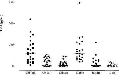
Another cytokine involved in the control of leishmaniasis is IL-10. This cytokine downregu-lates Th1 type responses thus preventing proper healing. Thus, it was of interest to investigate whether IL-10 was being produced in cultures from patients treated by the two different protocols. The results shown in Fig. 5 indicate that a significant decrease in IL-10 production could be observed in both treatment protocols already after the second series of treatment, persisting until the end of treat-ment. Interestingly, IL-10 production at the end of treatment in the IC group (5.7 ± 13.2 pg/ml) was statistically lower than in the CH group (26.7 ± 32.1 pg/ml) (p<0.05).
Since the main difference in the two treatment protocols is the addition of Leishmania antigen, we decided to investigate whether this antigen could inhibit IL-10 production by PBMC stimu-lated with SAC. As shown in Fig. 6, when Leish-mania antigen was added to cultures stimulated with SAC a dose dependent decrease in IL-10 pro-duction was observed in two out of three patients tested, suggesting that antigen inoculation during treatment may have contributed to the lower level of IL-10 detected in the supernatants of patients treated by immunochemotherapy.
Fig. 5: IL-10 production by antigen-stimulated peripheral blood mononuclear cells from patients decreases with treatment. Symbols represent cytokine production by cells stimulated with Leishmania antigen for 72 h for each individual before treat-ment onset (bt), after the second series of treattreat-ment (dt), and after complete healing of lesions (at). Bars represent the mean value for each group ± 1 SD. Treatments - CH: chemotherapy; IC: immunochemotherapy; n = 25 (CH) and 22 (IC)
Fig. 6: Leishmania antigen decreases IL-10 production by SAC-stimulated peripheral blood mononuclear cells (PBMC). PBMC from patients with active lesions (before treatment) were stimulated with SAC in the presence of increasing con-centrations of Leishmania antigen for 72 h. Values are repre-sentative of each individual. Cultures were done in dupli-cates.
95 95 95 95 95 Mem Inst Oswaldo Cruz, Rio de Janeiro, Vol. 96(1), January, 2001
cells from patients in the CH group showed an in-creased percentage of IFN-γ producing cells. This increase in IFN-γ production was particularly noted in the CD8+ population (p<0.05), although CD4+ IFN-γ producing cells also showed a tendency to increase. On the contrary, no differences were de-tected in cells from patients belonging to the IC group. These results are in agreement with the
IFN-γ production in culture supernatants (Fig. 2). DISCUSSION
Treatment against leishmaniasis is based pri-marily on the administration of antimonium salts via the intramuscular route during an extensive period of time. These salts, however, are expen-sive, present considerable toxicity, and require sev-eral injections under supervised administration. These facts justify the search for alternative treat-ment protocols that could reduce the amount of antimonium administered to the patient. In this study we compared the combined administration of Glucantime® and Leishvacin® to conventional chemotherapy in regard to several aspects includ-ing its efficacy, healinclud-ing time, and, specially the immune response associated with each protocol.
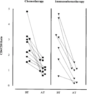
A total of 117 patients participated in the study (63 on chemotherapy and 54 on immunochemo-therapy) most of which had a single lesion usually located on the forearm or leg. Analysis of the patient’s data demonstrated that both treatment groups were comparable as to the demographic distribution of the patients as well as the clinical characteristics of the disease, reflecting the proper randomization of the study protocol. In addition, Fig. 7: phenotypic characterization of stimulated T cells
be-fore and after treatment. Peripheral blood mononuclear cells were stimulated with Leishmania antigen for 72 h. Cells were collected and analyzed by flow cytometry for CD3, CD4 and CD8 markers. Symbols represent data from each patient at different time points. (BT): before treatment; (AT): after com-plete healing of lesions
Fig. 8: characterization of IFN-γ producing cells – peripheral blood mononuclear cells were stimulated for 72 h with Leishmania antigen and then incubated with Brefeldin A to allow for accumulation of IFN-γ. Cells were collected and double stained for IFN-γ and CD4 or CD8 markers. Bars represent the mean value for each group ± 1 SD. (bt): before treatment; (at): after complete healing of lesions; n = 10 (CH) and 8 (IC)
96 96 96 96
96 Immunochemotherapy in Cutaneous Leishmaniasis VPCP Toledo
Montenegro’s skin test were positive in 88% of the cases, supporting previous observations from our group as well as from other studies (Carvalho et al. 1995, da Costa et al. 1996), thus confirming the validity of this test as a helpful diagnostic tool in leishmaniasis.
Although no statistical differences were observed in the time required for healing between the two treatment protocols, it is important to mention that both protocols were successful in inducing complete healing in all patients involved in the study.
The proliferative response of lymphocytes stimulated with L. braziliensis was analyzed be-fore, during and after the end of the treatment pe-riod. The great majority of patients presented a positive response to the stimulus. Patients treated with chemotherapy alone showed a decrease in the response at the end of the treatment, in accordance with results reported by others (da Cruz et al. 1994a). However, patients treated with immuno-chemotherapy showed a persistent lymphoprolif-erative response throughout the treatment. This difference in response to in vitro stimulation is probably related to the permanent presence of the antigen in vivo during the treatment. It is expected that in vivo stimulation of T cells decreases as treat-ment progresses toward healing in patients treated with Glucantime® alone, since the reduction in the parasite load would probably lead to a decreased number of responsive T cells. In immunochemo-therapy, however, the continual inoculation of Leishmania antigen may have kept these cells con-sistently stimulated in vivo even with a decreased parasite load. This was reflected by an unaltered in vitro response at the end of treatment. Also in agreement with previous studies (Convit et al. 1993, da Cruz et al. 1994a, Mendonça et al. 1995), no correlation between the intensity of the Montenegro’s reaction and the in vitro prolifera-tive response was observed.
As part of the evaluation of the immune re-sponse, we analyzed the production of several cytokines by stimulated PBMC. Our study focused primarily on the production of IFN-γ by these cells due to the involvement of this cytokine in resis-tance to and recovery from leishmanial infections (Carvalho et al. 1995, Coutinho et al. 1998, Ribeiro-de-Jesus et al. 1998). When analyzing the produc-tion of this cytokine before treatment onset it was observed that cells from patients with chronic le-sions produced more IFN-γ than those from pa-tients with recent infections. This fact has been reported previously (Ribeiro-de-Jesus et al. 1998) and is probably related to the elevated number of IFN-γ producing T cells that infiltrate these lesions (Melby et al. 1994). Comparison of IFN-γ pro-duction in supernatants of stimulated PBMC
ob-tained at different time points during the course of the treatment, revealed a significant increase in IFN-γ from patients that recovered from infection after conventional treatment as compared to be-fore treatment. Again, this observation was in agreement with data previously reported (da Cruz et al. 1994a). This increased IFN-γ production is probably associated with an increased expression of CD8+-IFN-γ producing cells as shown in Figs 7 and 8. On the other hand, in patients treated by immunochemotherapy, a marked decrease in
IFN-γ production was observed when cultures from re-covered patients were compared to those from the same patients before treatment.
The differences in IFN-γ production at the end of the two protocols could not be related to differ-ences in the production of IL-12. Although we were not able to detect IL-12 p40 in culture super-natants under the conditions used to stimulate the cells (data not shown), we indirectly demonstrated the presence of this cytokine through its ability to induce IFN-γ production. Even though around 50% of IFN-γ production was dependent on the pres-ence of IL-12 (Fig. 4), no differpres-ences in IL-12-induced IFN-γ production could be detected be-tween the two experimental protocols.
IL-10 is involved in the downregulation of Th1 responses (Fiorentino et al. 1991, Mosmann & Moore 1991, Howard & O’Garra 1992, D’Andrea et al. 1993). As shown in Fig. 5, PBMC from in-fected patients produced significant levels of this cytokine before treatment. After therapy was initi-ated we observed a marked decrease in the produc-tion of this cytokine in both treatment protocols. The decreased production of IL-10 probably allowed for a more efficient action of IFN-γ, leading to healing of the lesions. It is interesting to observe that addi-tion of Leishmania antigen to PBMC cultures stimu-lated with SAC decreased IL-10 production by these cells (Fig. 6). It is conceivable that in patients treated by immunochemotherapy the continuous adminis-tration of Leishmania antigen may have contributed to a greater decrease in IL-10 production in these patients (Fig 5). This could lead to a faster elimina-tion of the parasites by IFN-γ-activated macroph-ages. Even though we did not detect a faster heal-ing of the lesions in patients treated by immuno-chemotherapy we can not exclude the possibility that parasite elimination was faster in these patients. Moreover, it is also important to note that patients treated with the combined therapy were able to con-trol disease progression even with a decreased
97 97 97 97 97 Mem Inst Oswaldo Cruz, Rio de Janeiro, Vol. 96(1), January, 2001
The search for alternative protocols for treat-ment of leishmaniasis is justified by the increase in the number of cases of resistance to conventional chemotherapy. Thus, Cunha et al. (1994) used the protocol described here to successfully treat five HIV+ patients with cutaneous leishmaniasis, four of which were resistant to conventional anti-monium therapy and one to antianti-monium and Am-photericin B. In another study, an HIV+ patient with disseminated cutaneous leishmaniasis resis-tant to antimonial therapy showed a significant improve in lymphoproliferative response and
IFN-γ production after treatment and successful heal-ing of over 250 lesions (da Cruz et al. 1994b). This protocol offers an alternative treatment to cutane-ous leishmaniasis cases where conventional che-motherapy is not effective justifying the in-creased cost of adding the vaccine to the treatment. Further studies should better clarify the mecha-nisms associated with this therapeutic protocol.
ACKNOWLEDGEMENTS
To Dr Evaldo Nascimento for access to the flow cytometer facility, Mr Jair Cecílio de Paula for techni-cal assistance, Dr Ivan BM Sampaio for assistance with the statistical analysis, and Dr Leda Q Vieira for care-fully reviewing the manuscript.
REFERENCES
Afonso, LCC, Scharton TM, Vieira LQ, Wysocka M, Trinchieri G, Scott P 1994. The adjuvant effect of Interleukin-12 in a vaccine against Leishmania ma-jor. Science 263: 235-237.
Antunes CMF, Mayrink W, Magalhães PA, Costa CA, Melo MN, Michalick MSM, Williams P, Oliveira Lima A, Vieira JBF, Schettini APM 1986. Controlled field trials of a vaccine against New World cutane-ous leishmaniasis. Int J Epidemiol 15: 572-580. Bacellar O, Brodskyn C, Guerreiro J, Barral-Netto M,
Costa CH, Coffman RL, Johnson WD, Carvalho EM 1996. Interleukin-12 restores interferon-gamma pro-duction and cytotoxic responses in visceral leish-maniasis. J Infect Dis 173: 1515-1518.
Berger BJ, Fairlamb AH 1992. Interactions between immunity and chemotherapy in the treatment of the trypanosomiases and leishmaniases. Parasitology 105 (Suppl.): S71-S78.
Camargo EP 1964. Growth and differentiation in Try-panosoma cruzi. I. Origin of metacyclic trypano-somes in liquid medium. Rev Inst Med Trop São Paulo 6: 43-100.
Carvalho EM, Correia FD, Bacellar O, Almeida RP, Lessa H, Rocha H 1995. Characterization of the immune response in subjects with self-healing cutaneous leish-maniasis. Am J Trop Med Hyg 53: 273-277. Convit J, Castellano PL, Rondón AJ, Pinardi ME, Ulrich
M, Castés M, Bloom B, Garcia L 1987. Immuno-therapy versus chemoImmuno-therapy in localized cutaneous leishmaniasis. Lancet 1: 401-405.
Convit J, Ulrich M, Fernandez CT, Tapia FJ,
Caceres-Dittmar G, Castes M, Rondon AJ 1993. The clinical and immunological spectrum of American cutane-ous leishmaniasis. Trans R Soc Trop Med Hyg 87: 444-448.
Coutinho SG, da-Cruz AM, Bertho AL, Santiago MA, de Luca P 1998. Immunologic patterns associated with cure in human American cutaneous leishma-niasis. Braz J Med Biol Res 31: 139-142.
Coutinho SG, Oliveira MP, da Cruz AM, de Luca PM, Mendonça SC, Bertho AL, Soong L, McMahon-Pratt D 1996. T-cell responsiveness of American cutane-ous leishmaniasis patients to purified Leishmania pifanoi amastigote antigens and Leishmania braziliensis promastigote antigens: immunologic pat-terns associated with cure. Exp Parasitol 84: 144-155.
Cunha RMC, Hallack KA, Mayrink W 1994. Leishmaniose disseminada em paciente infectado pelo vírus HIV. Evolução clínico-morfológica atípica. Rev Soc Bras Med Trop 27: 389-401. D’Andrea A, Aste-Amezaga M, Valiante NM, Ma X,
Kubin M, Trinchieri G 1993. Interleukin-10 inhib-its human lymphocyte IFN-γ production by suppress-ing natural killer cell stimulatory factor/interleukin-12 synthesis in accessory cells. J Exp Med 178: 1041-1048.
da Costa CA, Toledo VPCP, Genaro O, Williams P, Mayrink W 1996. Montenegro skin test-evaluation of the composition and stability of the antigen prepa-ration. Mem Inst Oswaldo Cruz 91: 193-194. da Cruz AM, Conceição-Silva F, Bertho AL, Coutinho
S 1994a. Leishmania - Reactive CD4+ and CD8+ T cells associated with cure of human cutaneous leish-maniasis. Infect Immun 62: 2614-2618.
da Cruz AM, Mayrink W, Filgueiras D, Coutinho S 1994b. Imunoquimioterapia em um paciente com leishmaniose mucocutânea e SIDA: Estudos imunológicos. Rev Soc Bras Med Trop 27: 388-389.
Etges R, Müller I 1998. Progressive disease or protec-tive immunity to Leishmania major infection: the result of a network of stimulatory and inhibitory in-teractions. J Mol Med76: 372-390.
Fiorentino DF, Zlotnik A, Vieira P, Mosmann TR, Howard M, Moore KW, O’Garra A 1991. IL-10 acts on the antigen-presenting cell to inhibit cytokine production by Th1 cells. J Immunol 146: 3444-3451. Genaro O, Toledo VPCP, Costa CA, Hermeto MV, Afonso LCC, Mayrink W 1996. Vaccine for pro-phylaxis and immunotherapy, Brazil. Clin Dermatol 14: 503-512.
Grogl M, Thomason TN, Franke ED 1992. Drug resis-tance in leishmaniasis: its implication in systemic chemotherapy of cutaneous and mucocutaneous dis-ease. Am J Trop Med Hyg 47: 117-126.
Heinzel FP, Sadick MD, Mutha SS, Locksley RM 1991. Production of interferon γ, interleukin 2, interleukin 4, and interleukin 10 by CD4+ lymphocytes in vivo during healing and progressive murine leishmania-sis. Proc Natl Acad Sci USA 88: 7011-7015. Heinzel FP, Schoenhaut DS, Rerko RM, Rosser LE,
98 98 98 98
98 Immunochemotherapy in Cutaneous Leishmaniasis VPCP Toledo
mice infected with Leishmania major. J Exp Med 177: 1505-1509.
Hermeto MV, Vieira-Dias D, Genaro O, Rotondo-Silva A, da Costa CA, Toledo VPCP, Michalick MSM, Williams P, Mayrink W 1994. Outbreak of cutane-ous leishmaniasis in the Rio Doce Valley, Minas Gerais, Brazil. Mem Inst Oswaldo Cruz 89: 519-521. Howard M, O’Garra A 1992. Biological properties of
interleukin 10. Immunol Today 113: 198-200. Kemp M 1997. Regulator and effector functions of
T-cell subsets in human Leishmania infections. APMIS 105 (Suppl. 68): 5-33.
Locksley RM, Heinzel FP, Sadick MD, Holaday BJ, Gardner Jr KD 1987. Murine cutaneous leishmania-sis: susceptibility correlates with differential expan-sion of helper T-cell subsets. Ann Inst Pasteur/ Immunol 138: 744-749.
Lowry OH, Rosebrough NJ, Farr AL, Randall RJ 1951. Protein measurement with the folin phenol reagent. J Biol Chem 193: 265-275.
Marsden P 1985. Pentavalent antimonials: old drugs for new diseases. Rev Soc Bras Med Trop18: 187-187. Mattner F, Magram J, Ferrante J, Launois P, Di Padova
K, Behin R, Gately MK, Louis JA, Alber G 1996. Genetically resistant mice lacking interleukin-12 are susceptible to infection with Leishmania major and mount a polarized Th2 cell response. Eur J Immunol 26: 1553-1559.
Mayrink W, Antunes CMF, da Costa CA, Melo MN, Dias M, Michalick MSM, Magalhães P, Oliveira Lima A, Williams P. 1986. Further trials of a vac-cine against American cutaneous leishmaniasis. Trans R Soc Trop Med Hyg 80: 1001.
Mayrink W, da Costa CA, Magalhães PA, Melo MN, Dias M, Oliveira Lima A, Michalick MS, Williams P 1979. A field trial of a vaccine against American dermal leishmaniasis. Trans R Soc Trop Med Hyg 73: 385-387.
Mayrink W, Magalhães PA, Michalick MSM, da Costa CA, Oliveira Lima A, Melo MN, Toledo VPCP, Nascimento E, Dias M, Genaro O, Hermeto MV, Williams P 1992. Immunotherapy as a treatment of American cutaneous leishmaniasis: preliminary stud-ies in Brazil. Parassitologia 34: 159-165.
Mayrink W, Michalick MSM, Melo MN, Williams P, Nascimento E, Magalhães PA, da Costa CA, Oliveira Lima A, Dias M 1991. Tratamento da leishmaniose tegumentar americana utilizando vacina. An Bras Dermatol 66: 55-59.
Mayrink W, Williams P, Magalhães PA, Melo MN, Dias M, Oliveira Lima A, Michalick MSM, Ferreira Carvalho E, Barros GC, Sessa PA, Alencar JTA 1985. An experimental vaccine against American dermal leishmaniasis: experience in the state of Espírito Santo, Brazil. Ann Trop Med Parasitol 79: 259-269.
Melby PC, Andrade-Narvaez FJ, Darnell BJ, Valencia-Pacheco G, Tryon VV, Palomo-Cetina A 1994. In-creased expression of pro-inflammatory cytokines in chronic lesions of human cutaneous leishmania-sis. Infect Immun 62: 837-842.
Mendonça SC, Coutinho SG, Amendoeira RR, Marzochi MCA, Pirmez C 1986. Human cutaneous leishma-niasis (Leishmania b. braziliensis) in Brazil: lymphoproliferative responses and influence of therapy. Clin Exp Immunol 64: 269-276.
Mendonça SCF, de Luca PM, Mayrink W, Restom TG, Conceição-Silva F, da Cruz AM, Bertho AL, da Costa CA, Genaro O, Toledo VPCP, Coutinho SG 1995. Characterization of human T lymphocyte-mediated immune responses induced by a vaccine against American tegumentary leishmaniasis. Am J Trop Med Hyg 53: 195-201.
Mosmann TR, Moore KW 1991. The role of IL-10 in crossregulation of TH1 and TH2 responses. Immunol Today: A49-A53.
Murray HW, Oca MJ, Granger AM, Schreiber, RD 1989. Requirement for T cells and effect of lymphokines in successful chemotherapy for an intracellular in-fection. Experimental visceral leishmaniasis. J Clin Invest 83: 1253-1257.
Nabors GS, Afonso LCC, Farrell JP, Scott P 1995. Switch from a type 2 to a type 1 T helper cell re-sponse and cure of established Leishmania major infection in mice is induced by combined therapy with interleukin 12 and Pentostam. Proc Natl Acad Sci USA 92: 3142-3146.
Nascimento E, Mayrink W, da Costa CA, Michalick MSM, Melo MN, Barros GC, Dias M, Antunes CMF, Lima MS, Taboada DC, Liu TY 1990. Vacci-nation of humans against cutaneous leishmaniasis: cellular and humoral immune responses. Infect Immun 58: 2198-2203.
Peters BS, Fish D, Golden R, Evans DA, Bryceson AD, Pinching AJ 1990. Visceral leishmaniasis in HIV infection and AIDS: clinical features and response to therapy. Q J Med 77: 1101-1111.
Prussin C, Metcalfe DD 1995. Detection of intracyto-plasmic cytokine using flow cytometry and directly conjugated anti-cytokine antibodies. J Immunol Methods 188: 117-128.
Ribeiro-de-Jesus A, Almeida RP, Lessa H, Bacellar O, Carvalho EM 1998. Cytokine profile and pathology in human leishmaniasis. Braz J Med Biol Res 31: 143-148.
Scharton TM, Scott P 1993. Natural killer cells as a source of interferon-γ that drives differentiation of CD4+ T cells subsets and induces early resistance to Leishmania major in mice. J Exp Med 178: 567-577.
Scott P 1989. The role of TH1 and TH2 cells in experi-mental cutaneous leishmaniasis. Exp Parasitol 68: 369-372.
Scott P 1991. IFN-γ modulates the early development of Th1 and Th2 responses in a murine model of cu-taneous leishmaniasis. J Immunol 147: 3149-3155. Scott P, Trinchieri G 1995. The role of natural killer cells in host-parasite interactions. Curr Opinion Immunol 7: 34-40.