INTRODUCTION
Recurrent corneal erosion syndrome (RCES) was first described in 1872 by Hansen(1). It is a relatively common condition and may be
clas-sified as either dystrophic (dystrophic-RCES) where it occurs associated with an anterior corneal dystrophy, non-dystrophic (nd-RCES) where it occurs following a sudden, sharp, abrading injury (fingernail, paper cut, tree branch), or unrelated to any pre-existing corneal disease or past history of trauma (idiopathic-RCES)(2).
Clinically, patients experience recurrent episodes of acute symp-toms of pain, tearing, redness and photophobia usually at night or upon first awakening(2). Physical examination during acute attacks reveals
areas of eroded or loosely attached epithelium(3). Treatment with
epi-thelial scraping followed by eye patching with lubricants, hypertonic saline, antibiotics, or therapeutic contact lenses, resolves symptoms in most cases. However, there is a small group of patients who suffer
recurrent attacks at weekly or monthly intervals despite conventional treatments(2,3).
Several methods of surgical treatment have been shown to be help-ful including epithelial basement membrane debridement(3,4), super ficial
keratectomy with a diamond burr(5), anterior stromal puncture by sharp
needles(4), and Nd:YAG laser(6). Also, several authors have reported the
use of excimer phototherapeutic keratectomy (PTK) for this
condi-tion(7-18). Reported success rates with PTK have varied from 69% to
86%(7-18). Recurrence rates are likely to be higher when PTK is used for
dystrophic-RCES(8,12).
Issues such as recurrence of the pathology after PTK and ultimate need for additional surgery are important to patients and the sur geons counseling them.
The aim of this study was to assess the morphologic and functio-nal very long-term results of minimal invasive subepithelial PTK for
Ten-year results of phototherapeutic keratectomy on recurrent corneal erosions
Resultados após dez anos da ceratectomia fototerapêutica para erosões recorrentes de córnea
Belquiz RodRiguesdo AmARAl NAssARAllA1, João JoRge NAssARAllA JuNioR2
Submitted for publication: April 27, 2011 Accepted for publication: December 2, 2011
Study carried out at Goiânia Eye Institute, Department of Cornea and Refractive Surgery, Goiânia (GO), Brazil.
1 Physician, Goiânia Eye Institute, Department of Cornea and Refractive Surgery, Goiânia (GO), Brazil. 2 Physician, Faculty of Health Sciences, Universidade de Brasília - UnB - Brasília (DF), Brazil, Goiânia
Eye Institute, Department of Retina and Vitreous, Goiânia (GO), Brazil.
Funding: No specific financial support was available for this study.
Disclosure of potential conflicts of interest: B.R.A.Nassaralla, None; J.J.Nassaralla Jr., None. Correspondence address: Belquiz A. Nassaralla. Instituto de Olhos de Goiânia. Rua L, 53 - 12o andar, Setor Oeste, Goiânia (GO) - 74120-050 - Brazil - E-mail: [email protected]
ABSTRACT
Purpose: To determine the ten-year visual results and outcomes of excimer laser pho totherapeutic keratectomy (PTK) for recurrent corneal erosions.
Methods: Twenty-six eyes of 23 patients with recurrent corneal erosions were trea-ted by PTK from 1996 to 2000 at the Goiania Eye Institute, Brazil. All eyes had failed to respond to conventional therapy. Data regarding preoperative and postopera ti ve best-spectacle-corrected visual acuity (BSCVA), spherical equivalent (SE), sympto matic relief, incidence of recurrence, and complications arising from the laser treatment were analyzed. The mean duration of symptoms prior to PTK was 18 months (ran-ge, 8 to 36 months). The corneal epithelium was debrided, and laser ablation was performed to a depth of 5 micron with an ablation zone of 7 to 9 mm, using the Technolas 217C Plano Scan excimer laser. Mean postoperative follow-up was 12 years (range, 10 to 14 years).
Results: At the last follow-up visit, 15 eyes (57.69%) were symptomsfree. Five eyes (19.2%) had occasional mild symptoms of irritation and photophobia upon awakening. Recurrence of painful corneal erosions occurred in six eyes (23.07%), which required a PTK retreatment. Twenty-four eyes had a preserved or improved BCVA, while 2 eyes showed deterioration of 1 line on Snellen test. Eleven eyes (42.3%) had no change in SE, and the others (57.69%) had a change of less than +/-0.75 diopters (D). There were no major complications during the follow-up period.
Conclusion: Ten-year data show that PTK is a safe, fast, effective and minimal invasive choice of treatment for recurrent corneal erosions in patients who do not respond to conventional treatments.
Keywords: Photorefractive keratectomy; Corneal diseases/surgery; Corneal surgery, laser/methods; Lasers, excimer
RESUMO
Objetivo: Determinar os resultados visuais após dez anos de seguimento da ceratecto mia fototerapêutica (PTK) com excimer laser para erosões recorrentes de córnea. Métodos: Vinte e seis olhos de 23 pacientes portadores de erosões recorrentes de córnea foram tratados com PTK entre 1996 e 2000 no Instituto de Olhos de Goiânia, Brasil. Nenhum olho havia respondido às terapias convencionais. Dados pré-operatórios e pós-operatórios referentes à melhor acuidade visual corrigida (MAVC), equivalente esférico (EE), alívio dos sintomas, incidência de recorrência e complicações oriundas do tratamento a laser, foram analisadas. A média de duração dos sintomas antes do PTK foi de 18 meses (variando entre 8 a 36 meses). O epitélio da córnea foi debridado e a ablação realizada a uma profundidade de 5 µm e diâmetro de 7 a 9 mm, usando o excimer laser Technolas 217C Plano Scan. O seguimento médio foi de 12 anos (variando entre 10 e 14 anos).
Resultados: No último exame, 15 olhos (57,69%) estavam livres dos sintomas. Cinco olhos (19,2%) apresentavam sintomas, discretos e ocasionais, de irritação e fotofobia ao levan tar. Recorrência de erosões dolorosas ocorreu em 6 olhos (23,07%), o que ne cessitou de retrata-mento com PTK. Vinte e quatro olhos mantiveram ou melhoraram sua MAVC, enquanto que 2 olhos perderam 1 linha de visão pela tabela de Snellen. Onze olhos (42,3%) mantiveram o mesmo EE e os outros (57,69%) apresentaram alterações inferiores a +/-0,75 dioptrias (D). Nenhuma complicação significativa foi observada durante o período de seguimento. Conclusões: Os dados de 10 anos mostram que o PTK é uma opção segura, rápida, eficaz e pouco invasiva para o tratamento de erosões recorrentes de córnea em pacientes que não respondem bem às terapias convencionais.
the treatment of recurrent corneal erosions (RCE) in patients who do not respond to conservative treatments.
METHODS
Twenty-six eyes of 23 patients (17 women and 9 men) were trea-ted by PTK for RCE from 1996 to 2000. Mean patient age was 45.07 ± 13.19 years (range, 26 to 72 years). All procedures were performed at the Goiania Eye Institute, Goiania, GO, Brazil, by a single surgeon (BAN).
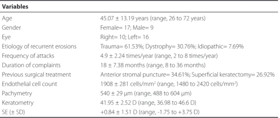
Sixteen patients (61.53%) could recall a sharp corneal injury (nd-RCES), 8 eyes (30.76%) had map-dot dystrophy (dystrophic-RCES), and 2 eyes (7.69%) had no pre-existing corneal disease or past his tory of trauma (idiopathic-RCES). All patients had been treated at our institu tion with extensive conventional therapy before PTK. Six teen eyes (61.53%) had also received a surgical treatment: nine eyes (34.61%) were treated with anterior stromal puncture by sharp needles (avoiding the visual axis) and 7 eyes (26.92%) were treated with superficial keratectomy with a diamond burr. However, in all cases clinical symp toms were refractory to such treatments. Mean duration of complaints before PTK was 18 ± 7.38 months (range, 8 to 36 months). Mean frequency of attacks was 4.9 ± 2.24 times a year (ranged, 2 to 8 times a year). All eyes had corneal erosions involving the visual axis. In all cases, PTK was performed during the acute state. Table 1 shows the mean preoperative data.
All patients underwent complete ophthalmologic examination including personal medical ocular history, cause and duration of RCE, treatments given before PTK, number of recurrences before and after PTK, best spectacle corrected visual acuity (BSCVA), computerized corneal topography, keratometry, ultrasonic pachymetry, manifest and cycloplegic refraction, tonometry, slit-lamp microscopy, specular mi croscopy, fundus examination and anterior photography. Data re-garding symptomatic relief and complications arising from PTK laser treatment were also analyzed. Three patients had PTK treatment in both eyes.
Informed consent was obtained from all patients after the pro-cedure had been fully explained. All patients were pretreated with a mild oral sedative (diazepam 5 mg) 30 minutes before surgery and two drops of topical 0.5% proparacaine 2 to 5 minutes before surgery. Patients were taken to the laser and reclined to a supine position. A sterile drape and a wire eyelid speculum were placed in the operative eye. The central 9.0 mm zone of the corneal epithelium was gently removed using a wet Merocel® sponge (Xomed, Jacksonville, FL) and a blunt Paton spatula (Storz Instrument, St. Louis, MO). The tightly adherent peripheral corneal epithelium was left intact.
Laser ablation was performed using the Technolas 217C Plano Scan excimer laser (Bausch & Lomb, Rochester, NY). Laser ablation was performed on a 7 to 9 mm central corneal epithelial defect using a
computer-controlled area ablation program. In this PTK program, only two parameters were entered: the diameter of the area to be treated and the desired depth of ablation. The ablation depth was 5 µm for all eyes. The laser treatment was centered on the patient’s pupil.
At the end of the procedure, a bandage soft contact lens was applied, and one drop each of tobramicin-dexamethasone and 0.5% ketorolac tromethaminewas instilled into the inferior conjunctival cul-de-sac. Oral analgesics were prescribed for the first 24 hours. To bramycin-dexamethasone and ketorolac tomethamine were pres-cribed four times a day until the epithelial defect was completely healed. After epithelial closure the contact lens was removed. All eyes were then treated with 1% fluorometholone acetate four times a day for 1 month. Unpreserved artificial tears were recommended for mild irritation.
Follow-up visits were scheduled daily for the first week or until complete re-epithelialization, once a week for 2 weeks, once the follo-wing month, and every 3 to 6 months thereafter. The last follow-up time after PTK was recorded in years. Mean postoperative follow-up was 12 ± 1.52 years (range, 10 to 14 years).
Besides frank macroerosions, we defined the “recurrence of cor neal erosion” either as the presence of clinical symptoms, such as pain, pho tophobia, or tearing, or as microscopically observable reparative signs or microerosions of the cornea. Patients were asked to return should they experience any recurrence of symptoms.
Haze was evaluated according to Hanna’s grading scale using a slit lamp. It was graded as follows: 0= clear cornea, no haze; 0.5= barely perceptible, seen only by tangential illumination; 1= trace haze of mi nimal density seen with difficulty using direct illumination; 2= mo-derate haze easily visible with direct slit illumination; 3= marked haze that partially obscures anterior chamber observation or iris detail; 4= severe haze that obscures anterior chamber or iris details(19).
Statistical analysis was performed with the paired t test using Mi crosoft Office Excel 2007 (Microsoft Corp, Seattle, Wash). The signi-ficance level wasP<0.05.
RESULTS
Patients experienced ocular pain for 24 to 48 hours after laser treatment. Mean re-epithelialization time was 4.61 ± 1.13 days (range, 3 to 6 days).
After a mean of 12 ± 1.52 years (range, 10 to 14 years), the overall success rate after only one treatment was significant. Twenty eyes (76.92%) presented no recurrence episode. Among these, fifteen eyes (57.69%) were free of symptoms and five (19.2%) had occasional mild stinging and photophobia upon awakening, which was relieved by lubricants. Slit-lamp microscopy was normal in all these twenty eyes. Recurrence of painful corneal erosions occurred in six eyes (23.07%) in the first 6 months after PTK that lead to a second PTK treat ment.
Table 1. Preoperative data
Variables
Age Gender Eye
Etiology of recurrent erosions Frequency of attacks Duration of complaints Previous surgical treatment Endothelial cell count Pachymetry Keratometry SE (± SD)
45.07 ± 13.19 years (range, 26 to 72 years) Female= 17; Male= 9
Right= 10; Left= 16
Trauma= 61.53%; Dystrophy= 30.76%; Idiopathic= 7.69% 4.9 ± 2.24 times/year (range, 2 to 8 times/year) 18 ± 7.38 months (range, 8 to 36 months)
Anterior stromal puncture= 34.61%; Superficial keratectomy= 26.92% 1908 ± 281 cells/mm2 (range, 1480 to 2420 cells/mm2)
540 ± 29 µm (range, 488 to 604 µm) 41.95 ± 2.52 D (range, 36.98 to 46.6 D) +0.84 ± 1.51 D (range, -1.75 to +3.75 D)
All these six eyes had map-dot dystrophy (dystrophic-RCES). Slit-lamp microscopy showed that the recurrence occurred in the same area that was previously treated by PTK in two eyes, and in a second area in the other four eyes. They were retreated using the same technique described above. After a second PTK, two eyes (7.69%) had another recurrence at 8 and 12 months after retreatment. Both patients had been treated with superficial keratectomy for dystrophic-RCES, with a diamond burr before PTK. They were treated for the third time and were symptom free thereafter. Both eyes developed central corneal haze grade 2 in the early postoperative stage, which decreased to grade 0.5 during the follow-up. No eye had more than 2 recurrent events after the first PTK.
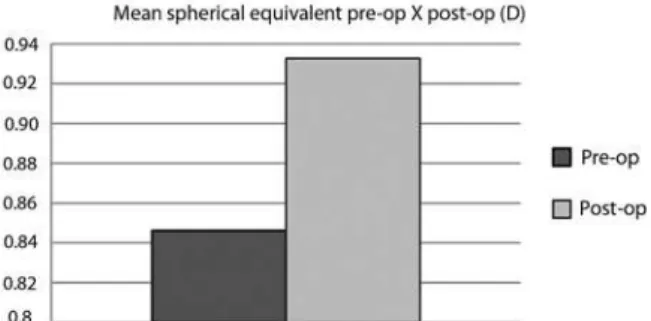
Before PTK, the mean spherical equivalent (SE) was +0.84 ± 1.51 D (range, -1.75 to +3.75 D). At the last follow-up visit, the mean SE was +0.93 ± 1.32 D (range, -1.50 to +3.50). The mean spherical equiva-lent manifest refraction change was +0.08 ± 0.30 D (range, -0.50 to +0.75 D) in the hyperopic direction compared to the preoperative refraction. Figure 1 shows the mean manifest spherical equivalent re fraction changes over time. Eleven eyes (42.3%) had no change in SE and the others (57.69%) had a change of less than +/-0.75 diopters. Despite this slight trend toward hyperopia, the difference between the preoperative and the last follow-up visit manifest refraction was not statistically significant (p=0.32).
At the last follow-up visit, BSCVA was unchanged in twenty eyes (76.92%) and improved 1 line in four eyes (15.38%). However, two eyes (7.69%) that required three PTK treatments for recurrence of painful erosions lost one line each of the BSCVA. Both of them presented haze grade 0.5 at the last examination. No eye had any other recur-rence episode thereafter. Statistical analysis did not show significant difference in BSCVA before and after PTK (p=0.19). Figure 2 shows the BSCVA before and 10 years after PTK treatment.
No statistically significant difference was also noted in the mean endothelial cell count before (1908 ± 281 cells/mm2; range, 1480 to
2420), and after PTK (1885 ± 262 cells/mm2; range, 1250 to 2430). Mean
keratometric central power remained unchanged, with 41.95 ± 2.52 D (range, 36.98 to 46.6 D) preoperatively and 41.77 ± 2.81 D (range, 36.8 to 46.4 D) postoperatively. Also there was not significantly change in the mean central corneal pachymetry before (540 ± 29 µm; range, 488 to 604 µm), and after PTK (545 ± 30 µm; range, 480 to 610 µm). All measurements were taken during a symptom-free interval.
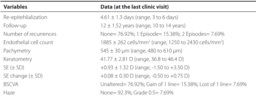
There were no major complications during the follow-up. No pa-tient reported visual symptoms such as glare or halos. Table 2 shows the mean postoperative data.
DISCUSSION
Excimer laser, with its facility to ablate corneal tissue with extreme precision and minimal adjacent tissue damage has been a useful tool in the management of RCES(7-18). Studies have shown that partial
ablation of Bowman’s layer gives a smooth bed for migrating epithe-lium, and results in new hemidesmosomal adhesion complexes(20,21).
Histological studies of excimer laser ablated monkey corneas have shown increases amounts of type 7 collagen, a major component of anchoring fibrils and hemidesmosomes along the basement mem-brane of the basal epithelial cells(21). Human studies have shown that
the basal epithelial layer forms hemidesmosomes and new basement membrane within 2 weeks of photoablation(22). In the treatment of
recalcitrant erosions, it is possible that both the removal of the ab-normal epithelium and the basement membrane are important for a successful outcome. The former results in regeneration of the basal epithelial cells and the latter will allow the epithelium to come into direct contact with stromal elements, stimulating the synthesis of new anchoring fibrils and hemidesmosomes(22).
A literature review was completed to look at long term follow-up of PTK for RCES. In this study, with a mean follow-up of 12 ± 1.52 years (range, 10 to 14 years), we report the longest follow-up after PTK for RCES compared to other studies(7-18). Overall our results may
contribu-te to establish the very long contribu-term success of this contribu-technique. Using a PTK it is possible to treat affected areas that encroach on the visual axis with excellent visual rehabilitation. This is in contrast with other forms of interventional treatment, which also aims at creating a firm bond between the epithelium and Bowman’s layer-notably, anterior stromal puncture and superficial keratectomy, where pos-toperative scarring may limit treatment of the central cornea. These methods have recurrence-free rates of 80 to 100% during follow-ups ranging from 4 to 33 months(3-6). Although less costly, surgical
me-thods that penetrate Bowman’s layer and the stroma carry the risk of creating corneal scars or perforating the cornea(3), as opposed to the
extreme safety reported for PTK(7-18).
In this case series, all patients underwent extensive conventional therapy at our institution before PTK. However, the clinical symptoms were refractory to such treatments.
The recurrence of corneal erosions after PTK in various studies was reported within the first 6 to 9 months following laser
treat-ment(10-14). It is speculated that completely normal reformation of the
adhesion complexes, including normal density of hemidesmosomes, basement membrane, and anchoring fibrils, may take months to years(19,23). Experimental animal studies showed that the extent of
he mi desmosome formation was subnormal up to 3 months after
epithelial debridement, with or without PRK(23). Histological studies of
human corneal buttons demonstrated that restoration of epithelial adhe sion complexes was not complete as late as 15 months after ex cimer laser PTK(21). It is not known to what extent the decrease in
hemidesmosomes and anchoring fibrils formation can cause clini-cally significant corneal erosion. However, this may contribute to symptoms like foreign body sensation on awakening and the recur-rence of painful corneal erosions in some of the eyes after PTK(9,17,18).
Figure 1. Mean manifest spherical equivalent refraction changes (diopter) before and 10 years after PTK on recurrent corneal erosions.
Besides effectiveness of the laser ablation, large beam cross-sec-tions, typically several millimeters in diameter, allow simultaneous treatment of wider areas. Our concern, even with a PTK treatment profile, is the possibility of inducing refractive change. In contrast to hyperopic shift after PTK in other corneal pathologies, no significant refractive shift occurred in our patients, what is in accordance to other studies(7-18). This is likely to be due to the very shallow depth of
ablation, leaving at least one-half of the Bowman’s layer intact, since it is approximately 10 to 12 µm thick(23). The surgical plan in this study
was to remove 5 µm from Bowman’s membrane by diffuse PTK, using a computer controlled area ablation program. We preferred not to perform a localized focal laser treatment to the area of the erosion to avoid inducing irregular astigmatism or visual symptoms when the pupil becomes dilated.
In accordance to others studies(7-18), most subjects (76.92%) in our
series had a disease-free survival after only one treatment. It shows that excimer laser ablation to this depth promotes adhesion com-plex formation, which would lead to a better and stable epithelial adhesion(22). However, six eyes (23.07%) had recurrence of corneal
erosions within 6 months of the initial PTK and required a second PTK treatment. Anecdotally and as previously described(8,12), all these
six eyes had dystrophic-RCES and had been treated with superficial keratectomy before PTK. Dinh et al.(8) have found that, in patients with
anterior membrane dystrophy, RCES reoccurred in 42%, with a mean follow-up of 19.5 months. Baryla et al.(13) reported a greater
propor-tion of RCES caused by corneal dystrophies rather than trauma. After a second PTK, two of these eyes (7.69%) had another recurrence at 8 and 12 months after retreatment. Both developed central corneal haze grade 2 in the early postoperative stage which decreased to grade 0.5 during follow-up. This could explain the lost of one line of their BSCVA. Probably, the association of both procedures may have increased the postoperative scarring of the central cornea.
Recurrences of corneal erosions after PTK were also shown by Baryla et al.(13) where recurrences were noted up to 21 months after initial
PTK. In a single center study by Seitz et al.(14) with a mean follow-up
of 2.2 ± 1.6 years (maximum of 5.6 years), they reported a recurrence rate of 6.5%, 11.5%, and 13.6% after 1, 2, and 5 years, respectively, following PTK.
Differently from other studies in which patients were referred from other centers for PTK, in this series all eyes had been followed at our institution for a mean of 18 ± 7.38 months prior to PTK. So, their refractive measurements were taken during a symptom-free interval before PTK. In this way, we could precisely address the issue of refrac-tive changes before and after PTK. After a mean of 12 ± 1.52 years, BSCVA was unaltered in twenty eyes (76.92%) and gained one line in four eyes (15.38%). Mean keratometry and corneal topography did not show any induced irregularities of the corneal surface. The slight improvement in the BSCVA is most likely the result of a smoother epithelial surface after excimer laser ablation. It is speculated that the
Table 2. Postoperative data
Variables Data (at the last clinic visit)
Re-epitehlialization Follow-up
Number of recurrences Endothelial cell count Pachymetry Keratometry SE (± SD) SE change (± SD) BSCVA Haze
4.61 ± 1.3 days (range, 3 to 6 days) 12 ± 1.52 years (range, 10 to 14 years)
None= 76.92%; 1 Episode= 15.38%; 2 Episodes= 7.69% 1885 ± 262 cells/mm2 (range, 1250 to 2430 cells/mm2)
545 ± 30 µm (range, 480 to 610 µm) 41.77 ± 2.81 D (range, 36.8 to 46.4 D) +0.93 ± 1.32 D (range, -1.50 to +3.50 D) +0.08 ± 0.30 D (range, -0.50 to +0.75 D)
Unaltered= 76.92%; Gain of 1 line= 15.38%; Lost of 1 line= 7.69% None= 92.3%; Grade 0.5= 7.69%
SE= spherical equivalent; SD= standard deviation; D= diopters; BSCVA= best spectacle-corrected visual acuity
possible causes for improved BSCVA in some patients with recurrent corneal erosions after PTK include decrease of irregular astigmatism, decrease of epithelial edema, reduced photophobia, improved epi-thelial stability and smoothing of the corneal surface(8,9,18).
In conclusion, PTK using a 193-nm excimer laser can be conside-red a safe, fast, effective and minimal invasive choice of treatment for recurrent corneal erosions in patients refractory to conventional treatments. A repeat PTK is effective in those patients who fail a pri -ma ry treatment. The success rate observed over a mean of twelve years of follow-up is very high and complications are rare.
REFERENCES
1. Hansen E. Om den intermitterende keratitis vesiculosa neuralgica af traumatisk o prind -se le. Hosp Stidende. 1872;15:201-3.
2. Thygeson P. Observations on recurrent erosion of the cornea. Am J Ophthalmol. 1959; 47(5 Pt 2):48-52.
3. Kenyon KR. Recurrent corneal erosion: pathogenesis and therapy. Int Ophthalmol Clin. 1979;19(2):169-95.
4. McLean EN, MacRae SM, Rich LF. Recurrent erosion. Treatment by anterior stromal punc ture. Ophthalmology. 1986;93(6):784-8.
5. Soong HK, Farjo Q, Meyer RF, Sugar A. Diamond burr superficial keratectomy for recur rent corneal erosions. Br J Ophthalmol. 2002;86(3):296-8.
6. Geggel HS. Successful treatment of recurrent corneal erosion with Nd:YAG laser an terior stromal puncture. Am J Ophthalmol. 1990;110(4):404-7. Comment in Am J Ophtha lmol. 1991;111(2):252-5.
7. Cavanaugh TB, Lind DM, Cutarelli PE, Mack RJ, Durrie DS, Hassanein KM, Graham CE. Phototherapeutic keratectomy for recurrent erosion syndrome in anterior basement membrane dystrophy. Ophthalmology. 1999;106(5):971-6.
8. DinhR, Rapuano CJ, Cohen EJ, Laibson PR. Recurrence of corneal dystrophy after ex ci mer laser phototherapeutic keratectomy. Ophthalmology. 1999;106(8):1490-7. 9. Jain S, Austin DJ. Phototherapeutic keratectomy for treatment of recurrent corneal ero
-sion. J Cataract Refract Surg. 1999;25(12):1610-4.
10. Maini R, Loughnan MS. Phototherapeutic keratectomy re-treatment for recurrent corneal erosion syndrome. Br J Ophthalmol. 2002;86(3):270-2.
11. Chow AM, YiuEP, Hui MK, Ho CK. Shallow ablations in phototherapeutic keratectomy: long-term follow-up. J Cataract Refract Surg. 2005;31(11):2133-6.
12. Fagerholm P, Fitzsimmons TD, Orndahl M, Ohman L, Tengroth B. Phototherapeutic ke-ra tectomy:long-term results in 166 eyes. Refke-ract Corneal Surg. 1993;9(2 Suppl):S76-81. 13. Baryla J, Pan YI, Hodge WG. Long-term efficacy of phototherapeutic keratectomy on
recurrent corneal erosion syndrome. Cornea. 2006;25(10):1150-2.
14. Seitz B, Langenbucher A, Hafner A, Kus MM, Naumann GO. [Phototherapeutic kera-tectomy for recurrent corneal erosion syndrome (e-PTK). Report on 116 consecutive interventions]. Ophthalmologe. 2002;99(9):703-8. German.
15. Pogorelov P, Langenbucher A, Kruse F, Seitz B. Long-term results of phototherapeutic keratectomy for corneal map-dot-fingerprint dystrophy (Cogan-Guerry). Cornea. 2006; 25(7):774-7.
16. Fagerholm P. Phototherapeutic keratectomy: 12 years of experience. Acta Ophthal-mol Scand. 2003;81(1):19-32. Review.
17. Morad Y, Haviv D, Zadok D, Krakowsky D, Hefetz L, Nemet P. Excimer laser photo-therapeutic keratectomy for recurrent corneal erosion. J Cataract Refract Surg. 1998;24(4): 451-5. Comment in J Cataract Refract Surg. 1998;24(11):1418-9. 18. Rashad KM, Hussein HA, El-Samadouny MA, El-Baha S, Farouk H. Phototherapeutic
keratectomy in patients with recurrent corneal epithelial erosions. J Refract Surg. 2001; 17(5):511-8.
after excimer laser refractive surgery: objective measurements and functional impli-cations. Eur J Ophthalmol. 1991;1(4):173-80.
20. Gipson IK. Adhesive mechanisms of the corneal epithelium. Acta Ophthalmol Suppl. 1992;(202):13-7.
21. Aitken DA, Beirouty ZA, Lee WR. Ultrastructural study of the corneal epithelium in the re current erosion syndrome. Br J Ophthalmol. 1995;79(3):282-9.
22. Marshall J, Trokel SL, Rothery S, Krueger RR. Long-term healing of the central cornea after photorefractive keratectomy using and excimer laser. Ophthalmology. 1988;95(10): 1411-21.