ABSTRACT
http://dx.doi.org/10.1590/1678-775720150451
Comparative
in vit r o
evaluation of CAD/CAM vs
conventional provisional crowns
Adil Othman ABDULLAH1, Effrosyni A. TSITROU2, Sarah POLLINGTON3
1- Erbil Polytechnic University, Department of Dental Prevention, Erbil, Kurdistan Region, Iraq.
2- Aristotle University of Thessaloniki, Department of Operative Dentistry, School of Dentistry, Thessaloniki, Greece. 8QLYHUVLW\RI6KHI¿HOG6FKRRORI&OLQLFDO'HQWLVWU\$FDGHPLF8QLWRI5HVWRUDWLYH'HQWLVWU\6KHI¿HOG8QLWHG.LQJGRP
Corresponding address: Adil O. Abdullah - Department of Dental Prevention, Erbil Polytechnic University, Erbil, Kurdistan Region, Iraq - Phone: +964(0)7504604246 - e-mail: [email protected]
6XEPLWWHG2FWREHU0RGL¿FDWLRQ0DUFK$FFHSWHG$SULO
O
mode of fracture of CAD/CAM provisional crowns with that of direct provisional crowns. ceramic crown following tooth preparation guidelines. The materials tested were: VITA CAD-Temp®, Polyetheretherketone “PEEK”, Telio CAD-Temp, and Protemp™4 (control group).The crowns were divided into four groups (n=10), Group1: VITA CAD-Temp®, Group 2:
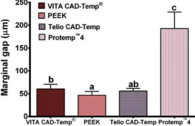
PEEK, Group 3: Telio CAD-Temp, and Group 4: Protemp™4. Each crown was investigated was performed using GraphPad Prism software version 6.0. Results: The average marginal gap was: VITA CAD-Temp® 60.61 (±9.99) μm, PEEK 46.75 (±8.26) μm, Telio CAD-Temp
56.10 (±5.65) μm, and Protemp™4 193.07(±35.96) μm (P<0.001). The average internal
® 124.94 (±22.96) μm, PEEK 113.14 (±23.55) μm, Telio CAD-Temp
110.95 (±11.64) μm, and Protemp™4 143.48(±26.74) μm. The average fracture strength was: VITA CAD-Temp® 361.01 (±21.61) N, PEEK 802.23 (±111.29) N, Telio CAD-Temp
719.24 (±95.17) N, and Protemp™4 416.40 (±69.14) N. One-way ANOVA test showed between all groups (p<0.001). However, the mode of fracture showed no differences between the groups (p>0.05). Conclusions: CAD/CAM fabricated provisional crowns
Ke y w or ds: Temporary dental restoration. Dental marginal adaptation. CAD-CAM. Dental impression technique. Dental crowns. Dental prosthesis.
I N TROD UCTI ON
Provisional restorations are used as an intermediate stage for short or long-term placement on teeth between the time of tooth preparation until
and placed4,23. The demand for tooth-colored
years because of improved techniques and patient’s demand. Therefore, using various new restorative materials, which have excellent mechanical properties, are essential for both provisional and
1,9.
The emergence of computer-aided design/ c o m p u t e r - a i d e d m a n u f a c t u r e ( C A D / C A M )
technology in dentistry has allowed the successful use of different materials7. Using these systems to
in comparison with conventional techniques1. In addition, this technology permits shaping of materials with high precision that cannot be easily carried out via a traditional method to make a dental restoration, and this technology now includes the fabrication of provisional restorations13.
maintaining esthetics and oral functions, such as mastication and speech, is paramount1,16.
There are two major approaches for fabrication of provisional crowns: direct and indirect methods. A variety of new provisional blocks are available to use with the CEREC3 CAD/CAM system (Sirona, Bensheim, Germany). These materials can withstand the milling process because of their high strength7. However, conventional provisional materials cannot be prepared via the milling process, but require to be fabricated manually.
manual technique, for example, inadequate surface
1. It has been reported that CAD/ CAM provisional crowns were stronger and exhibited better marginal accuracy than directly fabricated bis-acryl composite crowns, especially following thermal cycling. Therefore, using CAD/CAM may resolve these issues26.
The aim of this study was to compare the
mode of fracture of provisional CAD/CAM crowns compared with chair side directly made provisional crowns. The null hypothesis tested was twofold: the CAD/CAM provisional crowns will have a better
In addition, the CAD/CAM provisional crowns will provide superior strength to that of the directly fabricated crowns.
M ATERI AL AN D M ETH OD S
(Frasaco A3, Frasaco Franz Sachs & Co., GmbH, Germany) was prepared for a full ceramic crown with 1.5 mm occlusal reduction, the convergence angle of the wall was prepared to be approximately 6° and a round shoulder of 1 mm using a high speed hand piece operating with water coolant. A paralleling device (Nesor product LTD, Britain) was used during the preparation to enhance reproducibility of the preparation. An impression of the preparation was fabricated with vinyl-polysiloxane (Dublisil 15, Dreve Dentamid GmbH, Germany) to fabricate a master die from polyurethane base resin (AlphaDieTMMF,
Schütz Labortechnik, Germany).
The CEREC InEos system (Sirona, Bensheim, Germany) was used for scanning the preparation. The preparation was powdered with scan spray (Vita Zahnfabrik, Germany) using a propellant to provide a thin, even layer of powder. The preparation was then scanned and the crown designed using the CEREC 3D v3.60 software. The three CAD/CAM provisional blocks were used:
Acrylate polymer material VITA CAD-Temp® (VITA
Zahnfabrik, Germany), Polyethertherketone PEEK (Invibio Biomaterial Company, UK), and Polymethyl
methacrylate material Telio CAD-Temp (Ivoclar Vivadent, Liechtenstein, Germany). Ten provisional crowns were milled for each group. The correlation mode was used with the spacer set at 10 microns for the three CAD/CAM groups and the default milling burs (1.2 mm cylinder bur, step bur) were used for the milling of the crowns. Following milling, the restorations were examined for the presence of any defects or cracks.
A fourth group consisted of ten provisional crowns made from ProtempTM4 (3M ESPE, Germany), which
comprised the direct fabrication technique. An
tooth was initially taken before any preparations were made to serve as an index impression for the fabrication of the provisional crowns. The materials used were: heavy bodied materials (Aquasil putty, blue and orange colour, Dentsply-Detrey GmbH, Germany) and light bodied material (Aquasil LVTM,
green colour, Dentsply-Detrey GmbH, Germany); the manufacturer’s instructions were followed. The components of ProtempTM4 restorative material
were mixed through a self-mixing gun and injected into the indexed impression. The indexed impression was placed on the master die until the mixed material completely set. Thus, ten direct provisional crowns were made. The crowns were
Lex™ Disc 3M ESPE, Germany) and were examined to detect any defects circumferentially.
0HDVXUHPHQWRIPDUJLQDO¿W
The marginal gap was evaluated by means of a replica technique and a luting agent. A light-bodied silicone rubber impression material (Aquasil LVTM,
green colour, Dentsply-Detrey GmbH, Germany) was used for the purpose of cementation. Each
and placed on the corresponding replica with a constant force of 40 N for 3 minutes using a universal tensometer (Lloyd Universal Testing Machine, LRX 2K5, Hants, UK). After setting of the silicone material, the crowns were carefully
light bodied impression material was adhered to the inner surface of the crown in all cases. In order
of contrasting colours (Aquasil putty, blue and orange colour, Dentsply-Detrey GmbH, Germany) was used. After setting, an index was marked on a heavy-bodied material to provide a consistent series of locations (buccal, lingual, mesial, and distal) for sectioning, and each silicone replica was sectioned into smaller segments for microscopic examination.
M e a su r e m e n t of fr a ct u r e st r e n gt h
The crowns were cemented to the master die using TempBond NE (Kerr, CA, USA) with a standardized time of six minutes to allow for complete setting of the cement. The cement was mixed on a paper pad, following the manufacturer’s
and seated on its corresponding master die. The cemented crowns were subjected to a static loading test after storage in water for 24 hours, without any thermal or load cycling fatigue.
Samples were loaded under a standard compression load at a crosshead speed of 1mm/ minute and the force recorded using the universal testing machine (Lloyd Universal Testing Machine, LRX 2K5, Hants, UK) with a 2500 Newton loaded cell for three minutes. A plunger with a steel ball (4.24 mm diameter) was used to transmit the compressive force until fracture occurred. The ball was positioned in the middle of the occlusal plane, between the buccal and palatal cusps. A piece of rubber dam was placed as a stress breaker between each crown sample and the steel ball in order to remove any potential stress concentration during applying the load. Loading was continued until fracture occurred and fracture loads were recorded.
was fabricated from AlphaDie and represented the abutment, was microscopically examined (Wild M3Z, Heerbrugg, Switzerland) to detect any deformation or cracks from occlusal surface and/
The mode of fracture detected was also recorded for all samples. This was recorded depending on Burke’s classification, which comprises the following categories according to the pattern of crown fracture3.
Class I – Minimal fracture or crack in crown Class II – Less than half of crown lost
Class III – Crown fracture through midline; half of crown displaced or lost
Class IV – More than half of crown lost
Class V – Severe fracture of tooth and/or crown
St a t ist ica l a n a ly sis
All data about the different fabrication technique and materials used were analyzed. The average of
standard deviations were calculated using one-way analysis of variance (ANOVA) followed by post hoc
tests (Tukey’s test). However, the data of mode of fracture were compared using Kruskal-Wallis non-parametric test. The p-value less than 0.05
All the graph, calculation, and statistical analyses were performed using GraphPad Prism software version 6.0 for Windows (GraphPad Software, San Diego, California, USA).
RESULTS
Mean values and standard deviations (M±SD) of the marginal gap, internal gap, and fracture strength for all groups are graphically shown in Figures 1-3.
The average marginal gap for each group was: VITA CAD-Temp®
and Protemp™
analysis of the results indicated that there was a statistically significant difference (p<0.001) between the groups. The average internal fit for each group was: VITA CAD-Temp® group
Protemp™
Figure 1- Bar graph showing the mean values and
standard deviation (SD) of the marginal gap for each group of material. Means with the same letter are not
VLJQL¿FDQWO\ GLIIHUHQW 0HDQV ZLWK GLIIHUHQW OHWWHUV DUH VLJQL¿FDQWO\GLIIHUHQW
Figure 2- Bar graph showing the mean values and
standard deviation (SD) of the internal gap for each group of material. Means with the same letter are not
average fracture strength of each group was: VITA CAD-Temp® 361.01±21.61 N, PEEK 802.23±111.29
N, Telio CAD-Temp 719.24±95.17 N, and Protemp™4
416.40±69.14 N. The results of the fracture strength between all groups showed a statistically
One-way ANOVA test indicated that for the variables marginal gap, internal gap, and fracture strength, the p-value was lower than 0.05 for all the materials. Therefore, there was a statistically
However, the outcome of mode of fracture in the current study showed that there was no statistically
as shown in Figure 4.
D I SCUSSI ON
The purpose of this study was to investigate the difference in performance of provisional crowns that were fabricated either by a traditional direct technique or with the more sophisticated indirect CAD/CAM approach. The importance of precise provisional restorations is generally accepted in
properly. In addition, there is certain evidence that the CAD/CAM provisional restorations may be superior to their direct counterparts22. The
introduction of provisional CAD/CAM restorations promises a certainly easier method of fabrication for the clinician, but also offers potentially stronger provisional restorations. However, there may be an impetuous to use CAD/CAM provisional materials, these restorations comprise a more expensive alternative to conventional directly made provisional restorations. If the performance of these restorations is similar to conventionally fabricated
provisional chairside restorations, then their use may be regarded as excessive.
as successful when it exhibits a good marginal
the oral environment. Regarding the marginal adaptation, this is critical in the case of provisional
literature there are different approaches to measure the marginal gap of restorations. Two common techniques to measure the marginal and internal gap are measurement of embedded and sectioned specimens, and measurement of the replica of the marginal and internal gap2,10. The replica technique
is a non-invasive and valid technique to measure the adaptation of a restoration to the tooth structure14.
In this study a replica technique that was used in a previous study was applied in order to evaluate the effectiveness of each material and method of provisional crown fabrication25.
The mean marginal gap of the CEREC provisional crowns reported in the current study ranged
μ
the theoretically based requirements according to
between 25-40 μm15. However, in the literature, the
marginal gaps reported for provisional crowns are well above the gap reported in this study8. In this
study the marginal gap reported for ProtempTM4 was
lower to that previously reported6,24. However, this
could be due to the different methodology followed in the current study, since a replica technique was used to measure the marginal gap.
One of the problems reported for the directly made provisional restorations is the marginal discrepancies that occur due to polymerization shrinkage6,17. This problem is significantly
greater with PMMA provisional materials and is comparatively less with bis-acryl composite resin materials27, but still poses a problem. CAD/CAM
provisional materials do not face this issue as the restoration is milled from pre-polymerized blocks
Figure 3- Bar graph showing the mean values and
standard deviation (SD) of the fracture strength for each group of material. Means with the same letter are not
VLJQL¿FDQWO\ GLIIHUHQW 0HDQV ZLWK GLIIHUHQW OHWWHUV DUH VLJQL¿FDQWO\GLIIHUHQW
Figure 4- Bar graph showing the mode of failure for each
of the provisional material, in such a way that any degree of polymerization shrinkage has taken place during processing of the block. In this study, the CAD/CAM provisional crowns demonstrated lower marginal gaps compared with the direct counterpart. This result was consistent with a study by Yao, et al.26 (2014), in which it was found that
the CAD/CAM provisional crowns had lower marginal gaps compared with direct provisional crowns26.
However, when comparing the values reported, the marginal gap value in this study for Telio CAD-Temp and VITA CAD-Temp® were lower. This could be due
to the different methodology followed by Yao, et al.26 (2014), in which the crowns were cemented
with glass ionomer cement26. Another interesting
the CAD/CAM provisional materials, with the VITA CAD-Temp® demonstrating the highest average
marginal gap.
difference between the groups, with the Telio CAD-Temp group demonstrating the lower internal gap.
CAM groups, the greater average internal gap was found at the occlusal part of the restorations, while for the direct provisional material a more universal gap was found internally. This result was consistent with a previous study25 and may be due to the machining process of fabricating the crowns and the shape of the milling burs.
In order to measure the fracture strength of the provisional restorations, provisional cement
situation. The mean fracture strength reported in this study ranged from 361-802 N. The PEEK material demonstrated the highest fracture resistance, while VITA CAD-Temp® demonstrated
found between all groups (p<0.001). However,
material19.
The fracture strength exhibited by all tested materials occurred at loads above the maximum loads that can occur in the mouth in normal situations. It has been found that the produced force of a human is approximately 40 N during swallowing; 170–881 N during chewing nuts, and 39–788 N for corresponding mastication loads. In addition, this amount of force increases to optimum range from 200-540 N in the posterior (molar) region5,18,24. Therefore, these materials may not
be able to withstand extreme occlusal forces in the oral environment.
Very little data exist in the literature regarding
the fracture strength of these modern provisional materials. Ivoclar Vivadent in an in vit r o study has reported that the fracture strength for Telio CAD-Temp and VITA CAD-CAD-Temp® were approximately 1170 N and 605 N respectively12. However, it has
been found that the fracture strength was about 300 N for anterior and 600 N for posterior crowns using PEEK material11. The results reported in this study
differed from the above, which could be attributed to the different methodology. Regarding the fracture mode, the Kruskal-Wallis non-parametric
analysis showed that there was no statistically
traditional provisional materials in the current study (p>0.05). The highest number of Type II fracture (less than half of crown fracture) was seen with PEEK and Protemp™4. One of the limitations of this study was that no fatigue loading was applied to the restorations. The average cyclic load during an individual life in an actual oral environment may exceed 107 cycles, which can lead to up to
50% strength reduction, particularly in ceramic restorations because of fatigue14,20,21. The intention
was to give a primary indication of the performance of the currently available CAD/CAM materials. However, in a study by Yao, et al.26 (2014), in which
provisional materials were tested for their marginal adaptation prior to and after thermocycling, the results reported prior to thermocycling were similar to the results found in this study26. The next plan
of the work in assessing these restorations is to subject them to more complex durability tests.
CON CLUSI ON
of the null hypothesis was partly accepted. The CAD/CAM provisional crowns demonstrated superior
crowns. The mean internal gap was lower for the CAD/CAM groups; however, the internal gap was more uniform for the direct provisional crowns. Finally, the last part of the null hypothesis was rejected because not all CAD/CAM provisional crowns demonstrated superior fracture strength to that of the direct provisional material.
REFEREN CES
1- Alt V, Hannig M, Wöstmann B, Balkenhol M. Fracture strength
fabricated restorations. Dent Mater. 2011;27(4):339-47. 2- Beschnidt SM, Strub JR. Evaluation of the marginal accuracy of
mouth. J Oral Rehabil. 1999;26(7):582-93.
4- Burns DR, Beck DA, Nelson SK, Committee on Research in Fixed Prosthodontics of the Academy of Fixed Prosthodontics. A review
prosthodontic treatment: report of the Committee on Research in Fixed Prosthodontics of the Academy of Fixed Prosthodontics. J Prosthet Dent. 2003;90(5):474-97.
5- De Las Casas EB, Almeida AF, Cimini Junior CA, Gomes PT, Cornacchia TP, Saffar JM. Determination of tangential and normal components of oral forces. J Appl Oral Sci. 2007;15(1):70-6. 6- Ehrenberg D, Weiner GI, Weiner S. Long-term effects of storage and thermal cycling on the marginal adaptation of provisional resin crowns: a pilot study. J Prosthet Dent. 2006;95(3):230-6. 7- Fasbinder DJ. Clinical performance of chairside CAD/CAM restorations. J Am Dent Assoc. 2006;137(Suppl):22S-31S. 8- Givens EJ Jr, Neiva G, Yaman P, Dennison JB. Marginal adaptation and color stability of four provisional materials. J Prosthodont. 2008;17(2):97-101.
9- Hamza TA, Rosenstiel SF, Elhosary MM, Ibraheem RM. The
2004;91(3):258-64.
10- Holmes JR, Bayne SC, Holland GA, Sulik WD. Considerations in
11- Invibio Biomaterial Solutions. New material options for innovations in restorative and prosthetic dentistry [online]. 2011 [cited 2015 Oct. 2]. Available from: http://www.lastruttura.it/ Contents/Documents/15%20PEEK/Letteratura%20%20PEEK.pdf.
April 2010. [cited 2015 Oct. 2]; Schaan. Available from: https:// www.ivoclarvivadent.com/zoolu-website/media/document/4541/ Telio+CAD.
13- Jedynakiewicz NM, Martin N. CEREC: science, research, and clinical application. Compend Contin Educ Dent. 2001;22(6 Suppl):7-13.
14- Jung YG, Peterson IM, Kim DK, Lawn BR. Lifetime-limiting strength degradation from contact fatigue in dental ceramics. J Dent Res. 2000;79(2):722-31.
thickness by an in vivo technique. Br Dent J. 1971;131(3):107-11. 16- Mormann WH. The evolution of the CEREC system. J Am Dent Assoc. 2006;137 (Suppl):7S-13S.
restorations fabricated from four interim autopolymerizing resins. J Prosthet Dent. 2006;95(5):364-7.
18- Pallis K, Griggs JA, Woody RD, Guillen GE, Miller AW. Fracture resistance of three all-ceramic restorative systems for posterior applications. J Prosthet Dent. 2004;91(6):561-9.
19- Peñate L, Basilio J, Roig M, Mercadé M. Comparative study of interim materials for direct fixed dental prostheses and their fabrication with CAD/CAM technique. J Prosthet Dent. 2015;114(2):248-53.
20- Peterson IM, Wuttiphan S, Lawn BR, Chyung K. Role of microstructure on contact damage and strength degradation of micaceous glass-ceramics. Dent Mater. 1998;14(1):80-9. 21- Powers JM, Sakaguchi RL. Craig’s restorative dental materials. 13th ed. St Louis: Mosby Elsevier; 2012. p. 135-46.
22- Rayyan MM, Aboushelib M, Sayed NM, Ibrahim A, Jimbo R. Comparison of interim restorations fabricated by CAD/CAM with those fabricated manually. J Prosthet Dent. 2015;114(3):414-9. 23- Strassler HE, Lowe RA. Chairside resin-based provisional
Educ Dent. 2011;32(9):10,12,14.
24- Strub JR, Beschnidt SM. Fracture strength of 5 different all-ceramic crown systems. Int J Prosthodont. 1998;11(6):602-9. 25- Tsitrou EA, Northeast SE, van Noort R. Evaluation of the
using CAD/CAM. J Dent. 2007;35(1):68-73.
strength and marginal accuracy of traditional and CAD/CAM interim materials before and after thermal cycling. J Prosthet Dent. 2014;112(3):649-57.