ABSTRACT
Photographic assessment of nasal morphology
Omar Gabriel da SILVA FILHO1, Tulio Silva LARA2, Priscila Vaz AYUB3, Amanda Sayuri Cardoso OHASHI4, Francisco
Antônio BERTOZ5
1- DDS, MSc in Orthodontics, Araçatuba Dental School, UNESP - Univ. Estadual Paulista, Araçatuba, SP, Brazil; Orthodontist, Hospital for Rehabilitation of Craniofacial Anomalies, University of São Paulo (HRAC-USP), Bauru, SP, Brazil.
2- DDS, MSc in Orthodontics, Araçatuba Dental School, UNESP - Univ. Estadual Paulista, Araçatuba, SP, Brazil; Orthodontist, FUNCRAF, Hospital for Rehabilitation of Craniofacial Anomalies (HRAC-USP), University of São Paulo, Bauru, SP, Brazil.
3- DDS, Federal University of Mato Grosso do Sul, Campo Grande, MS, Brazil; Student, Orthodontic Course, Hospital for Rehabilitation of Craniofacial Anomalies (HRAC-USP), University of São Paulo, Bauru, SP, Brazil.
4- DDS, Federal University of Pará, Belém, PA, Brazil; Resident, Hospital for Rehabilitation of Craniofacial Anomalies, University of São Paulo (HRAC-USP), Bauru, SP, Brazil.
5- PhD, Orthodontics, Araçatuba Dental School, UNESP - Univ. Estadual Paulista, Araçatuba, SP, Brazil; Professor, Postgraduate Program in Orthodontics, Araçatuba Dental School, Araçatuba, SP, Brazil.
Corresponding address: Omar Gabriel da Silva Filho - Rua Rio Branco, 20-81 - 17014-037 - Bauru, SP - Brasil - Phone: 55 14 32343348 / 55 14 32343239 - e-mail: [email protected]
5HFHLYHG-XO\0RGL¿FDWLRQ0DUFK$FFHSWHG0D\
O
effects of rapid maxillary expansion (RME) on nasal morphology in children in the ! " #$ %& ' * + *, , - " . $ / ! 0 # 1 2%34 - 35*4 2 6 %7%4 8 /9 dentition did not have any impact on nasal morphology, as assessed using facial analysis.Key words: < = > 1
INTRODUCTION AND LITERATURE
REVIEW
, expansion (Figure 1)12. During this procedure, the halves of the maxilla are pushed apart in a pendulous manner in both the horizontal and frontal directions. Studies involving anteroposterior radiography have revealed important skeletal alterations attributed to the orthopedic expansion of the maxilla, such as
zygomatic process18,22,24. Hence, rapid maxillary expansion (RME) causes a variable increase in the
3,6,8,12,13,16,18,20,24.
Figure 1- Rapid maxillary expansion procedure with Haas appliance; (A) Frontal oral photograph in pre-expansion phase; (B) Occlusal view with Hass appliance installed in the pre-expansion phase; (C) Occlusal view, immediate post-expansion; (D) Occlusal view, one year following expansion; (E) Frontal oral photograph in the post-expansion phase
A B C
D E
resistance10,13,25-27. Studies employing acoustic rhinometry corroborate the increase in volume /9 # surgical assistance (LeFort I) in adult patients1,5,7,9,19.
Subjective and objective data suggest the possibility of structural and functional changes /9 change could contraindicate RME in children in the primary dentition stage due to the risk of producing undesirable clinical changes to the nose. The case report of a possible fracture of nasal bones caused by maxillary disjunction21 ' a fracture of nasal bones occurs in children, it does not lead to medium- or long-term functional and morphological problems. The only implication is a temporarily altered nasal morphology and increased sensitivity to touch. The assessment of the behavior of the intercanthal distance (measured on a frontal facial photograph)4 # # of the base of the skull during maxillary expansion.
The only article to assess facial alterations induced in the soft tissue by orthopedic expansion of the maxilla used measurements taken on standardized frontal photographs4 " $ * for at least 12 months after removing the expansion appliance. There are no other data in the literature W /9
using clinical facial analysis.
X # photographs to determine the immediate and short-term effects of RME on nasal morphology in children posterior cross-bite.
MATERIAL AND METHODS
%& "2Y 3+ $ Z Y months (maximum patient age: 12 years 10 months and minimum patient age: 6 years 1 month), # \ - Orthodontics of the Hospital for Rehabilitation of Craniofacial Anomalies of the University of São < _ \< _X 1 expansion RME using a Haas appliance (Figure 1). The RME protocol involved one complete turn of the = maxillary morphology. The parameter to expansion *+
Figure 4- Frontal facial photograph for assessment of nasal structures; nasal width of the middle third (A); nasal base (B); ! " (parents signed informed consent authorizing the publication of these pictures)
Structure Agreement (%)
Examiner 1
Kappa Agreement (%)
Examiner 2
Kappa Agreement (%)
Examiner 3
Kappa
Nasal width of middle third 100% 1 91.11% 0.60 100% 1
Nasal base 100% 1 93.33% 0.33 97.78% 0.33
Dorsum of nose 98.89% 0.67 97.78% 0.33 100% 1
Nasolabial angle 92.22% 0 95.55% 0.31 85.55% 0.53
Alar base 100% 1 100% 1 78.88 0
Table 1- Percentage of agreement and Kappa values for the intra-examiner analysis
period, immediate post-expansion period and one
w# << *&&Y expansion, immediate post-expansion and 1-year expansion, immediate post-expansion and 1-year # ' each screen to be analyzed and compared by three examiners.
by three independent experienced orthodontists, , the determination of the error of the method. On the second evaluation, only half of the sample (selected $ , facial structures: dorsum of the nose, nasal-labial "! 3$ " $ recorded on a standardized chart. If any alteration
perceived alteration.
the Kappa test17 for analysis of the degree of incidence of the alterations.
RESULTS
The results of the agreement percentages and Kappa values for the intraexaminer and interexaminer analyses are displayed in Tables 1 and 2, respectively.
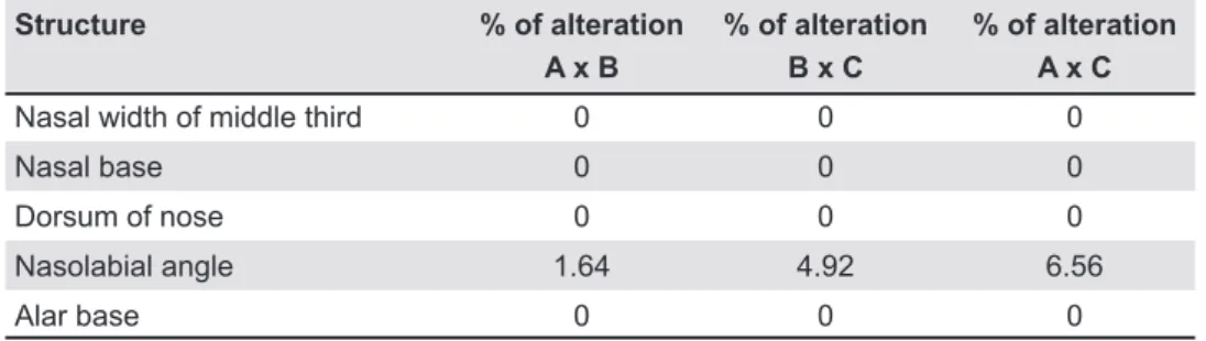
Structure % of alteration A x B
% of alteration B x C
% of alteration A x C
Nasal width of middle third 0 0 0
Nasal base 0 0 0
Dorsum of nose 0 0 0
Nasolabial angle 1.64 4.92 6.56
Alar base 0 0 0
Table 3-#$&
(A), immediate post-expansion (B) and 1-year post-expansion (C) photographs
Structure Agreement (%)
Ex1 X Ex2
Kappa Agreement (%)
Ex2 X Ex3
Kappa Agreement (%)
Ex1 X Ex3
Kappa
Nasal width of middle third 88.88% 0 88.88% 0 100% 1
Nasal base 95.55% 0 93.33 -0.08 97.78% 0.33
Dorsum of nose 97.78% 0.33 100% 1 97.78% 0.33
Nasolabial angle 97.78% 0.33 74.44% -0.12 76.66% 0
Alar base 100% 1 78.88% 0 78.88% 0
Table 2- Percentage of agreement and Kappa values for the inter-examiner analysis
Ex = examiner
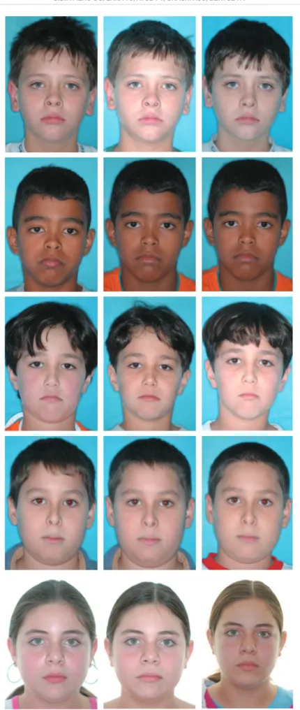
Figure 5- '*
expansion period (C,D) and 1 year following expansion (E,F) of patient in whom an increased nasolabial angle was detected (parents signed informed consent authorizing the publication of these pictures)
A
B
C
D
E
Figure 6- '* expansion period (C,D) and 1 year following expansion (E,F) of patient in whom a decreased nasolabial angle was detected (parents signed informed consent authorizing the publication of these pictures)
A
B
C
D
E
F
DISCUSSION
~ # /9 has not been a novelty since the 1960s12. Since # , /9 repercussions on respiration5,7-11,13,16,19,20,24-27. RME thus have a variable orthopedic effect capable of causing favorable morphological changes to the
For orthodontists, the positive results of RME are mainly evidenced through occlusal analysis. /9 of establishing upper morphology and transversal occlusion.
# = the purpose of evaluating the potential effects /9 of children in the stages of primary and mixed , , considering the buccolingual slant of the posterior teeth (“torque”). The patients had posterior
lingual slant of the posterior teeth. This implied = This characteristic is a reference for the diagnosis of skeletal atresia of the upper dental arch. Therefore, /9 ' complete turn per day (2/4 in the morning and 2/4 at night) until the correction of the posterior
Although it has an indisputable dental effect corresponding to the slant of the anchorage teeth in the vestibular direction, RME causes an cavity3,6,8,11,12,13,16,18,20,22,24. The part of the maxilla /9 closest to the appliance and farthest from the base of the skull. Thus, transversal alterations are more expressive at the point of the occlusal plane and
,24.
numerical sagittal changes in the soft tissue, such as anterior displacement of the tip of the nose, and an increase in the H angle and facial convexity as a consequence of the immediate maxilla advancement
,14,15.
No changes in nasal prominence, thickness of the , /9 15. These results reveal that RME is W - W hard tissues.
Frontal radiographs have proven the orthopedic of the nasal cavity from 1 to 3 mm, 1.7 to 2.5 mm, around 2.08 mm, from 2 to 4.5 mm and of 3.47 mm3,12,13,16,24. One study employing computed =# *74 in the area and 15% in the volume of the nasal cavity20. Do such alterations imply changes in facial morphology? If so, are these changes positive or negative?
One report in the literature stimulated the development of the present investigation. This report discusses an undesirable change in the nasal morphology of a 5-year-old girl after activation of 2& frailty of the inter-nasal suture at this age2. In the /9 dentition, starting at 5 years of age23. Therefore, of the face of children subjected to orthopedic using photographs of the patients prior to RME, immediately after the active phase of RME and 12 /9 anatomical changes caused by RME in the primary and mixed dentition stages, above all, on the nasal # and nasal base. Based on the results of the Kappa test for intraexaminer and interexaminer reliability (Tables 1 and 2), subjective facial analysis is a reliable method for the assessment of morphological alterations. According to the assessment of the three experienced orthodontists, only the nasolabial angle proved altered in 6% of the patients. The # X the clinic: RME does not alter nasal morphology in children in the stages of primary and mixed dentition.
a minimal potential to alter facial morphology in # - # - # the maxilla.
CONCLUSIONS
! X # that RME in children in the primary and mixed dentition stages does not lead to changes in the mid face or nasal morphology.
REFERENCES
1- Babacan H, Sokucu O, Doruk C, Ay S. Rapid maxillary expansion and surgically assisted rapid maxillary expansion effects on nasal volume. Angle Orthod. 2006;76:66-71.
* _ \ 1 <# ~/ 8 --- - <# ~/ ~ /< \ 8 dentofacial deformity. St. Louis: Mosby; 2003. p.507-73. 3- Basciftci FA, Mutlu N, Karaman AI, Malkoc S, Küçükkolbasi H. Does the timing and method of rapid maxillary expansion have an effect on the changes in nasal dimensions? Angle Orthod. 2002;72:118-23.
4- Berger JL, Pangrazio-Kulbersh V, Thomas BW, Kaczynski R. < expansion. Am J Orthod Dentofacial Orthop. 1999;116:563-71. 5- Bicakci AA, Agar U, Sökücü O, Babacan H, Doruk C. Nasal 1 Orthod. 2005;75:1-6.
6- Cameron CG, Franchi L, Baccetti T, McNamara JA Jr. Long term effects of rapid maxillary expansion: a posteroanterior cephalometric evaluation. Am J Orthod Dentofacial Orthop. 2002;121:129-35.
7- Ceroni Compadretti G, Tasca I, Alessandri-Bonetti G, Peri S, D’Addario A. Acoustic rhinometric measurements in children undergoing rapid maxillary expansion. Int J Pediatr Otorhinolaryngol. 2006;70:27-34.
8- Cross DL, McDonald JP. Effect of rapid maxillary expansion on skeletal, dental, and nasal structures: a postero-anterior cephalometric study. Eur J Orthod. 2000;22:519-28.
9- Doruk C, Sökücü O, Sezer H, Canbay EI. Evaluation of nasal rhinometry. Eur J Orthod. 2004;26:397-401.
10- Enoki C, Valera FC, Lessa FC, Elias AM, Matsumoto MA, Anselmo-Lima WT. Effect of rapid maxillary expansion on the dimension of the nasal cavity and on nasal air resistance. Int J Pediatr Otorhinolaryngol. 2006;70:1225-30.
11- Franchi L, Baccetti T, Cameron CG, Kutcipal EA, McNamara JA. Thin-plate spline analysis of the short and long-term effects of rapid maxillary expansion. Eur J Orthod. 2002;24:143-50. 12- Haas AJ. Rapid expansion of the maxillary dental arch and nasal cavity by opening the midpalatal suture. Angle Orthod. 1961;31:73-90.
2+ ' ' \ _ ~ ~ 8 1 J Orthod. 1976;69:274-84.
14- Karamman AI, Basciftci FA, Gelgor I, Demir A. Examination of soft tissue changes after rapid maxillary expansion. World J Orthod. 2002;3:217-22.
16- Krebs A. Expansion on the midpalatal suture, studied by means of metallic implants. Acta Odontol Scand. 1959;92:491-501. 17- Landis JR, Koch GG. The measurement of observer agreement for categorical data. Biometrics. 1977;33:159-74.
18- Oliveira NL, Silveira ACS, Kusnoto B, Viana G. Three-dimensional assessment of morphologic changes of the maxilla: a comparison of 2 kinds of palatal expanders. Am J Orthod Dentofacial Orthop. 2004;126;354-62.
19- Oliveira De Felippe NL, Silveira ACS, Viana G, Kusnoto B, Smith _ 9 81 / X effects. Am J Orthod Dentofacial Orthop. 2008;134:370-82. 20- Palaisa J, Ngan P, Martin C, Razmus T. Use of conventional rapid palatal expansion. Am J Orthod Dentofacial Orthop. 2007;132:458-66.
*2 <# ~/ 8 3th ed. St. Louis:
Mosby; 2007.
** \, 'X \ \, after maxillary expansion in the mixed dentition. Am J Orthod Dentofacial Orthop. 1997;111:321-7.
*+ \ ! w ! ! 1 81 > Correction of posterior crossbite in the primary dentition. J Clin Pediatr Dent. 2000;24:165-80.
24- Silva Filho OG, Montes LA, Torelly LF. Rapid maxillary expansion in the deciduous and mixed dentition evaluated through posteroanterior cephalometric analysis. Am J Orthod Dentofacial Orthop. 1995;107:268-75.
25- Timms DJ. The effect of rapid maxillary expansion on nasal _ w 25Z%62+**2Z
*% ~ ~ ' ' 1 ' 1 '# ~ 1 w Dentofacial Orthop. 1987;91:111-6.