Follicle Viability after Vitri
fi
cation of Bovine
Ovarian Tissue
Viabilidade folicular após a vitri
fi
cação de tecido ovariano
bovino
Janaína de Souza Guedes
1,2Jhenifer Kliemchen Rodrigues
3,4Ana Luisa Menezes Campos
1,2Camila Cruz de Moraes
1,2João Pedro Junqueira Caetano
1,2,3Ricardo Mello Marinho
1,2,31Clínica Pró-Criar - Reproductive Medicine, Belo Horizonte, MG, Brasil 2Post-graduation, Faculdade de Ciências Médicas de Minas Gerais,
Belo Horizonte, MG, Brasil
3Rede Brasileira de Oncofertilidade
–Brazilian Oncofertility Consortium, Belo Horizonte, MG, Brazil
4In Vitro Consultoria, Belo Horizonte, MG, Brazil
Rev Bras Ginecol Obstet 2017;39:614–621.
Address for correspondenceJanaína de Souza Guedes, BSc, Pró-Criar Medicina Reprodutiva, Rua Bernardo Guimarães, 2.063, Lourdes, Belo Horizonte, MG, Brazil - CEP: 30140-082
(e-mail: [email protected]).
Keywords
►
in vitro follicular
maturation
►
fertility preservation
►
vitri
fi
cation
Abstract
Purpose
The present study aimed to evaluate the impact of vitri
fi
cation on the
viability of follicles using a three-dimensional (3D) in vitro culture.
Methods
Bovine ovarian tissue samples (
n
¼
5) obtained from slaughterhouses were
utilized. The cortex was cut into small fragments of 2
3
0.5 mm using a tissue
slicer. From these fragments, secondary follicles were
fi
rst isolated by mechanical and
enzymatic methods, then encapsulated in alginate gel and individually cultured for
20 days. Additional fragments of the same ovarian tissue were vitri
fi
ed in a solution
containing 25% glycerol and 25% ethylene glycol. After warming, the follicles
under-went the same follicular isolation process that was performed for the fresh follicles.
Results
A total of 61 follicles were isolated, 51 from fresh ovarian tissue, and 10 from
vitri
fi
ed tissue. After the culture, the vitri
fi
ed and fresh follicles showed 20% and 43.1%
survival rates respectively (
p
¼
0.290), with no signi
fi
cant differences. At the end of the
culture, there were no signi
fi
cant differences in follicular diameter between the vitri
fi
ed
(422.93
85.05 µm) and fresh (412.99
102.55 µm) groups (
p
¼
0.725). Fresh
follicles showed higher mean rate of antrum formation when compared with vitri
fi
ed
follicles (47.1% and 20.0% respectively), but without signi
fi
cant difference (
p
¼
0.167).
Conclusions
The follicles were able to develop, grow and form antrum in the 3D
system after vitri
fi
cation, despite the lower results obtained with the fresh tissue.
Resumo
Objetivo
O presente estudo teve como objetivo avaliar o impacto da vitri
fi
cação na
viabilidade dos folículos utilizando a cultura in vitro tridimensional (3D).
Métodos
Foi utilizado tecido ovariano bovino (
n
¼
5) obtido de abatedouros. O
córtex foi cortado em pequenos fragmentos de 2
3
0,5 mm, utilizando o
tissue
slicer
e a partir destes fragmentos foram isolados folículos secundários por meio de
received
November 22, 2016 accepted
June 9, 2017 published online August 31, 2017
DOI https://doi.org/ 10.1055/s-0037-1606129. ISSN 0100-7203.
Copyright © 2017 by Thieme Revinter Publicações Ltda, Rio de Janeiro, Brazil
Introduction
Taking into account the recent advances in cancer diagnosis and treatment, young women diagnosed with cancer present a better prognosis nowadays than a few years ago, with a good chance of cure or prolonged survival. A large portion of these women still desire to gestate. Nevertheless, the che-motherapy, the radiotherapy or even the surgery performed in the treatment may compromise their future fertility.1–4
Cryopreservation of oocytes and embryos are alternatives for young adult women who can wait 2–3 weeks for ovarian stimulation and oocyte collection.
The only option for the preservation of the fertility of the patients that require immediate treatment, such as children and prepubescent adolescents, is the cryopreservation of ovarian tissue.1–6
Studies have shown that freezing ovarian tissue fragments may preserve the gametes and also the ability to produce hormones.2,7,8The collection procedure can be readily pro-grammed at any phase of the cycle, and the patient’s expo-sure to hormone treatment is not required.9
When the patient is allowed to conceive, thawed frag-ments could be transplanted again so that the pregnancy may occur spontaneously or after performing assisted repro-duction (AR) techniques. Studies show that retransplantation is a promising technique, since 60 births have been reported to date.10,11
Apart from the non-standardization of ovarian tissue cryopreservation techniques, there are problems such as the local ischemia that occurs after retransplantation,7which currently limit the results.
One of the risks of the retransplantation would be the reintroduction of malignant cells that might have remained in the cryopreserved ovarian tissue.1Although to date there are no reports in humans, this is considered a theoretical risk.4 The maturation of existing ovarian follicles in cryopre-served ovarian tissue fragments in the laboratory to obtain
mature oocytes to be fertilized in vitro would avoid the risk of cancer recurrence.3–5
In vitro follicular maturation techniques have been devel-oped for humans, but their improvement becomes difficult due to the enormous difficulty in obtaining tissue and due to ethical issues.12
The results of human studies are still limited to the study by Amorim et al,13 in which pre-antral ovarian follicles survived for a period of 7 days after cryopreservation. Rodrigues et al14reported thefirst in vitro culture of fresh human pre-antral follicles using the three-dimensional sys-tem (3D) in Brazil. The authors obtained follicle growth for a period of 28 days. Regarding large animals, antrum forma-tion has already been obtained in cows,15,16and embryos were produced in sheep17and goats.18In non-human pri-mates, isolated secondary follicles reached antral stage and were able to produce mature oocytes for fertilization and embryos.19Rodrigues et al20obtained embryos of primates in the morula stage from mature oocytes of secondary ovarian follicles cultured in vitro.
The regulation of the development of the pre-antral follicle is highly complex, and involves many intra-ovarian and endocrine factors. In this context, more studies are needed to better understand the mechanisms that control the initiation and development of follicular growth.20
Bovines are widely used as models for the development of these techniques,12 since their ovaries have large follicle stocks. Furthermore, access to slaughterhouses is easy, with no ethical restrictions. Similarities in ovarian physiolo-gy and dynamics of the reproductive cycle between humans and bovines make this an excellent experimental model for reproductive studies.
The main objective of this study was to compare the development of follicles after vitrification and fresh culture, evaluating the impact of the vitrification in this process, to contribute to the improvement of the technique and to its future application in human ovarian follicles.
método enzimático e mecânico, encapsulados em gel de alginato e cultivados
individualmente durante 20 dias. Outros fragmentos do mesmo tecido ovariano foram
vitri
fi
cados em solução contendo 25% de glicerol e 25% de etilenoglicol. Após
aquecimento, os folículos passaram pelo mesmo processo de isolamento folicular
realizado a fresco.
Resultados
Foram isolados 61 folículos, sendo 51 originários de tecido ovariano a
fresco, e 10 de tecido vitri
fi
cado. Após a cultura, os folículos vitri
fi
cados apresentaram
taxa de sobrevida de 20%, e o grupo a fresco apresentou taxa de 43,1% (
p
¼
0,290). O
diâmetro folicular ao
fi
nal da cultura também não apresentou diferença signi
fi
cativa
entre o grupo vitri
fi
cado (422,93
85,05 µm) e a fresco (412,99
102,55 µm)
(
p
¼
0,725). Os folículos a fresco apresentaram maior taxa média de formação de antro
do que os folículos vitri
fi
cados (47,1% e 20,0%, respectivamente), mas sem diferença
signi
fi
cativa (
p
¼
0,167).
Conclusões
Os folículos foram capazes de se desenvolver, crescer e formar antro em
sistema 3D após a vitri
fi
cação.
Palavras-chave
►
maturação folicular
in vitro
►
preservação da
fertilidade
Methods
We performed a prospective, controlled study approved by the Ethics and Animal Use Committee of one of our institutions.
Ovary Collection
Bovine ovaries (n¼5) were obtained at a local slaughter-house in the city of Belo Horizonte, in the state of Minas Gerais, Brazil. Immediately after the animals were slaugh-tered, the ovaries were collected by a properly instructed person and sent to the laboratory of another of our institu-tions. The ovaries were placed into a vessel containing glucose serum and gentamicin (10μm/mL, Sigma-Aldrich G1264, Sigma-Aldrich, St. Louis, MO, US) at 4°C. The ovaries were cleaned with water for injection and transferred into a Petri dish containingαMinimum Essential Medium (αMEM, Sigma-Aldrich M4526) supplemented with Serum Substitute Supplement (SSS, Irvine 99193, Irvine Scientific, Santa Ana, CA, US), and gentamycin (10μm/mL).
Ovariy Processing, Follicle Isolation, Encapsulation and Culture
Each ovary was cut in half with the aid of a Solidor scalpel blade #24 (Lamedid, Osasco, SP, Brazil). One half of each ovary was placed into a tissue slicer, in which it was possible to remove a thin layer of the ovarian cortex. The medulla was discarded. Using scalpels, curve scissors and tweezers, we obtained 24 fragments of 230.5 mm from the ovarian cortex.
Total 12 of these fragments were separated and kept in a holding medium (containingαMEM supplemented with SSS, and gentamycin 10μm/mL) inside an incubator at 37°C and 5% carbon dioxide (CO2) until the time of vitrification. The
remaining twelve fragments were subjected to the enzymatic and mechanical follicle isolation procedures.
These twelve fragments were separated on four conical tubes (BD Falcon 352099, Becton, Dickinson and Company [BD], Franklin Lakes, NJ, US) of 15 mL (3 fragments per tube) containing the holding medium and collagenase (1 mg/mL type IA, Sigma-Aldrich C5894). The tubes were placed into a water bath for 20 minutes. Every 5 minutes the tubes were removed from the bath and vortexed for 3 minutes. After 20 minutes, the tubes were centrifuged for 5 minutes at 1,500 rpm. After removing the supernatant, 3 mL of a manipulation medium at 4°C was added to stop digestion. The centrifugation process was repeated once again, the supernatant was removed, and 1 mL of the medium was added.
The fragments were then kept incubated at 37°C, with 5% CO2 and pH between 7.2–7.4. The strips were removed
individually and placed into a new plate with the holding medium to perform mechanic follicle isolation under a stereomicroscope magnifying glass.
With the aid of 25 g needles, secondary follicles with diameters between 125μm and 400μm were isolated from each fragment. These follicles were transferred into another dish with the holding medium, and placed in the incubator at 37°C for a period of 2 to 6 hours, during the processing of all ovarian strips. The isolation of a total of 51 fresh secondary follicles was possible.
Only secondary follicles with the following characteristics were selected for encapsulation: absence of antral cavity; an intact basement membrane, with attached stroma; a visible rounded and centrally-located oocyte within the follicle.
Drops of 10 µL of sodium alginate 0.25% (FMC Biopoly-mers, Philadelphia, PA, US) were prepared. Each follicle was washed individually in two of these drops, and kept into the third and final drop. With a 10-μL tip, the alginate drop containing the follicle was pipetted and gently placed on a cross-linking calcium solution. The process was repeated for all of the isolated follicles.
The cross-linking solution (50 mM CaCl2, 140 mM NaCl,
and 10 mM HEPES solution) is responsible for the chemical reaction that allows the liquid sodium alginate 0.25% to be transformed into gel.
After one minute in the solution, the alginate gel struc-tures where the follicles were located were transferred onto individual wells of a 48-well culture dish (BD Falcon 353872) containing 300μL ofαMEM supplemented with 1,050 ng/mL of follicle stimulating hormone (FSH, Sigma-Aldrich F2293), 3 mg/mL of albumin (Irvine Scientific 9988), 100 mg/mL of fetuin Aldrich F6131), 1 mg/mL of insulin (Sigma-Aldrich), 1 mg/mL of transferrin (Sigma-Aldrich T8158), and 50 ng/mL sodium selenite (Sigma-Aldrich S5261).
The encapsulated follicles were cultured for a period of 20 days.Every 2 days, 150 uL of the culture medium of each well of the plate was replaced with fresh medium. During the culture, images of each of the follicles were obtained to measure their diameter using the ImageJ software 1.33U (National Institutes of Health, Bethesda, MD, US).
Vitrification and Warming
The fragments were vitrified using the method described by Ting et al (2011).21Initially, the fragments were equilibrated sequentially in solutions containing 1.2 M of glycerol (10% glycerol v/v, Sigma-Aldrich G2025) for 3 minutes, 1.2 M of glycerolþ3.6 M of ethylene glycol (10% glycerolþ20% ethylene glycol Sigma-Aldrich 102466) for 3 minutes and 3 M of glycerolþ4.5 M of ethylene glycol (25% glycerol
þ25% ethylene glycol) for 1 minute. This whole procedure was performed at room temperature. Following the last solution, the fragments were placed individually onto a piece of aluminum foil (measuring 84 mm2) and immediately
submerged into liquid nitrogen ( 196°C), transferred into cryovials, and stored until the moment of thawing. For warming, the cryovials were removed from the liquid nitro-gen, and the fragments were individually placed immediate-ly for 5 minutes into solutions containing 0.5 M of sucrose (Sigma-Aldrich S1888), then 0.25 M of sucrose, 0.125 M of sucrose and, to finish, only on equilibrium solution or holding medium by 2 times of 10 minutes, and the whole procedure was performed at a temperature of 37°C. After warming, the fragments were kept in the holding medium solution supplemented with 15% v/v SSS and 29 mg/mL of ascorbic acid (Sigma-Aldrich A4403) to isolate its secondary follicles and individually encapsulated into the alginate.
enzyme isolation, mechanical procedure, encapsulation in alginate gel, and culture for 20 days.
Follicle Survival and Growth
The survival and diameters of the follicles were evaluated using images taken with an Eclipse Ti-S inverted microscope (Nikon, Tokyo, Japan) and an attached digital camera (OCTAX Micro-science, Bruckberg, Bavaria, Germany). Images were obtained from each follicle, as well as images from a micrometer (1 mm with 0.01 mm divisions, Fisher Micromaster, Fisher Scientific, Fair Lawn, NJ, US) for calibration purposes. For the measure-ment, the images were imported to the ImageJ software 1.33U. Each follicle diameter was determined in units of pixels and converted to micrometers based on the conversion determined by measuring the image of the calibrated micrometer.
The follicles were measured from the outer layer of the cells, and the measurements included the largest follicle diameter and a second measurement perpendicular to thefirst. The mean of these values was then calculated and considered as the follicle diameter.Antrum formation was observed upon a mor-phological analysis of the follicles. The follicles were considered to be degenerating when the oocyte was no longer surrounded by a layer of granulosa cells, and when the oocyte became dark and the granulosa cells became dark and fragmented.
Statistical Analysis
The statistical analysis consisted of absolute and relative frequencies for categorical variables and meanstandard deviation (SD) for continuous variables. The comparison of two proportions was performed by proportions tests; the comparison of the percentage of antrum formation, by Fisher exact test; and the comparison of two means, by nonpara-metric Wilcoxon test.
In order to evaluate the growth of the ovarian follicles, we used the percentage variation between a start timeT0and an end timeT1, calculated by the formula: V¼(T1-T0)/T0x 100.
The Kaplan-Meier method and log-rank test were used to estimate the curves until the infeasibility of the follicles, and to compare them according to their groups (fresh and vitri-fied), and we calculated the median survival time of the follicles considering the follicle survival time, and the num-ber of days between the isolation and their infeasibility. The analyses were performed using the free R software (R Foundation for Statistical Computing, Vienna, Austria), version 3.1.3. The significance level was set to 5%.
The power of the sample was of 95%, and the calculation was based on the size of the sample analyzed and the proposed objective.
Results
In the present study, 61 follicles were recovered after treat-ment with collagenase. A total of 51 follicles were isolated from fresh tissue, and 10 from vitrified tissue.
The survival rate at the end of 20 days of culture was 43.1% for the fresh group, and 20% for the vitrified group, without any significant difference.
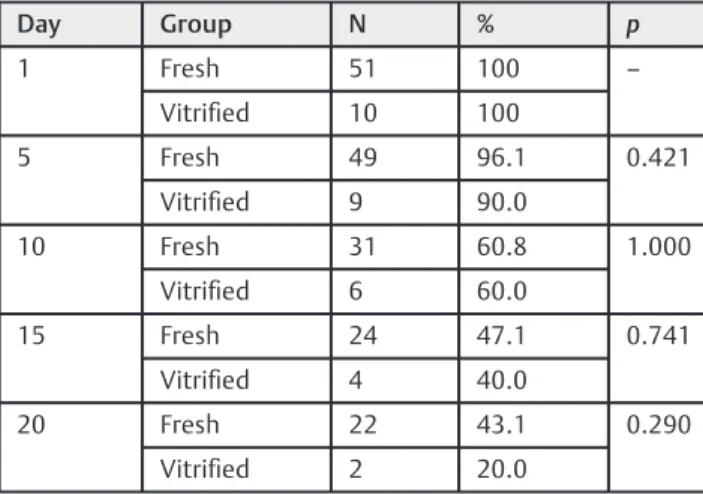
►Table 1shows the distribution of the surviving follicles on days 1, 5, 10, 15 and 20, regarding the fresh and vitrified groups, and the comparison test between the proportions of survivors per day. In absolute terms, the proportions of fresh surviving follicles were higher than those of the follicles after vitrification in all evaluated days, but without any significant difference. We were able to observe a gradual reduction in survival in the two groups, mainly in the vitrified group, though it was not significant.
►Fig. 1shows Kaplan-Meier curves for follicle survival time regarding both groups. We observed that the fresh follicles survived longer; however, the difference was not significant (p¼0.205).
►Table 2presents the meanSD of the diameters (in µm) of the two groups of follicles per day. Considering the follicles that survived, Table 2 also shows the mean diameters of both groups on thefirst and last days of culture. We observed a significant difference between the two groups on thefirst day of culture.
During culture, the vitrified group showed a variation in growth of 8.8% compared with 21.5% for the fresh group (►Table 3).
We observed a significant difference in variation of follicle growth between the groups from thefirst to thefifth days (p¼0.000). The fresh follicles showed a higher growth during this period. After vitrification, the follicles showed a signifi -cantly higher growth during the period from the tenth to the fifteenth days (p¼0.042). In general, the difference in growth between the two groups was not significant.
►Fig. 2 shows the distribution of antrum formation in both groups. We observed that the fresh follicles showed a higher mean antrum formation (47.1%) when compared with the vitrified follicles (20%), but this difference was not significant (p¼0.167).
Discussion
The results obtained demonstrated that the follicles cultured in vitro after vitrification are able to survive, develop and form antrum.
Table 1 Distribution of surviving follicles per day and comparison between the groups
Day Group N % p
1 Fresh 51 100 –
Vitrified 10 100
5 Fresh 49 96.1 0.421
Vitrified 9 90.0
10 Fresh 31 60.8 1.000
Vitrified 6 60.0
15 Fresh 24 47.1 0.741
Vitrified 4 40.0
20 Fresh 22 43.1 0.290
Vitrified 2 20.0
These are thefirst data (to the best of our knowledge) of isolation and follicle maturation obtained from vitrified bovine ovarian tissue.
Despite the sample size, we obtained a survival rate and antrum formation of 20% of these follicles following 20 days of
in vitro culture in an alginate matrix, which, for an initial study, is a great advance. However, we must highlight the need for further studies in order to reach a consistent conclusion.
The viability of both follicle groups, overall, showed no significant differences. As described by Bulgarelli et al,22 macaque secondary follicles isolated from vitrified ovarian tissue showed similar survival rates when compared with the fresh group, and showed no significant difference in the diame-ter and antrum formation afdiame-ter in vitro culture in the 3D system. In a study by Amorim et al13that evaluated the survival of human pre-antral follicles after ovarian tissue cryopreserva-tion, follicle isolation and in vitro culture into an alginate matrix, the authors did not observe any morphological or viability changes among the groups: follicles included in fresh ovarian tissue fragment, follicles included in the cryopreserved
Table 3 Mean variation (in %) of the fresh and vitrified follicle diameters
Days Fresh Vitrified p
MeanSD MeanSD
1–5 41.739.6 7.67.5 0.000
5–10 8.912.7 13.78.4 0.186
10–15 3.29.4 9.94.0 0.042
15–20 1.313.0 5.98.3 0.587
Total 21.521.4 8.86.3 0.074
Abbreviation: SD, standard deviation.
Notes:pvalue refers to the Wilcoxon test.Represents statistical difference (p<0.05).
Fig. 1 Kaplan-Meier curves and log-rank test for the survival time of the fresh and vitrified follicles in days; (A) only the curves and (B) curves with confidence intervals.
Table 2 Mean fresh and vitrified follicle diameters (in µm) according to the days following isolation
Day Fresh Vitrified p
MeanSD MeanSD
1 244.5580.57 348.7888.73 0.002
5 327.9487.84 375.35115.83 0.209
10 385.4377.98 426.59104.29 0.385
15 411.9687.77 485.70132.73 0.262
20 412.99102.55 422.9385.05 0.725
Abbreviation: SD, standard deviation.
Notes:pvalue refers to the Wilcoxon test.Represents statistical difference (p<0.05).
tissue, enzymatically-isolated follicle fragments after cryo-preservation, and fresh follicles cultured in an alginate matrix. This study proved to be thefirst in which the in vitro culture of human small pre-antral follicles in cryopreserved ovarian tissue after isolation and culture for 7 days was possible.
In our study, a progressive growth in follicle diameter during the 20 days of culture was observed. This suggests that the use of a 3D culture system effectively simulates the physiological conditions, promoting interactions between somatic cells, which may be an adequate support for the in vitro maturation of isolated pre-antral follicles.4,23
Xu et al24showed that, from fresh human ovarian tissue, they obtained the survival and growth of isolated secondary follicles using a 3D matrix. Therefore, they were able to create an environment that enabled oocyte growth.
In a study by Silva et al,25 higher survival rates in goat secondary follicle cultures using the 3D system were obtained when compared with the two-dimensional (2D) system.
Another important factor to support follicular develop-ment was the use of a culture medium with an adequate composition to aid the development.
In the present study, a culture medium containing FSH was used for both groups. The FSH is considered important for the in vitro maturation of follicles. Its absence may contribute to low antrum formation rates, as demonstrated by Luz et al26 and Barberino et al.27In a study published by Matos et al,28a concentration of 50 ng/mL of FSH was able to promote the growth of activated pre-antral follicles, demonstrating that the FSH plays a vital role in the culture of goat ovarian follicles.
However, to date, there are no conclusive studies on the effectiveness of different media for the in vitro culture of bovine ovarian tissue, especially after cryopreservation.29
In the present study, we also found that the diameters of the vitrified follicles on thefirst day of culture were signifi -cantly higher than those of the fresh follicles (p¼0.002). We believe that this fact may be related to the use of collagenase. This enzyme in fresh ovarian tissue assists in the isolation of pre-antral follicles, leaving the tissue less dense, and the follicles more accessible within the stroma, which makes the removal of the stromal cells surrounding the teak easier.
On the other hand, the vitrified follicles showed a higher fragility, which hampered their removal, leaving a thicker layer of stromal cells on their surfaces, making it difficult to measure their diameters on the first day of culture. This difficulty in isolating the follicles after vitrification also explains the lower number of follicles collected in this group. During our culture, the fresh follicles showed a higher growth from days 1 to 5 when compared with the vitrified follicles during the same period. On the other hand, the vitrified follicles grew more between days 10 to 15 of culture. We obtained a variation in diameter growth of 21.5% for the fresh group versus 8.8% for the vitrified group (p¼0.074), and a mean diameter of 412.99μm and 422.93μm respec-tively (p¼0.725).
The reason for this variation in growth is unknown; however, Gutierrez et al,15 in an in vitro culture of fresh bovine pre-antral ovarian follicles, indicated a rapid
in-crease in follicle diameter during thefirst week of culture, followed by a delay in growth after 8–10 days of culture. This finding is in agreement with the results found in the present study.
We can also assume that the higher growth of vitrified follicles within 10 to 15 days could be justified since these follicles were the most resistant follicles selected in the vitrification and isolation processes: they were able to maintain their structural integrity during vitrification and isolation, and had higher survival capacity.
Moniruzzaman et al30observed a slowdown in the rate of development of vitrified primordial follicles of pigs. Ting et al5 also observed a delay in the growth of vitrified isolated secondary follicles of primates.
Another possibility would be the alleviation of the effect of stromal cells in the measurement of follicular diameter during culture. Thus, the growth rate could have been simi-lar, but confused by the larger initial diameter.
After warming, the tissue was exposed to digestion by collagenase, followed by mechanical isolation as performed in the fresh method. After it was digested, the vitrified tissue showed greater sensitivity to the mechanical isolation, re-sulting in easy follicle rupture. Thesefindings can be related to the enzymatic method of isolation of ovarian follicles in vitrified tissue.
The enzymatic isolation is used to facilitate the isolation of follicles with dense andfibrous ovarian cortices, such as those of humans.31 The enzymatic isolation of pre-antral follicles improves the recovery rate of the isolated follicles; however, the integrity of the follicle is not always preserved, damaging the theca cells and the basal membrane.31–33
Rupture in the follicular membrane and the destruction of other intrafollicular components during the enzymatic isolation represent major problems in the development of pre-antral follicles in vitro.34The potential follicle develop-ment may be hindered if the integrity of the follicle is compromised.4
The sensitivity of the ovarian cortex fragments demon-strated that the use of collagenase after vitrification could damage intact follicles for in vitro culture in the 3D matrix systems, but the small sample size is a limitation. Moreover, vitrification has a direct effect on the quality of the tissue after the process.
Vitrification is a challenging process due to the complex structure and different cell types present in the ovarian tissue,35 even though it is considered an effective, faster and less expensive process.
Ting et al21 demonstrated that macaque secondary fol-licles are better preserved after vitrification when compared with slow freezing.
In a recent study, Lunardi et al,36comparing the effect of vitrification in the development of sheep secondary follicles included in the ovarian tissueversusfollicles isolated using only mechanical isolation, verified that follicles that were vitrified included in the tissue showed significantly larger rate of follicles that regressed its growth.
Another important factor that is related to the number of isolated follicles is that each tissue fragment presents a different number of follicles, which makes the comparison, in terms of numbers of follicles, of the vitrified and fresh tissue groups very difficult.
Even though the effects of collagenase and/or vitrification may impair the isolation of pre-antral follicles, the growth and development in an in vitro culture was possible, confirming the viability of the vitrified follicles after enzymatic isolation. The follicles that have survived until the last day of culture, in 100% of the cases, reached the antral stage. Antrum formation was observed from the 4th day of culture in most follicles. This ability to develop and form antrum can be considered a follicular functionality indicator.16
In the present study, the ovarian tissue was subjected to several different procedures, such as vitrification, warming, follicular isolation and encapsulation in alginate gel. These procedures may potentially damage the follicles, as dis-cussed by Amorim et al37and Dolmans et al.38
Based on our results, we can assume that the isolation and culture of bovine ovarian follicles after vitrification is a possible approach. We believe more studies are necessary to improve the in vitro culture of follicles, especially for tissues previously submitted to cryopreservation. These preliminary results are important to increase the under-standing and the chances of future applicability in patients requiring immediate treatment for cancer, as well as in children and prepubescent adolescents.
Conclusions
In conclusion, the present study demonstrates, for thefirst time, the feasibility of in vitro follicular maturation of bovine secondary pre-antral follicles after vitrification and enzymatic and mechanical isolation from ovarian tissue fragments in a 20-day period in an alginate matrix. Despite the lower results obtained with the fresh tissue, the cultured follicles of vitrified tissue were able to grow and, at the end of the culture, 100% of the survivors formed antrum.
The enzymatic isolation with collagenase may have im-paired the efficiency in the isolation of a higher number of follicles, which indicates the need for further studies to evaluate the best technique to be used.
Despite the difficulty with the technique, we can conclude that the 3D culture system is potentially viable, and provides a new opportunity to study the optimization of in vitro follicle maturation, as well as the future possibility of clinical application.
Conflicts of Interest
Authors have no conflicts of interest to disclose.
Acknowledgments
We would like to thank Clínica Pró-Criar Medicina Reprodutiva, which enabled one doing this job, and Isabel Cristina Gomes for statistical support.
References
1 Salama M, Isachenko V, Isachenko E, Rahimi G, Mallmann P. Updates in preserving reproductive potential of prepubertal girls with cancer: Systematic review. Crit Rev Oncol Hematol 2016;103:10–21
2 Donnez J, Dolmans MM. Ovarian tissue freezing: current status. Curr Opin Obstet Gynecol 2015;27(03):222–230
3 Carvalho BR, Rodrigues JK, Marinho RM, Caetano JPJ, Rosa-e-Silva ACJS. Visão geral sobre preservação da fertilidade feminina depois do câncer. Reprod Clim 2014;29(03):123–129
4 Rodrigues JK, Campos JR, Marinho RM, Xu J, Zelinski MB, Stouffer RL. Desenvolvimento folicular e maturação oocitária in vitro. In: Marinho RM, Rosa e Silva ACJS, Caetano JPJ, Rodrigues JK, editores. Preservação da fertilidade: uma nova fronteira em medicina reprodutiva e oncologia. Rio de Janeiro: Medbook; 2015:161–169
5 Ting AY, Yeoman RR, Lawson MS, Zelinski MB. Synthetic polymers improve vitrification outcomes of macaque ovarian tissue as assessed by histological integrity and the in vitro development of secondary follicles. Cryobiology 2012;65(01):1–11
6 Dolmans MM, Jadoul P, Gilliaux S, et al. A review of 15 years of ovarian tissue bank activities. J Assist Reprod Genet 2013;30(03): 305–314
7 Marinho RM, Rodrigues JK, Lamaita RM, et al. Fertility preserva-tion in women with cancer: update and perspectives. Rev Med Minas Gerais 2013;23(04):510–517
8 Campos JR, Rodrigues JK, Bulgarelli DL, Zelinski MB. Aspectos laboratoriais da criopreservação de tecido ovariano. In: Marinho RM, Rosa e Silva ACJS, Caetano JPJ, Rodrigues JK. , editores. Preservação da fertilidade: uma nova fronteira em medicina reprodutiva e oncologia. Rio de Janeiro: Medbook; 2015:155–160
9 Campos JR, Rosa-e-Silva JC, Carvalho BR, Vireque AA, Silva-de-Sá MF, Rosa-e-Silva AC. Cryopreservation time does not decrease follicular viability in ovarian tissue frozen for fertility preserva-tion. Clinics (Sao Paulo) 2011;66(12):2093–2097
10 Donnez J, Dolmans MM. Fertility preservation in women. Nat Rev Endocrinol 2013;9(12):735–749
11 Donnez J, Dolmans MM. Ovarian cortex transplantation: 60 reported live births brings the success and worldwide expansion of the technique towards routine clinical practice. J Assist Reprod Genet 2015;32(08):1167–1170
12 Jorssen EPA, Langbeen A, Marei WF, et al. Morphologic character-ization of isolated bovine early preantral follicles during short-term individual in vitro culture. Theriogenology 2015;84(02):301–311
13 Amorim CA, Van Langendonckt A, David A, Dolmans MM, Donnez J. Survival of human pre-antral follicles after cryopreservation of ovarian tissue, follicular isolation and in vitro culture in a calcium alginate matrix. Hum Reprod 2009;24(01):92–99
14 Rodrigues JK, Marinho RM, Cota AMM, Wainstein AJA, Caetano JPJ. In vitro maturation of human preantral ovarian follicles in a tridimentional culture system. JBRA Assist Reprod 2014;18(03):92
15 Gutierrez CG, Ralph JH, Telfer EE, Wilmut I, Webb R. Growth and antrum formation of bovine preantral follicles in long-term culture in vitro. Biol Reprod 2000;62(05):1322–1328
16 Rossetto R, Saraiva MVA, dos Santos RR, et al. Effect of medium composition on the in vitro culture of bovine pre-antral follicles: morphology and viability do not guarantee functionality. Zygote 2013;21(02):125–128
17 Luz VB, Araújo VR, Duarte AB, et al. Eight-cell parthenotes originated from in vitro grown sheep preantral follicles. Reprod Sci 2012;19(11):1219–1225
18 Magalhães DM, Duarte AB, Araújo VR, et al. In vitro production of a caprine embryo from a preantral follicle cultured in media supplemented with growth hormone. Theriogenology 2011;75 (01):182–188
20 Rodrigues JK, Navarro PA, Zelinski MB, Stouffer RL, Xu J. Direct actions of androgens on the survival, growth and secretion of steroids and anti-Müllerian hormone by individual macaque follicles during three-dimensional culture. Hum Reprod 2015; 30(03):664–674
21 Ting AY, Yeoman RR, Lawson MS, Zelinski MB. In vitro develop-ment of secondary follicles from cryopreserved rhesus macaque ovarian tissue after slow-rate freeze or vitrification. Hum Reprod 2011;26(09):2461–2472
22 Bulgarelli DL, Ting AY, Zelinsk MB. Vitrificação de folículo secundário isolado: uma nova opção para preservação de ferti-lidade em pacientes com câncer. JBRA Assist Reprod 2013;17 (04):240
23 Xu M, Kreeger PK, Shea LD, Woodruff TK. Tissue-engineered follicles produce live, fertile offspring. Tissue Eng 2006;12(10): 2739–2746
24 Xu M, Barrett SL, West-Farrell E, et al. In vitro grown human ovarian follicles from cancer patients support oocyte growth. Hum Reprod 2009;24(10):2531–2540
25 Silva GM, Rossetto R, Chaves RN, et al. In vitro development of secondary follicles from pre-pubertal and adult goats cultured in two-dimensional or three-dimensional systems. Zygote 2015; 23(04):475–484
26 Luz VB, Araújo VR, Duarte ABG, et al. Kit ligand and insulin-like growth factor I affect the in vitro development of ovine preantral follicles. Small Rumin Res 2013;115(1–3):99–102
27 Barberino RS, Barros VR, Menezes VG, et al. Amburana cear-ensis leaf extract maintains survival and promotes in vitro development of ovine secondary follicles. Zygote 2016;24(02): 277–285
28 Matos MH, Lima-Verde IB, Luque MC, et al. Essential role of follicle stimulating hormone in the maintenance of caprine preantral follicle viability in vitro. Zygote 2007;15(02):173–182
29 Castro SV, Carvalho AA, Silva CM, et al. Fresh and vitrified bovine preantral follicles have different nutritional requirements during in vitro culture. Cell Tissue Bank 2014;15(04):591–601
30 Moniruzzaman M, Bao RM, Taketsuru H, Miyano T. Development of vitrified porcine primordial follicles in xenografts. Theriogen-ology 2009;72(02):280–288
31 Abedelahi A, Rezaei-Tavirani M, Mohammadnejad D. Fertility preservation among the cancer patients by ovarian tissue cryo-preservation, transplantation, and follicular development. Iran J Cancer Prev 2013;6(03):123–132
32 Demeestere I, Delbaere A, Gervy C, Van Den Bergh M, Devreker F, Englert Y. Effect of preantral follicle isolation technique on in-vitro follicular growth, oocyte maturation and embryo develop-ment in mice. Hum Reprod 2002;17(08):2152–2159
33 Rossetto R, Lima IMT, Saraiva MVA, Lima-Verde IB, Sales ET, Figueiredo JR. Avanços no isolamento e sistemas de cultivo de folículos pré-antrais. Acta Vet Bras 2011;5(01):15–23
34 Gosden RG, Mullan J, Picton HM, Yin H, Tan SL. Current perspective on primordial follicle cryopreservation and culture for reproductive medicine. Hum Reprod Update 2002;8(02):105–110
35 Bagchi A, Woods EJ, Critser JK. Cryopreservation and vitrification: recent advances in fertility preservation technologies. Expert Rev Med Devices 2008;5(03):359–370
36 Lunardi FO, de Aguiar FL, Duarte ABG, et al. Ovine secondary follicles vitrified out the ovarian tissue grow and develop in vitro better than those vitrified into the ovarian fragments. Therio-genology 2016;85(07):1203–1210
37 Amorim CA, Rondina D, Lucci CM, Gonçalves PB, Figueiredo JR, Giorgetti A. Permeability of ovine primordial follicles to different cryoprotectants. Fertil Steril 2006;85(Suppl 1):1077–1081