w w w . j c o l . o r g . b r
Journal
of
Coloproctology
Original
Article
Changes
in
the
proportions
of
types
I
and
III
collagen
in
hemorrhoids:
the
sliding
anal
lining
theory
Carlos
Sardi ˜nas
a,∗,
Dilia
Diaz
Arreaza
b,
Héctor
Osorio
caHospitalUniversitariodeCaracas,UnidaddeColoproctología,LaboratoriodeFisiologíaAnorrectal,Caracas,Venezuela
bUniversidadCentraldeVenezuela,InstitutoAnatomopatológico“Dr.JoséAntonioO’Daly”,Caracas,Venezuela
cInstitutoVenezolanodeInvestigacionesCientíficas(IVIC),Venezuela
a
r
t
i
c
l
e
i
n
f
o
Articlehistory:
Received12February2016 Accepted6April2016 Availableonline27April2016
Keywords: Hemorrhoids Anal Canal Collagen TypeI TypeIII Fetus Deterioration
a
b
s
t
r
a
c
t
Objective:ThisstudyaimstodeterminechangesintheproportionsoftypesIandIIIcollagen inhemorrhoidsandtoverifytheslidinganalcanalliningtheory.
Patientsandmethod:Thestudyisfocusedonasampleof17patients,9femalesand8males (agerange:30–70years),withgradeIIIandgradeIVhemorrhoids.Tissuefrom4fetuses(age: 16weeksofgestation)wasusedascontrolsample.Alltheparticipantsgavetheirinformed consent.Samplesweregatheredin2014.Allpatientsunderwentopenhemorrhoidectomyby usingthetechniquedescribedbyMilliganandMorgan,publishedinLancetjournalin1937. Thehemorrhoidsampleswerestainedwithhematoxylin–eosinforthehistologicstudyto confirmthehemorrhoidaltissuediagnosis.Thepicrosiriusredstainingprotocolwasused afterthehistologicanalysis.Themethodusedforimageprocessingisdescribedinthetext. ImageswereimportedtotheImageToolforWindowssoftware.Thesameprocesswasused ontheembryonictissue.Dataresultingfromtheanalysisofimageswereprocessedusing STATISTICA,asoftwareforstatisticalanalysis.
Results:Whencompared,itwasfoundthatthetwotissuespresentedverydifferentvalues, withhemorrhoidscontainingthehighesttypeIIIcollagenvalues.
Conclusion:Ourresultsseemtoimplythathemorrhoidshavealargerproportionoftype IIIcollagenthanfetaltissue.Theyalsosuggestapossibleage-relateddeteriorationofthe tissue.
©2016SociedadeBrasileiradeColoproctologia.PublishedbyElsevierEditoraLtda.This isanopenaccessarticleundertheCCBY-NC-NDlicense(http://creativecommons.org/
licenses/by-nc-nd/4.0/).
∗ Correspondingauthor.
E-mails:[email protected],[email protected](C.Sardi ˜nas).
http://dx.doi.org/10.1016/j.jcol.2016.04.003
Mudanc¸as
nos
percentuais
do
colágeno
dos
tipos
I
e
III
em
hemorroidas:
teoria
do
revestimento
anal
deslizante
Palavras-chave: Hemorroidas Anal Canal Colágeno TipoI TipoIII Feto Deteriorac¸ão
r
e
s
u
m
o
Objetivo: Esseestudotemporobjetivodeterminarmudanc¸asnospercentuaisdocolágeno dostiposIeIIIemhemorroidaseverificarateoriadorevestimentodecanalanaldeslizante. Pacientesemétodo: Oestudoestáfocadoemumaamostrade17pacientes(9mulherese8 homes;faixaetária:30-70anos),comhemorroidasdegrausIIIeIV.Utilizamostecidode quatrofetos(idade:16semanasdegestac¸ão)comoamostradecontrole.Todosos partic-ipantesderamconsentimentoinformado.Asamostrasforamreunidasem2014.Todosos pacientespassaramporumahemorroidectomiaaberta;paratanto,foiempregadaa téc-nicadescritaporMilliganeMorgan,publicadanoperiódicoLancetem1937.Asamostrasde hemorroidaforamcoradascomhematoxilina-eosinacomvistasaoestudohistológicopara confirmac¸ãododiagnósticodetecidohemorroidal.Apósaanálisehistológica,omaterialfoi coradocomoprotocolodepicrosiriusred.Ométodoempregadoparaoprocessamentodas imagensestádescritonotexto.AsimagensforamimportadaspelosoftwareImageToolfor Windows.Omesmoprocessofoiempregadonotecidoembrionário.Osdadosresultantes daanálisedasimagensforaprocessadoscomoprogramaSTATISTICA,umsoftwarepara análiseestatística.
Resultados: Porcomparac¸ão,constatamosqueosdoistecidosapresentavamvaloresmuito diferentes,eashemorroidascontinhamosmaisaltosvaloresdecolágenodotipoIII.
Conclusão: Nossosresultadosparecemimplicarquehemorroidaspossuemumpercentual
maiselevadodecolágenodotipoIIIversustecidofetal.Osresultadostambémsugeremuma possíveldeteriorac¸ãodotecido,relacionadaàidade.
©2016SociedadeBrasileiradeColoproctologia.PublicadoporElsevierEditoraLtda.Este ´eumartigoOpenAccesssobumalicenc¸aCCBY-NC-ND(http://creativecommons.org/
licenses/by-nc-nd/4.0/).
Introduction
In1950GassandAdamsrevealedthathemorrhoidsresulted fromdegenerationofsupportivetissueintheanalcanalafter observing connective tissue fragmentation in hemorrhoids specimens,andbelievedthattheirprotrusionwasrelatedto alaxanus.1Later,Hughes(1957)andPatey(1972)supported thatideabecauseitwasinkeepingwiththepresenceofthe hemorrhoiddescent.2,3
Thepresenceofanimportantlayerofsmoothmuscle tis-sueintheanalsubmucosaisrelevant.Itwasfirstdescribed in1853byTreitz,whonoticedthatapartofitarisesfromthe internalsphincter,andtheotherfromtheconjoint longitudi-nalmuscle,knowntodayasTreitz’smuscle.Itisresponsible forhemorrhoidsretractionandelevationduringdefecation, andforthereturnofthesestructurestotheirnormalposition, togetherwiththeconnectivetissue.4
Oneoftheargumentsthathavebeendebatedthemostis howthedeteriorationofconnectivetissuethatsupports hem-orrhoidsfacilitatesitsprolapse,asproposedbyThomsonin 1975.5AresearchbyHaasin1984showedthedeterioration oftheconnectivetissueand,consequently,theslidingofthe analstructureinpatientswithhemorrhoids,aprocesswhich increasesgraduallywithaging.6 Otherfactorsthat produce alterationsintheeliminationhabitsandtraumasthatcause suchsymptomsmustbeaddedtothat.Hemorrhoidsunder constantlocalstressproducedbythepatient’seffortwould eventuallyleadtoruptureoftheTreitz’smuscleandprolapse ofthehemorrhoidalbundles.
In1988Morgadostudiedthemicroscopicanatomyofthe anal canalinagroup offetuses withanaverageage of32 weeksofintrauterinelife.7 Hefoundthatthe muscular tis-suewasclusteredineithergroovedorsmoothbundleswith collagenfiberagrupationsofhomogeneous,regularand non-fragmented appearance. This confirmed that hemorrhoids wereconnectedtotherestoftheanalcanalwallbyathick, homogeneousandwell-defendedbundleoffibersthatarenot fragmented,producingfirmadherencebetweenthemandthe wallthatsurroundsthem.Thisallowstoestablishthathuman fetusesarethecomparativepatternfortheevaluationof pos-sibledeteriorationofmuscularstructureandcollageninadult patientswithhemorrhoidproblems.
However,in2009Willispresents astudy wherehe com-pares quantityto qualityofcollagen amongadultpatients withorwithouthemorrhoidprolapse,findingnocorrelation withage orsex, aswellas withendogenousor exogenous causesforalterationsofcollagenconcentrations.8
Thisstudyaimstodeterminechangesintheproportions oftypesIandIIIcollageninpatientswithhemorrhoidsusing humanfetusesascomparisongroup.
Materials
and
methods
Patientsandmethods
16weeksofgestation)wasusedascontrolsample.Allthe par-ticipantsgavetheirinformedconsent.Samplesweregathered in2014.Allpatientsunderwentopenhemorrhoidectomyby usingthetechniquedescribedbyMilliganandMorgan, pub-lishedinLancetjournalin1937.9
Histologicalstudyandpicrosiriusredstaining
Thehemorrhoidsampleswerefixedin10%formalinfor24h, then processed and embedded in paraffin using standard histologicaltechniques. 5-Micronthicksessions weretaken and placed on glass plates. The cuts were stained with hematoxylin–eosintodoahistologicalstudyusingan Olym-pusCX31microscopeandtoconfirmthehemorrhoidaltissue diagnosis(Fig.1).
Afterthehistologicalanalysis, apicrosiriusred staining protocol was performed. The selected cuts were deparaf-finized and hydrated by immersion in xylene (twice), descendinggradedalcoholsolutions(100%,95%,70%,50%), andthenwashedwithdistilledwater4times.Next,thecuts werestainedwithpicrosiriusredandlefttorestfor1h.The excessstainwasremovedwithtworinsesofdistilledwater andthentheyweredehydrated,firstwithethanolat100%(3 changes)andthenwithaxylenetreatment.
The picrosirius red stained cutswere examined with an
OlympusCX31microscope(usingapolarizedlightanalyzer). Theobservationofstained tissueswiththismethodallows differentiatingtypeIandtypeIIIcollagen(Figs.2and3). Bear-ingthisinmind,manyphotographsofeverycutweretaken withan8.0megapixelHPPhotosmartR927camera.
ThemethodusedforimageprocessingisdescribedinFig.4. ImageswereimportedtotheImageToolforWindowssoftware.10 Thenimageswereturnedintograyscalefigures.Objectsof interestwerechosendependingontheirbrightnessinthegray scale.Afterthat,thepixelsoftheresultingbinaryimageswere counted.Thederivedvaluesrepresenttheassessmenton pro-portionalitybetweencollagentype1andtype3(CIII/CI).Data resultingfromtheanalysisofimageswereprocessedusing STATISTICA,softwareforstatisticalanalysis.11
Fig.1–Hemorrhoids(hematoxylin–eosin).Stratifiedflat epitheliumofthemucoustypecorrespondingtotheanus andtheglandularepitheliumoftherectum.The
submucosaevidencesdilated,thin-walledandthick-walled vesselscorrespondingtovenousplexusesintheregion.
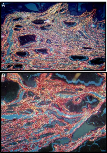
Fig.2–(AandB)Hemorrhoids(picrosiriusred).Stratifiedflat epitheliumofthemucoustypecorrespondingtotheanus. ThesubmucosashowstypeIandtypeIIIcollagenfibers stainedinredandgreenwiththepolarizedlight,together withvenousplexusesintheregion.
Results
Fig.5showsacomparisonbetweentheCIII/CIvaluesforfetal tissueandhemorrhoidaltissue.Asitcanbeobserved,when thetwotissuesarecompared,theypresentverydifferent val-ues,withhemorrhoidscontainingthehighesttypeIIIcollagen values.
Discussion
Fig.3–Embryo(picrosiriusred).(A)Immaturepubicboneis observedatalowerincrease,followedbygenitalroutes fibrousseptumandrectumwithvenousplexusesinthe region.(B)Fibersoftheseptumandrectumwithcollagen fibersareobservedatahigherincrease,whichinthedark fieldseemtobeyellow.
Thevascularcushionsofthesubmucosaaregenerally sup-portedby the pectinate line and by the muscular layer of thesubmucosa.Duringdefecation,theinternalsphincteris relaxedand thereisaneversionofthevasculartissueand thepectinateline.Thiseversionisproducedattheanorectal union,whileprobablyadisruptionofthisnaturaleversionand thelowerrectumreturnisthefundamentalmechanismfor theproductionofhemorrhoids,asstatedbyGassandAdams in 1950, when they considered that hemorrhoids resulted fromdegenerationofsupportivetissueintheanalcanal.This isknown astheslidinganal liningtheory.Thefactorsthat disrupt the normal eversion and return can be related to endocrinedisruptions,ageandconstipation.Asfor constipa-tion,nodataareavailablesofarastoconsiderthefrequency
andtimespentintheevacuationoffecalmatterasacauseof hemorrhoidaldisease.
Prolapse through the anus is considered a hemorrhoid from a folkloric point ofview.Thesymptom ofprotrusion withspontaneousreduction,orthroughdigitalcontrolofthe masses insidethe anal canal is one ofthe mostfrequent characteristicsofthehemorrhoidaldisease.Manytimesthis signaltendstobeconfusedwithahemorrhoidal thrombo-sis,orperianalfoldsareinterpretedasprolapsedirreducible hemorrhoids. Hypertrophicpapillae or polypsofthe lower thirdofthe rectum are rarelyconfusedwithhemorrhoidal prolapse because theycanprolapse through theanus, and becausetheycanbereduced.Dataonthenaturalhistoryof untreatedhemorrhoidaldiseasearescarce.Therefore,thereis noinformationavailableontheproportionofpatientswho,at somepoint,experiencehemorrhage,prolapse,painoritching, andthosewhopresentcomplications.Itisalsounknownhow thesecomplicationsaredeveloped.Inpatientsseeking con-sultationduetocomplications,prolapseaccountedfor77%, thrombosis45%,andbleeding27%(Morgado.1988).
Irregulareliminationhabitshavebeenassociatedwithhard andbulkystoolsthatwoulddemandasignificanteffort.This would meanpushingthe vascularcushionsout oftheanal canal, producing an increase in the stress and congestion ofthetissuesduringevacuation,andleadingtomuchmore intensesliding.Ifstretchedandsubmittedrepeatedlytosuch forces, the Treitz’smuscle would suffer an imbalance that would produceimminentor permanentprolapse. This evi-denceallowstostatethatthevascularcushionsprolapseis simplytheresultoftheanalcanalliningslidingdownward, whichsuggeststhatthetheoryproposedbyThomsonin1975 isprobablycorrect.
AninterestingelementintroducedbyHaasin1984isthat thevascularcushionsareformedduringembryoniclifeand contributetotheanalcanalclosuremechanism.Withthisin minditcanbestatedthatthehumanembryoisthebest com-parisonsubjectforstudieslikethis,sinceithasbeenproved byThomsonand Haasthatvascularcushionsareanchored totheanalcanalbycollagenfibersoftheconnectivetissue. Suchfibersaredense,strongandundamagedinembryos,but weak,disruptedandbrokeninadults,asshownbyMorgado inhis1988comparativestudybetweenembryosandadults. Thesame processtakesplace inother partsofthehuman bodyduetoaging.Therefore,itisnecessarytoaddthetheory ofagingproposedbyStrehlerin1963andBornsteinin1976 towhatwassaidabove.Itwouldhelpexplainthe deteriora-tionoftheanchorasthedisruptionexperiencedbycollagen fiberswithage,whichleadstoanalterationoftheirfunctions, addedtoalterationsinthecollagensynthesisasaresultofthe individual’saging.
Fig.4–Imageprocessing.Step1.ImageswereimportedtotheImageToolforWindowssoftware,whereimages(16,24or 32bpp)weremodifiedto8bppgrayscalesusingtheprocessingoptions(A).Step2.Applyingthe“Threshold”setting functions(B),thekeyelementswerechosenontheimageaftermodifyingthewiderangeofthevaluespectrumpresented bytheprogram.Asaconsequence,theelementschosenontheimageareautomaticallymarkedinred.Step3.After pressingtheOKbutton,theprogramcreatedablackandwhitecopyoftheimage(C);init,theblackcolorcorrespondedto thechosenelementsonthepreviousimage.Step4.Usingthe“blackandwhitepixelcount”tool,whichwillallowtocount theamountofblackandwhitepixelsonthebinaryimage.Theresultswereshowninblackandwhitepixelnumbersand percentages,thereforequantifyingthechosenstructures.
0.50
0.45
0.40
0.35
0.30
0.25
0.20
CIII/CI
0.15
0.10
0.05
0.00
–0.05
Fetus Hemorrhoids
Sample
Mean Mean±SD
Fig.5–RangechartwithstandarddeviationforCIII/CI indexvs.fetusandhemorrhoidssamples.Wiskers, standarddeviation;marker:arithmeticmean.
experiencechangesintheirgeometricarrangementandthe fiberdiameter,whichcausestheirlossofmotionandreduces themechanicalstabilityoftheconnectivetissue,asproposed byWiedemannin1975andFleischmajer in1990.Similarly, Willis shows in his research a disruption in the collagen metabolism in patients with hemorrhoidal prolapses and statesthehypothesisofstabilityreductionasakeyfactorin theincidenceofhemorrhoidalprolapse.
Thisallowedto developastudy that takes into consid-erationsomedifferences,withembryos asthe comparison
subjectsfortherelationbetweentypeIandtypeIIIcollagens, followingresearchbyStrehlerin1963,Bornsteinin1976and Morgadoin1988.
Fig.5showsacomparisonbetweentheCIII/CIvaluesfor fetaltissueandhemorrhoidaltissue.Whencompared,itwas foundthathemorrhoidscontainedthehighesttypeIII colla-genvalues.Thisseemstoindicatethathemorrhoidshavea largerproportionoftypeIIIcollagenthan fetaltissue. Tak-ing this into account, it could be hypothesized that these changes inthecollagen proportionscouldbeassociatedto anage-relateddeteriorationoftissueand/ortothe process oftissuerepairthatislinkedtothedamageinflictedonthe collagenfibers thatanchorthevascularcushionsofpeople withirregularevacuationhabitsduetohardstool.Therefore, webelievethesefindings couldoffergreater supporttothe researchdoneinthissubject.
Conflicts
of
interest
Theauthorsdeclarenoconflictsofinterest.
r
e
f
e
r
e
n
c
e
s
1.GassOC,AdamsJ.Haemorrhoids:aetiologyandpathology.
AmJSurg.1950;79:40–3.
2.HughesESR.Surgeryoftheanus,analcanalandrectum.
Scotland:Edinburgh;1957.
3.PateyD.Aeteologyofvaricosity(LettertotheEditor).BrMedJ.
4. Treitz.UbereinenneuenMuskelamDuodenumdes
Menschen,uberelastischeSehnen,undeinigeandere
anatomischeVerhaltnisse.VjschrpraktHeilkPrag.
1853;37:113–44.
5. ThomsonWHF.Thenatureofhaemorrhoids.BrJSurg.
1975;62:542–52.
6. HaasPA,FoxTAJr,HaasGP.Thepathogenesisof
hemorrhoids.DisColonRectum.1984;27:442–50.
7. MorgadoPJ,SuarezJA,GomezLG,MorgadoPJJr.Histoclinical
basisforanewclassificationofhemorrhoidaldisease.Dis
ColonRectum.1988;31:474–80.
8.WillisS,JungeK,EbrahimiR,PrescherA,SchumpelickV.
Haemorrhoids–acollagendisease.ColorectalDis.
2009;12:1249–53.
9.MilliganETC,MorganNC,JonesL,OfficerR.Surgicalanatomy
oftheanalcanal,andtheoperativetreatmentof
haemorrhoids.Lancet.1937;230:1119–24.
10.UniversityofTexasHealthScienceCenter.ImageToolfor Windows(imageanalisissoftwaresystem),version1.28;1997.
www.cme.msu.edu/cmeias/