Characteristics of women with
breast cancer seen at reference
services in the North of Minas
Gerais
Características das mulheres com
câncer de mama assistidas em
serviços de referência do Norte de
Minas Gerais
Priscila Bernardina M. Soares
ISidinei Quirino Filho
IIWilliam Pereira de Souza
IIRenata Cristina R. Gonçalves
IIIDaniella Reis B. Martelli
IVMarise Fagundes Silveira
IVHercílio Martelli Júnior
IVI Departamento de Oncologia da Santa Casa de Misericórdia de Montes Claros,
MG.
II Iniciação Cientíica do Curso de Medicina da Universidade Estadual de Montes
Claros – Unimontes, MG.
III Departamento de Oncologia da Santa Casa de Misericórdia de Montes Claros,
MG.
IV Centro de Ciências Biológicas e da Saúde da Universidade Estadual de Montes
Claros – Unimontes, MG.
Correspondência: Priscila Bernardina Miranda Soares. Avenida Mestra Fininha, 1.951, CEP 39403-222 Montes Claros, MG. E-mail: [email protected]
Abstract
Objective: To describe the main
charac-teristics, including stage of disease and local treatment of patients admitted to two reference services for the treatment of breast cancer in the North of Minas Gerais. Methods: We conducted a cross-sectional descriptive study. We evaluated medical records of 288 female patients with breast cancer admitted between January 2006 and December 2009, referred from a public hospital and a private clinic. Variables were analyzed using the chi-square test and multinomial logistic regression. Results: 68.1% of patients were referred from the public hospital. There was a predominance of patients over 50 years old (54.5%), mar-ried (59%) and with children (87.8%). The mean age of the population studied was 63 years old. Time between suspected cancer and confirmation of diagnosis was over six months in 42.7% of patients. Cancer diag-nosis was late (stage III and IV) in 47.6% of patients. Family history of breast cancer was present in 20.1%, 20.8% of them had performed self-breast examination, and 41% had been submitted to a mammogram. Conclusion: There was a higher prevalence of stage III/IV patients from the public ser-vice when compared to the private sector. We found that the major factors associated with the late diagnosis of breast cancer were the delay between suspected and confirmed diagnosis, the absence of family history of breast cancer and not having a mammogram.
Keywords: Breast cancer. Epidemiology.
Resumo
Objetivos: Descrever as principais caracter-ísticas de pacientes com câncer de mama admitidas em dois serviços de referência para o tratamento desse tipo de câncer no norte de Minas Gerais, incluindo estágio da doença ao diagnóstico e local de tratamen-to. Métodos: Realizou-se estudo transversal e descritivo, avaliando 288 prontuários de pacientes do gênero feminino com câncer de mama, admitidas entre janeiro de 2006 a dezembro de 2009, oriundas de um serviço público e de um privado. As variáveis analisadas foram submetidas a tratamento estatístico por meio dos testes qui-quadrado e regressão logística multino-mial. Resultados: Observou-se que 68,1% da população analisada procediam do serviço público. Predominaram pacientes com mais de 50 anos (54,5%), casadas (59%) e com fil-hos (87,8%). Dentre a população estudada, a média de idade foi de 63 anos, sendo que em 42,7% dos casos prevaleceu o intervalo de tempo acima de 6 meses entre a suspeita clínica e a confirmação diagnóstica. Em 47,6% das mulheres o diagnóstico foi tardio (estágios III e IV). 20,1% tinham histórico familiar de câncer de mama; 20,8% faziam autoexame das mamas e 41% faziam ma-mografia. Conclusão: Verificou-se maior prevalência de pacientes nos estágios III e IV no serviço público quando comparado ao privado. O tempo prolongado entre a sus-peita clínica e a confirmação diagnóstica, a ausência de história familiar de câncer de mama e a não realização de mamografia de rastreamento são observados, neste estudo, como os principais fatores associados ao diagnóstico tardio.
Palavras-Chave: C â n c e r d e m a m a . Epidemiologia. Diagnóstico tardio. Serviços de saúde. Fatores de risco.
Introduction
Cancer is a relevant public health prob-lem worldwide, accounting for 7 million deaths annually1. Tumors in the following
organs are associated with the highest mortality rates: lungs, stomach, colon and breasts. It is estimated that by 2020 there will be 15 million new cancer cases annu-ally, of which 60% will occur in developing countries1.
Breast cancer is a malignant neoplasm more frequently found in women, total-ing 23% of all cancer cases worldwide2.
Annually, more than one million women are diagnosed with this disease in the world and more than 410,000 will die from it3. This
neoplasm is more frequent in developed countries and the highest incidences are found in the United Kingdom, Australia, the USA and Canada1. Although the mortality of
patients with breast cancer has still shown an increasing trend in several countries for several years, developed countries such as the USA, United Kingdom and Australia have already recorded a reduction in mor-tality4, which is attributed to the increasing
use of mammography and early disease treatment5. In general, the mean survival
rate of patients with breast cancer is higher than five years in developed countries such as the USA, Canada, Japan and certain Western European countries and lower in developing countries such as Algeria, Brazil and Eastern European countries6,7. Such
dif-ferences in survival rate can be explained by the greater development in diagnosis in developing countries8.
In Brazil, it was estimated that by 2010 there would be 49,240 new cases of breast cancer and an estimated risk of 49 cases per 100,000 women. In the Southeast region, breast cancer is more frequent among wom-en, with an estimated risk of 65 new cases per 100,000 women9. In the state of Minas
Gerais in particular, between 1998 and 2007, approximately 85,000 new cancer cases oc-curred, of which 14,363 were breast cancer, nearly 17% of the total10. In addition, it is the
in the Brazilian female population and it is likely that diagnosing this disease in a more advanced stage is the main responsible for the continuing high mortality rates9.
Studies have suggested that factors such as lack of access to health services and de-lays in the investigation of suspicious breast lesions and in disease treatment implemen-tation have contributed to late diagnosis and, consequently, to a high mortality from breast cancer11-17. Thus, the present study
aimed to describe the main characteristics of patients with breast cancer admitted to two cancer referral services, one public and the other private.
Methods
A cross-sectional descriptive study was conducted. Researchers assessed a total of 288 medical records of patients admitted to two cancer referral services, one public and the other private, located in the city of Montes Claros, north of Minas Gerais, Brazil. The services mentioned used the same medical reports and therapeutic protocol. All patients with a histopathological diagno-sis of breast carcinoma were included in this study, regardless of the clinical variables. Cases with a diagnosis of histological types of malignant breast neoplasm other than carcinomas and cases of breast cancer in males were excluded.
General and clinical information about patients were collected. General charac-teristics were as follows: age (categorized into three age groups), place of origin (Montes Claros and other cities), marital status (single, married, widowed, divorced/ separated), professional activity (employed/ self-employed, housewife/retired), religion (Catholic, Evangelical and others), number of children, smoking habit (yes or no), alcohol drinking (yes or no), and place of collection (public or private health service). With regard to clinical characteristics, the following variables were investigated: clini-cal stage of tumor (stage I, stage II, stages III/IV ), length of time between clinical suspicion and diagnostic confirmation (zero
to five months and more than five months), menopausal status (premenopausal and postmenopausal), presence of metastasis upon diagnosis (yes or no), mammogram performed (yes or no), breast self-exam performed (yes or no), surgical treatment (conservative surgery or mastectomy), family history of breast cancer (yes or no), chemotherapy performed (yes or no), ra-diotherapy performed (yes or no), hormone therapy performed (yes or no) and immu-nohistochemical profile of lesions (ER, PR, HER2 and triple-negative).
A descriptive analysis of general and clinical characteristics was performed us-ing frequency distributions. Chi-square test was used to compare the frequency of clinical stage of the disease between the two health services.
In addition, bivariate and multiple analyses were performed and crude and adjusted prevalence ratios were estimated to assess the association between the “clinical stage” and the remaining characteristics of patients. To achieve this, stages III and IV were grouped, so that the outcome variable was categorized into three levels (stage I, stage II and stages III/IV). Consequently, the multinomial logistic model was adopted, whose reference category was Stage I. The significance level was set at p<0.05. The database was constructed using SPSS® 17.0
(Statistical Package for Social Science for Windows, Inc., USA).
The present study was approved by the Research Ethics Committee of the
Universidade Estadual de Montes Claros, Minas Gerais, Brazil, and both cancer refer-ral services agreed to participate.
Results
health service; women aged more than 50 years (54.5%), married (59%), with children (87.8%) and postmenopausal (53.5%) pre-dominated. The interval of time between the clinical suspicion and diagnostic confirma-tion was longer than six months in 42.7% of women; a late diagnosis (stages III and IV)
was made in 47.6% and the percentages recorded in the public health service were higher than those found in the private ser-vice; 20.8% performed a breast self-exam; 41% had a mammogram; and 40.6% had a mastectomy. With regard to the immuno-histochemical profile of lesions, 69.4% were
Table 1 - General characteristics of women with breast cancer admitted to oncology reference services in Montes Claros, North of Minas Gerais, between 2006 and 2009.
Tabela 1 - Características gerais de mulheres com câncer de mama admitidas em Serviços de referência em Montes Claros, Norte de Minas Gerais, entre 2006 e 2009.
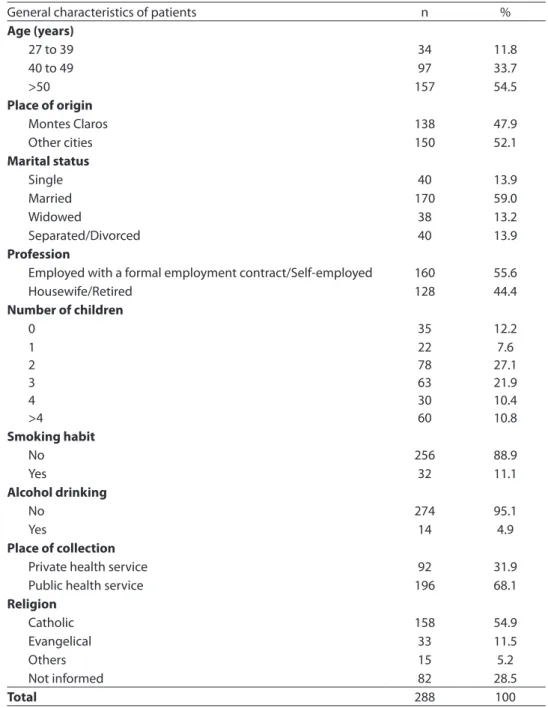
General characteristics of patients n %
Age (years)
27 to 39 34 11.8
40 to 49 97 33.7
>50 157 54.5
Place of origin
Montes Claros 138 47.9
Other cities 150 52.1
Marital status
Single 40 13.9
Married 170 59.0
Widowed 38 13.2
Separated/Divorced 40 13.9
Profession
Employed with a formal employment contract/Self-employed 160 55.6
Housewife/Retired 128 44.4
Number of children
0 35 12.2
1 2 3 4 >4
22 78 63 30 60
7.6 27.1 21.9 10.4 10.8
Smoking habit
No 256 88.9
Yes 32 11.1
Alcohol drinking
No 274 95.1
Yes 14 4.9
Place of collection
Private health service 92 31.9
Public health service 196 68.1
Religion
Catholic 158 54.9
Evangelical 33 11.5
Others 15 5.2
Not informed 82 28.5
positive ER, 26% had an overexpression of HER-2 and 18.4% were triple-negative.
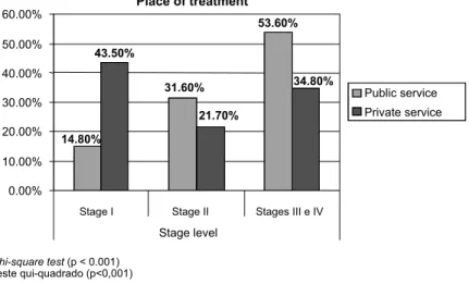
In terms of disease stages, the public health service was compared to the private one and a higher frequency of patients in clinical stages III and IV was observed in the public service (53.6% versus 34.8%). On the other hand, when the initial diagnosis was
analyzed (stage in situ/I), there was a higher frequency in the private service than in the public one (43.5% versus 14.8%), as shown in Graph 1. Table 3 shows an absence of associa-tion of age, menopausal status, occupaassocia-tion, immunohistochemical profile and breast self-exam with disease stages, using multino-mial logistic regression and comparing stages
Table 2 - Clinical characteristics of women with breast cancer admitted to oncology reference services in Montes Claros, North of Minas Gerais, between 2006 and 2009.
Tabela 2 - Características clínicas de mulheres com câncer de mama admitidas em Serviços de referência em oncologia em Montes Claros, Norte de Minas Gerais, entre 2006 e 2009.
Clinical characteristics of patients n %
Disease stage
Stage I 69 24
Stage II 82 28.5
Stages III and IV 137 47.6
Length of time between clinical suspicion and diagnostic conirmation (months)
0 to 5 165 57.3
≥ 6 123 42.7
Menopausal status
Postmenopausal 154 53.5
Premenopausal 134 46.5
Metastasis upon diagnosis
No 267 92.7
Yes 21 7.3
Mammogram performed
No 170 59
Yes 118 41
Breast self-exam performed
No 228 79.2
Yes 60 20.8
Surgical treatment
Conservative surgery 116 46.2
Mastectomy 101 40.6
Family history of breast cancer
No 230 79.9
Yes 58 20.1
Chemotherapy performed
No 67 23.3
Yes 221 76.7
Radiotherapy performed
No Yes
34 254
11.8 88.2
Hormone therapy performed
No Yes
125 163
43.4 56.6
II, III and IV with the initial stage (in situ/I). In a distinctive way, not having a mammogram (PRadjusted=5.10), the absence of a family his-tory of breast cancer (PRadjusted=2.23 and 2.43) and the length of time between the clinical suspicion and diagnostic confirmation ≥ 6 months (PRadjusted=2.97 and 3.04) were associ-ated with the more advanced clinical stages of disease.
Discussion
Differently from several developed countries, Brazil has had an increase in the
mortality rate from breast cancer in recent years, mainly because of late diagnosis and the delay in the implementation of adequate treatment, as this neoplasm is considered to be curable if diagnosed and treated early5-9. The present study enabled
researchers to know the profile of women with breast cancer admitted to public and private referral centers located in the city of Montes Claros, north of Minas Gerais, to receive cancer treatment. The mean age of these women was 63 years. The youngest one was 27 years and the oldest one was 100 years, while the majority (54.5%) were aged
Table 3 - Multinomial logistic regression for the association between mammogram, family history of breast cancer and time between suspected and conirmed diagnosis and clinical staging in women with breast cancer.
Tabela 3 - Regressão logística multinomial para associação entre mamograia, história familiar de câncer de mama e tempo entre suspeita e conirmação diagnóstica e estadiamento clínico em mulheres com câncer de mama.
Variable Stage II* Stages III and IV*
PRcrude (95%CI) PRadjusted (95%CI) PRcrude (95%CI) PRadjusted (95%CI)
Mammography Não 2.65 (1.35-5.18) 1.91 (0.87-4.18) 7.52 (3.92-14.20)5.10 (2.39-10.87)
Sim 1 1 1 1
Family history of breast cancer Não 2.56 (1.22-5.56) 2.23 (1.02-4.89) 3.13 (1.56-6.25) 2.43 (1.14-5.19)
Sim 1 1 1 1
Length of time between clinical suspicion and diagnostic conirmation
≥ 6 meses 3.71 (1.7-7.81) 2.97 (1.37-6.45) 4.76(2.39-9.52) 3.04 (1.46-6.37)
< 6 meses 1 1 1 1
*Regressão logística multinomial (referência = Estágio in situ/I) (p<0,05) *Multinomial logistic regression (reference = Stage in situ/I) (p <0.05) RPbruta=razão de prevalência bruta (crude prevalence ratio)
RPajustada=razão de prevalência ajustada (adjusted prevalence ratio)
IC-95% = intervalo de confiança de 95% (95% CI = 95% confidence interval)
Graph 1 -Stage of breast cancer in women according to treatment site (public or private service).
50 years and 53.5% were postmenopausal. Although age is a recognized risk factor for the development of breast cancer, this variable did not show an association between clinical stages of disease and diagnosis according to the results obtained. However, other studies suggest that breast cancer in younger women has a more aggressive physiopathology, con-tributing to late diagnosis, and the prognosis is worse when compared to breast tumors in older women18-23.
There was a predominance of married women (59%) and those with children (87.8%). In addition, marital status and the number of children did not interfere with the disease stages, although nulliparity is one of the risk factors associated with breast cancer24,25. Similarly, in a survival study
conducted with 1,022 women with breast neoplasm, marital status was not considered to be an important factor26, which confirms
the results from a systematic literature review performed by Ramirez et al13. Controversially,
another study with 540 American female patients revealed that the fact of having never been married increased the risk of having the advanced stage of this disease by almost three times27.
Family history of breast cancer, reported by 20.1% of women in the present study, was associated with the stage of the disease upon diagnosis, confirming the findings of Hoskins et al28, which stated that up to 20% of women
with breast cancer had a positive family history. According to cross-sectional stud-ies conducted with a population of adult women in the United States, from 5% to 10% had a family history of stage I of breast cancer, suggesting that these women in-herited a genetic mutation that puts them at an increased risk of developing breast and ovarian cancer28. A systematic review
of 14 selected studies on risk factors for breast cancer in Brazilian women concluded that little is known about the prevalence of family history of breast cancer in the Brazilian population and found prevalence rates varying between 3.7% and 13.10%29.
Another review of family history of breast cancer30 identified 74 published studies in
which authors revealed an estimated relative risk (RR) associated with such family history of 2.0 (CI = 1.8-2.1) for a mother, 2.3 (CI = 2.1-2.4) for a sister and 3.6 (CI = 2.5-5) for a mother and sister. The risks increased when a first degree relative had been diagnosed before the age of 50 years31.
With regard to immunohistochemistry, the present study found a 26% of overex-pression of HER-2 protein according to the medical records analyzed, whereas other studies32,33 confirmed the overproduction
of this protein between 25 and 30% of breast tumors. This protein is associated with worse prognosis, high histological grade, and the reduction in time without disease and overall survival. The estrogen receptor (ER) is expressed in approximately 65% of cases diagnosed before menopause and in nearly 80% of those diagnosed after menopause, and it is usually associated with more favorable prognoses34. Likewise, 69.4%
of all patients investigated in this study are ER positive. In addition, with regard to immunohistochemistry, Rakha et al35
identified triple-negative tumors, defined by the absence of expression of hormonal receptors and by the non-positivity of HER-2. It is believed that triple-negative breast cancer corresponds to approximately15% of cases, with a higher frequency in black women, those with BRCA1 mutations, and younger women36. Of all women analyzed
in this study, 18.4% were triple-negative. However, although hormonal receptors and the expression of the HER-2 protein are related to the prognosis33,34, these variables
were not associated with the stage level upon diagnosis in the present study.
A bivariate analysis of data was per-formed and found a higher percentage of women in stages III and IV, which was statistically more expressive in the public service than in the private one (53.6% versus 34.8%). In contrast, while 43.5% of women admitted to the private sector were consid-ered to be in stage I, this group totaled 14.8% in the public sector, confirming the results of Rezende et al16, who identified 51% of
Gonçalves et al37 emphasized that stage III
was present in one third of Brazilian women admitted to breast cancer services. Likewise, another study38 analyzed 43,442 cases of
breast cancer between 1995 and 2002 and revealed that 87.7% of women diagnosed with breast cancer were between stages II and IV (stage II=42.8%, stage III=32.6% and stage IV=12.3%). Whereas the standard mortality rates of breast cancer in developed countries decreased, Brazil had an increase in these rates during the same period (from 8.57 to 11.18/100,000 women). The median of percentage of patients between stages II and IV was 45.3% in Brazil and 12.1% in the United States38.
The monitoring method of breast self-examination, although not an appropriate technique for the early diagnosis of breast cancer, has been considered as an auxiliary method39. Several studies40-42 affirm that
there has not been scientific evidence that such practice contributes to the reduction in mortality from this type of cancer. In the present study, not performing a breast self-exam was not associated with more ad-vanced stages upon diagnosis. In contrast, having a mammogram as a more efficient method to monitor breast cancer had a posi-tive impact on the mortality rate43, which
could be reduced by 30% in the 40 to 69 year age group44. Types of cancer identified
in asymptomatic women are likely to have smaller sizes and to be in the first stages45.
Although there has been no consensus on guidance on breast cancer monitoring in the age groups of less than 50 years and more than 70 years46,47, since April 2009, the
Brazilian Sistema Único de Saúde (SUS – Unified Health System) has guaranteed that mammograms for all women aged 40 years and more will be performed9. Similarly, this
study pointed to mammography being more frequently performed in the private health service than in the public one and to not having a mammogram being associated with more advanced stages of breast cancer. Marchi et al48 conducted a cross-sectional
study in which 643 women submitted to mammography were interviewed and
observed that 472 of them were cared for in public health services and 171 in private health services. Among other characteristics, they assessed the use of mammography among users of public and private health services and concluded that the way they accessed these ser-vices influenced the proportion of women pre-viously monitored by mammography, which was higher in the private health network48.
The present study showed a strong as-sociation of the interval of time between the clinical suspicion and diagnostic confirmation with the stage level upon diagnosis of cancer (PRadjusted=2.97 and 3.04). This interval was longer than six months in almost half of the women (42.7%), indicating the slowness of the city’s health system in the period studied. These results confirm the conclusions of Rezende et al16, who conducted a study aimed at identifying
the causes of delay in caring for women diag-nosed with breast cancer in a tertiary hospital of the city of Rio de Janeiro, between January and July 2004, obtaining a median time of one month between the first sign or symptom of disease and the first consultation and of 6.5 months between this consultation and the diagnostic confirmation16. Similarly, while
studying a cancer service of a public hospital of the city of São Paulo, Trufelli et al17
empha-sized that the delay in the treatment of breast cancer cases was primarily associated with the length of time until the patient sought a health service to have a mammogram and biopsy of suspected lesions performed.
References
1. World Health Organization. International Agency for Research on Cancer. World Cancer Report. Lyon: IARC Press; 2008.
2. Jemal A, Bray F, Center MM, Ferlay J, Ward E, Forman D. Global cancer statistics. CA Cancer J Clin 2011; 61: 69-90.
3. Coughlin SS, Ekwueme DU. Breast cancer as a global health concern.Cancer Epidemiol2009; 33: 315-18.
4. Garcia M, Jemal A, Ward E.M et al. Global Cancer Facts
@ Figures 2007. Atlanta, GA: American Cancer Society;
2007.
5. Berry D.A; Cronin K.A; Plevritis S.K et al. Effect of screening and adjuvant therapy on mortality from breast cancer. N Engl J Med 2005; 353: 1784-92.
6. Coleman MP, Quaresma M, Berrino F, Lutz JM, De Angelis R, Capocaccia R et al. CONCORD Working Group. Cancer survival in five continents: a worldwide population-based study (CONCORD).Lancet Oncol
2008; 9: 730-56.
7. Coleman MP, Gatta G, Verdecchia A, Estève J, Sant M, Storm H et al. and the EUROCARE Working Group. EUROCARE-3 summary: cancer survival in Europe at the end of the 20th century. Annals of Oncology 2003; 14: 128-49.
8. Sant M, Allemani C, Capocaccia R et al. Stage at diagnosis is a key explanation of differences in breast cancer survival across Europe. Int J Cancer 2003; 106: 416-22.
9. Instituto Nacional Do Câncer – Inca (Brasil). Estimativa
2010: Incidência de Câncer no Brasil. Rio de Janeiro;
2009.
10. SisRHC. Sistema de Registro Hospitalar de Câncer (SisRHC). Disponível em http://irhc.inca. gov.br/ visualizaTabNetExterno.action [Acessado em 29 de Julho de 2010]
11. Richards MA, Westcombe AM, Love SB, Littlejohns P, Ramirez AJ. Influence of delay on survival in patients with breast cancer: a systematic review. Lancet 1999; 353: 1119-26.
12. Gullatte MM, Phillips JM, Gibson LM. Factors associated with delays in screening of self-detected breast changes in African-American women. J Natl Black Nurses Assoc 2006; 17: 45-50.
13. Ramirez AJ, Westcombe AM, Burgess CC. Factors predicting delayed presentation of symptomatic breast cancer: a systematic review. Lancet 1999; 353: 1127-31.
14. Olivotto IA, Gomi A, Bancej C, Brisson J, Tonita J, Kan L et al. Influence of delay to diagnosis on prognostic indicators of screen-detected breast carcinoma. Cancer
2002; 94: 2143-50.
15. Gebrim LH, Quadros LGA. Rastreamento do câncer de mama no Brasil. Rev Bras Ginecol Obstet 2006; 28: 319-23.
16. Rezende, MCR; Koch, HA; Figueiredo, JA; Thuler, LCS. Causas do retardo na confirmação diagnóstica de lesões mamárias em mulheres atendidas em um centro de referência do sistema único de saúde no Rio de Janeiro.
Rev Bras Ginecol Obstet 2009; 31: 75-81.
17. Trufelli DC, Miranda VC, Santos MBB, Fraile NMP, Pecoroni PG, Gonzaga SFR et al. Análise do atraso no diagnóstico e tratamento do câncer de mama em um hospital público. Rev Assoc Med Bras 2008; 54: 72-6.
18. Castiglione M, Aebi S. The enigma of young age. Ann
Oncol 2006; 17: 1475-7.
19. Nixon AJ, Neuberg D, Hayes DF, Gelman R, Connolly JL, Schnitt S et al. Relationship of patient age to pathological features of the tumor and prognosis for patients with stage I or II breast cancer. J Clin Oncol
1994; 12: 888-94.
20. Dubsky PC, Gnant MF, Taucher S, Roka S, Kandioler D, Pichler-Gebhard B et al. Young age as an independent adverse prognostic factor in premenopausal patients with breast cancer. Clin Breast Cancer 2002; 3: 65-72.
21. Aebi S, De Ridder M, Vlastos G, Vinh-Hung V, Storme G. Young age is a poor prognostic factor in women with stage I breast cancer. Eur J Cancer 2006; 4: 121.
22. Bonnier P, Romain S, Charpin C, Lejeune C, Tubiana N, Martin P et al. Age as a prognostic factor in breast cancer: relationship to pathological and biologic features. Int J Cancer 2006; 62: 138-44.
23. Chung M, Chang HR, Bland KI, Wanebo HJ. Younger women with breast carcinoma have a poorer prognosis than older women. Cancer 1996; 77: 97-103.
24. Lambe M, Hsieh CC, Chan HW, Ekbom A, Trichopoulos D, Adami HO. Parity, age at first and last birth, and risk of breast cancer: a population-based study in Sweden. Breast
Cancer Res Treat 1996; 38: 305-11.
25. Hulka BS, Stark AT. Breast cancer: cause and prevention.
Lancet 1995; 346: 883-7.
26. Palmer MK, Lythgoe JP, Smith A. Prognostic factors in breast cancer. Br J Surg 1982; 69: 697-8.
27. Lannin DR, Mathews HF, Mitchell J, Swanson MS, Swanson FH, Edwards MS. Influence of socioeconomic and cultural factors on racial differences in late-stage presentation of breast cancer. JAMA 1998; 279: 1801-7.
29. Pinho VF, Coutinho ES. Risk factors for breast cancer: a systematic review of studies with female samples among the general population in Brazil. Cad Saude Publica
2005; 21: 351-60.
30. Pharoah PD, Day NE, Duffy S et al.: Family history and the risk of breast cancer: a systematic review and meta-analysis. Int J Cancer 1997; 71: 800-9.
31. Meiser B, Butow P, Barratt A, Friedlander M, Kirk J, Gaff C et al. Breast cancer screening uptake in women at increased risk of developing hereditary breast cancer.
Breast Cancer ResTreat 2000; 59: 101-11.
32. Yaziji H, Goldstein LC, Barry TS et al. Her-2 testing using parallel tissue based methods. JAMA 2004; 291: 1972-7.
33. Wolff AC, Hammond ME, Schwartz JN et al. American Society of Clinical Oncology/college of American Pathologists guideline recommendations for human epidermal growth factor receptor 2 testing in breast cancer. J Clin Oncol 2007; 25: 118-45.
34. Anderson WF, Chatterjee N, Ershler WB, Brawley OW. Estrogen receptor breast cancer phenotypes in the surveillance, Epidemiology, and End Results database.
Breast Cancer Res Treat 2002; 76: 27-36.
35. Rakha EA, Reis-Filho JS, Ellis IO. Basal-like breast cancer: a critical review. J Clin Oncol 2008; 26: 2568-81.
36. Anders C, Carey LA. Understanding and treating triple-negative breast cancer. Oncology 2008; 22: 1233-9.
37. Gonçalves PHB, Gaui MF, Martins RG, Bines J. Padrão de tratamento cirúrgico do câncer de mama de acordo com a idade – Análise de 5 anos do Instituto Nacional do
Câncer (INCA). Trabalho apresentado no XVI Congresso
Brasileiro de Cancerologia e XIII Congresso Brasileiro de Oncologia Clínica, São Paulo, 26 a 30 de novembro de 2003.
38. Thuler LC, Mendonça GA. Estadiamento inicial dos casos de câncer de mama e colo do útero em mulheres brasileiras. Rev Bras Ginecol Obstet 2005; 27: 656-60.
39. World Health Organization. National cancer control
programmes: policies and managerial Guidelines. 2nd ed.
Geneva: WHO; 2002.
40. Ellman R, Moss SM, Coleman D, Chamberlain J. Breast self- examination programmes in the trial of early detection of breast cancer: ten year findings. Br J Cancer
1993; 68: 208-12.
41. Semiglazov VF, Moiseenko VM, Manikhas AG, Protsenko SA, Kharikova RS, Seleznev IK et al. A prospective randomized trial (St-Petersburg, WHO) of the role of self examination in early detection of breast cancer. Russ J
Oncol 2000; 2: 4-9.
42. Thomas DB, Gao DL, Ray RM, Wang WW, Allison CJ, Chen FL et al. Randomized trial of breast self-examination in Shanghai: final results. J Natl Cancer Inst 2002; 94: 1445-57.
43. Greenwald P, Kramer B, Weed D. Expanding horizons in breast and prostate cancer prevention and early detection. The 1992 Samuel C. Harvey Lecture. J Cancer Educ 1993; 8: 91-107.
44. Tabar L, Yen M, Vitak B, Chen HT, Smith RA, Duffy S. Mammography service screening and mortality in breast cancer patients: 20-year follow-up before and after introduction of screening. Lancet 2003; 361: 1405–10.
45. Jackson VP. Screening mammography: controversies and headlines. Radiology 2002; 225: 323-6.
46. Smith RA, Mettlin CJ, Davis KJ, Eyre H. American Cancer Society guidelines for the early detection of cancer. CA
Cancer J Clin 2000; 50: 34-49.
47. Morrison BJ; Canadian Task Force on Preventive Health Care. 1998 recommendation rewording: screening for breast cancer [Internet]. 1998. Disponível em http:// www.ctfphc. org/Tables/Ch65tab2.htm [Acessado em 10 de outubro de 2008]
48. Marchi AA, Gurgel MSC, Fonsechi-Carvasan GA. Rastreamento mamográfico em serviços de saúde público e privado. Rev Bras Ginecol Obstet 2006; 28: 214-9.