ABSTRACT
and diametral tensile strength of glass ionomer
cements
Rodrigo Borges FONSECA1, Carolina Assaf BRANCO2 4, Luciano de Souza GONÇALVES3, Carlos José SOARES45 !"6
1- DDS, MS, PhD, Professor at Federal University of Goiás, Dental School, Restorative Dentistry Area, Goiânia, GO, Brazil. 2- DDS, MS, PhD student, Ribeirão Preto Dental School, São Paulo University, Ribeirão Preto, SP, Brazil.
3- DDS, MS, PhD student, Department of Dental Materials, Piracicaba Dental School, State University of Campinas, Piracicaba, SP, Brazil.
4- DDS, MS, PhD, Professor, Department of Operative Dentistry and Dental Materials, Dental School, Federal University of Uberlândia, Uberlândia, MG, Brazil. 5- DDS, MS, PhD, Professor, Department of Restorative Dentistry, Dental School, Federal University of Pernambuco, Recife, PE, Brazil.
6- DDS, MS, PhD, Professor, Department of Dental Materials, Piracicaba Dental School, State University of Campinas, Piracicaba, SP, Brazil.
Corresponding address: Rodrigo B. Fonseca - Faculdade de Odontologia - Universidade Federal de Goiás - Praça Universitária esquina com 1ª. Avenida, s/n, Setor Universitário - Goiânia - GO - Brasil - 74605-220 - e-mail: [email protected] - Phone: +55-62-32096325 - Fax: +55-62-32096054
#$%&'()*+-//08%9#;& <;(=-//0##<;%&#;(0-//0
C
linicians tend to make reductions in glass ionomer power/liquid (P/L) ratios since some changing the P/L ratio decreases the physical and mechanical properties of conventional !"# $%!"#& '&*+, of P/L ratio on the radiodensity and diametral tensile strength (DTS) of glass ionomer cements. Material and Methods: There were 2 factors under study: P/L ratio (manufacturer’s recommended P/L ratio and a 50% reduced P/L ratio), and materials (Vitro Molar, Vitro Fil, Vitro Cem conventional GICs and Vitro Fil LC, Ortho Glass LC RMGICs. Five 1-mm-thick samples of each material-P/L ratio were produced for radiodensity evaluation. Samples were x-ray exposed onto Digora phosphor plate and radiodensity was obtained using the 6 7 89 $ ; < 6,= >;?; # of each material were tested (0.5 mm/min). Data were subjected to one- and two-way ANOVA (5x2) followed by Tukey’s HSD test, or Kruskal-Wallis and Dunn’s method. For paired comparisons, t-test or Mann-Whitney test were used (D@;;9# $+ ,B@;;;E# BJ # $BJ 6,= $%!"& affected for all materials (P<0.05). Conclusions: Reduced P/L ratio affected properties of the tested glass ionomer cements. RMGICs were more susceptible to lower values of DTS, but radiodensity decreased for all materials following P/L ratio reduction.
Key words: Tensile strength. Glass ionomer cements. Radiography. Microscopy, electron, scanning.
INTRODUCTION
Glass ionomer cements have wide spread clinical indications, being used as bases, liners, luting agents, and temporary and restorative dental materials17. The reasons for such a useful clinical
release6, adhesion to dental tissues and base
metals11,12,25 & &
of thermal expansion15,23,25 $Z
ionomer cements (RMGICs) extended these clinical applications since they present better mechanical properties due to the polymeric nature24, overcoming
some shortcomings of conventional GICs, such as surface crazing during dehydration, brittleness and low fracture strength22.
some interstitial spaces. According to Nicholson16
(1998), GICs consist of interpenetrating networks of inorganic and organic components forming a matrix in which particles of unreacted glass are embedded. In addition to this ionic cure, RMGICs have also a resinous polymer-based reaction due to the presence of visible light-polymerizable components14. Both reactions are processed at the
same time; however, light polymerization results in a rapid initial set while the acid-base reaction, which proceeds normally from the point of mixing, slows down or becomes virtually inhibited by network formation after irradiation26.
Several aspects have been studied in order to improve the mechanical and physical properties of GICs, such as composition25, viscoelastic
properties24, fracture toughness15,24, powder particle
size reduction15 radiodensity9,10,20, strength6,11 and
powder/liquid (P/L) ratio9. Composition seems to
be the most important factor which affects the radiopacity of dental materials9,10. In addition,
the material thickness9,10, x-ray beam angulation,
methodology21 Z
4, and alteration in P/L ratio9
, GICs were cermet-containing or metal reinforced cements, in which metals were responsible by high radiodensity levels16. After that, the addition
of radiopaque fillers resulted on sufficiently radiopaque GICs5, which could be detected against
a background of enamel and dentin, facilitating the evaluation of many clinical situations.
The diametral tensile strength (DTS) is one of the most common and useful mechanical properties of GICs, being extensively found in many studies18,23-25.
Depending on the clinical application, GICs should be strong enough to resist stresses generated within it when loaded with occlusal forces, as it is truth for luting materials11. The type of material,
filler size and proportion, addition of metals, composition and P/L ratio are factors which affect strength of GICs7,11,15,23-25. Increased P/L ratios tend
to increase DTS of GICs11. Mechanically, RMGICs
generally exhibit substantial plastic deformation in compression and conventional GICs display brittle fracture24. Clinicians tend to make reductions in
glass ionomer P/L ratios since some materials
13
grooves or pits2. In general, changing the P/L ratio
decreases the physical and mechanical properties of conventional GICs and RMGICs depending on the reduction of powder per volume15.
There is the chance of altering the DTS and radiodensity of GICs due to reduced P/L ratios, which could virtually lessen the longevity of clinical procedures. Then, the aim of this study was to 9;` P/L ratio on the DTS and radiodensity of different conventional GICs and RMGICs.
MATERIAL AND METHODS
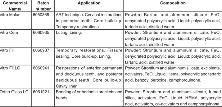
Five GICs (DFL, Rio de Janeiro, Brazil) with different clinical indications were employed in this study. Material applications, commercial names, batch numbers and composition are listed in Figure 1. Two tests were performed in order to analyze the effect of reducing the P/L ratio of different GICs: Radiodensity and DTS. Groups were formed according to 2 experimental factors under study:
Figure 1- Restorative materials used in the study Commercial
Name/
Batch number
Application Composition
Vitro Molar 6050868 ART technique. Cervical restorations
in posterior teeth. Core build-up. Temporary restorations.
Powder: Barium and aluminum silicate, FeO, dehydrated polyacrylic acid. Liquid: polyacrylic acid, tartaric acid, distilled water
Vitro Cem 6060935 Luting. Lining. Powder: Strontium and aluminum silicate, FeO,
dehydrated polyacrylic acid. Liquid: polyacrylic acid, tartaric acid, distilled water
Vitro Fil 6060987 Temporary restorations. Fissure
sealing. Core build-up. Lining.
Powder: Strontium and aluminum silicate, FeO, dehydrated polyacrylic acid. Liquid: polyacrylic acid, tartaric acid, distilled water
Vitro Fil LC 6060941 Restorations of anterior permanent
and deciduous teeth, and posterior deciduous teeth. Core build-up. Cavity liner.
Powder: Strontium and aluminum silicate, excipients, activators, FeO. Liquid: Hema, polyacrylic and tartaric acid, benzoyl peroxide, camphorquinone.
Ortho Glass LC 6061021 Bonding of orthodontic brackets and
bands.
materials, with 5 levels (Figure 1) and P/L ratio, with 2 levels, the manufacturer’s recommended P/L ratio and a 50% reduced P/L ratio. Samples were fabricated at room temperature according to each experimental method, as described below. Chemical cured materials were allowed to set during the period recommended by each manufacturer. Light-cured materials were photoactivated for 40 s with a halogen curing unit (XL3000; 3M ESPE, St. Paul, MN, USA) at 850 mW/cm2.
Radiodensity Test
Five 1-mm-thick ring-shaped standard samples were fabricated for each experimental group by mixing the materials according to the manufacturers’ instructions and inserting them in a 1-mm-thick stainless steel mold with diameter of 4.0 mm. After removal of the samples from the mold, the thickness E; mm (±0.1 mm). An aluminum stepwedge, ranging from 1.0 mm to 9.0 mm in thickness, served as a control.
The samples were positioned onto a phosphor plate and radiographic exposure was performed for 0.2 s at 70 kV and 10 mA, with a source-to-sample distance of 40 cm using an x-ray machine (GE 1000; General Electric, Milwaukee, WI, USA). Three exposures were done for each sample. The radiographs were transferred from the phosphor plate to the computer via a Digora scanner (Digora Optime, Soredex, Helsinki, Finland).
The radiodensity (in pixels) of the samples was determined with the resident software provided by the manufacturer. The Digora system has a windows-based software, Digora for Windows 2.5 Rev 0 (Soredex, Helsinki, Finland), which is capable to measure density curves of digital radiographies obtained by x-ray impregnation on the image phosphor plate. The radiodensity of each radiographed structure or material was obtained by clicking with the software cursor right above the digital image. Each digital image had it radiodensity measured immediately after scanning, without any &, shows data concerning the highest and the lowest radiodensity of the sample, and an average value, which was considered to be the sample’s initial radiodensity. Since each sample was subjected to considered to be the mean of those values.
DTS Test
Five cylindrical specimens of each experimental @9# 6,= 6 8>; long and 8.0 mm diameter aluminum mold was , mixed cements, samples were kept in 100%
humidity and 37oC for 1 h, followed by gentle
removal from the mold and immersion in distilled water for 23 h prior to testing. A compressive load was applied on the diametral surface of the samples to obtain the DTS at a crosshead speed of 0.5 mm/ min in a universal testing machine (Instron 4411; Instron Testing Instruments, Canton, MA, USA).
Scanning Electron Microscopy Analysis (SEM)
For observations of materials characteristics after complete setting, SEM analysis was accomplished in samples from Ortho Glass LC and Vitro Cem experimental groups. Fractured samples were sputter-coated with gold (MED 010; Balzers Union, Balzers, Liechtenstein) and observed with a scanning electron microscope (DSM 940A; Zeiss, Oberkoshen, Germany).
Statistical Analysis
Statistical analysis was performed in accordance with the results of Shapiro-Wilk test of normal distribution, with parametric or non-parametric tests, for each variable. One-way ANOVA followed & , = 6 =6# test was employed for DTS and the Kruskal-Wallis and Dunn’s method for radiodensity, using the SPSS 12.0 for Windows statistical software (SPSS " J =# , Z ' 98# followed by Tukey’s HSD test, with a general linear model procedure, was also used to analyze the interaction between materials and P/L ratio. For paired comparisons, independent samples t-test was used for DTS and Mann-Whitney Test for Radiodensity. The aluminum step was compared to each group by ANOVA and Dunnett’s 2-sided test. For all tests, groups were considered statistically different at Į@;;9
RESULTS
Tables 1 and 2 show the results of DTS test (in MPa) and radiodensity measurements (in pixels), respectively, together with the statistical analysis. Radiodensity means and standard deviations are presented only to facilitate the understanding. Since data were not normally distributed, the sum of the ranks (non-parametric analysis) is also provided. , P@;;;E# the factors under study (materials vs. P/L ratio). Thus, a separate analysis was accomplished for the interactions.
Groups DTS mean(SD) for man. P/L DTS mean(SD) for 0.5 P/L
Vitro Fil LC 20.94(3.34) Aa 11.20(2.84) Ab
Ortho Glass LC 15.0(2.12) Aa 10.94(1.87) Ab
Vitro Molar 7.73(3.24) Ba 4.08(1.32) Ba
Vitro Fil 7.01(3.21) Ba 4.19(1.57) Ba
Vitro Cem 2.83(0.71) Ba 2.37(0.66) Ba
!"!#$"% & comparison only – P<0.05). ‡ man. P/L: manufacturer’s recommended P/L ratio. § 0.5 P/L: 50% reduced P/L ratio Table 1- Diametral tensile strength (DTS) means and standard deviations (in MPa; n=5), and results of statistical analysis
of groups by ANOVA, Tukey’s HSD and T-test (D=0.05)
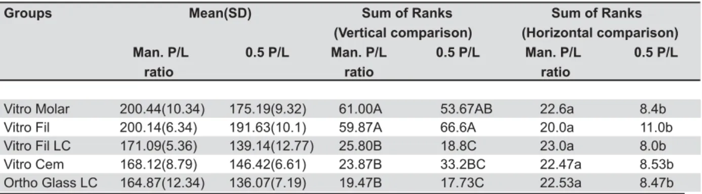
<8_ `k Pq Pq _Z;##<k_v;#<k 8xT/xyT8xT/xyT8xT/xyT ratio ratio ratio
Vitro Molar 200.44(10.34) 175.19(9.32) 61.00A 53.67AB 22.6a 8.4b
Vitro Fil 200.14(6.34) 191.63(10.1) 59.87A 66.6A 20.0a 11.0b
Vitro Fil LC 171.09(5.36) 139.14(12.77) 25.80B 18.8C 23.0a 8.0b
Vitro Cem 168.12(8.79) 146.42(6.61) 23.87B 33.2BC 22.47a 8.53b
Ortho Glass LC 164.87(12.34) 136.07(7.19) 19.47B 17.73C 22.53a 8.47b
!"!#$" % &
only – P<0.05).
‡ Man.P/L ratio: manufacturer’s recommended P/L ratio. § 0.5 P/L: 50% reduction in P/L ratio.
Table 2- Radiodensity means, standard deviations (SD) (in pixels; n=15) and sum of the ranks, and results of statistical analysis by Kruskal-Wallis test, Dunn’s method, and Mann-Whitney Test (D=0.05)
Figure 2- Comparison between experimental groups and aluminum stepwedge
the 50% reduced P/L groups, Vitro Fil and Vitro Molar were again the most radiopaque materials, but the last was similar to Vitro Cem, and this one
was similar to the RMGICs (Table 2).
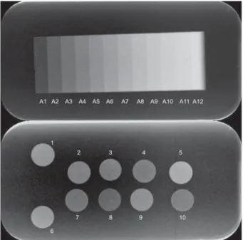
Within each material, pairwise DTS analysis by t-test showed that reducing the P/L ratio resulted * ' Glass LC and Vitro Fil LC, with 27.1% and 46.6% of reduction, respectively. Pairwise radiodensity analysis by the Mann-Whitney Test showed that BJ reduction in radiodensity for all materials. Figure 2 presents the comparison between the aluminum stepwedge and experimental groups. Figure 3 shows a radiographic image of the groups and Figure 3- Digital radiograph of the studied materials and
aluminum step wedge. 1: Vitro Molar - manufacturer’s recommended P/L ratio; 2: Vitro Molar – 50% reduced P/L ratio; 3: Vitro Cem – manufacturer’s recommended P/L ratio; 4: Vitro Cem – 50% reduced P/L ratio; 5: Vitro Fil – manufacturer’s recommended P/L ratio; 6: Vitro Fil – 50% reduced P/L ratio; 7: Ortho Glass LC – manufacturer’s recommended P/L ratio; 8: Ortho Glass LC – 0.5P/L; 9:Vitro Fil LC – manufacturer’s recommended P/L ratio; 10: Vitro Fil LC – 50% reduced P/L ratio; A1-A12: 1.0-12.0 mm thick aluminum
Figure 4- Scanning electron microscopy of Ortho Glass LC at manufacturer’s recommended P/L ratio showing a uniform interaction between resin matrix, salt matrix and unreacted powder particles, without clear signals of crack propagation. Note that the boundaries of powder particles cannot be clearly seen, which mean that the acid base reaction progressed in a free condition
Figure 5- Scanning electron microscopy Ortho Glass LC at 50% reduced P/L ratio showing a greater volume of matrix around powder particles; particle’s boundaries can be clearly seen, which mean that the acid-base reaction was partially inhibited by the greater presence of resin. Note the presence of crack propagation around the powder particle, which is related to a lower interaction between matrix-powder
the aluminum stepwedge. During mixing of the cements, it was noted that Vitro Molar and Vitro < recommended P/L and the mixed material resulted in a rough mass. At 50% reduced P/L ratio, it was easier to mix these materials and a smooth mass was obtained.
Figures 4 and 5 are SEM images of Ortho Glass LC at the manufacturer’s recommended P/L and the 50% reduced P/L ratio, respectively. Figures 6 and 7 of Vitro Cem at the manufacturer’s recommended P/L ratio and 50% reduced P/L ratio, respectively. For the RMGICs, reduction in liquid at 50% P/L resulted in poor interaction between matrix and powder particles with clear signs of crack development. For the conventional GICs, reduction in liquid at 50% P/L ratio resulted in a greater presence of salt matrix and its close interaction with powder particles without the development of cracks, which could be seen at the manufacturer’s recommended P/L.
DISCUSSION
Several factors can have an influence on conventional GIC and RMGIC properties, but the P/L ratio9,13 is the one that relies on the responsibility of
clinicians11. The hypothesis driven on this study was
& 6,= radiodensity of GICs and RMGICs due to changes in P/L ratio. The assumption that radiodensity depends more on the composition of dental materials than the
type of material5 6,=
are in general agreement with other studies23,24,
who reported that the RMGICs exhibited higher DTS than GICs. Composition, setting reactions, materials’ maturation and microstructures seemed
to be the principal reasons for these observations. Materials tested with the same thickness mostly
& 5.
X-ray beam interactions with the matter are always directly proportional to either the atomic number of the absorber or to its electric density8; then
depending on the atomic composition and density of each atom in the matter a radiographic image will &, elements with high atomic numbers such as zinc, strontium, zirconium, barium and lanthanum result in more radiopaque materials5,9,10. RMGICs
are not always radiopaque10 and GICs are usually
radioluscent9,10.
Vitro Molar and Vitro Fil, which present barium/ FeO and strontium/FeO, respectively, in their composition, were the most radiopaque materials, when mixed in the manufacturer’s recommended P/L ratio. However, even with the presence of strontium/FeO in the other materials, their radiodensity were similarly lower than the previous mentioned ones, which mean these components are present in relatively lower quantities. With the reduced P/L ratio, Vitro Fil and Vitro Molar still presented the highest radiodensity levels, but Vitro Cem was similar to Vitro Molar and the RMGICs were similar to Vitro Cem. Vitro Molar is a restorative GIC and is more viscous than Vitro Cem. The higher viscosity and the apparent presence of higher radiodensity, but with the reduction in P/L ratio, these differences disappeared. Since organic components do not seem to offer radiopaque characteristics to dental materials5, the observed
lower radiodensity for RMGICs at 50% reduced P/L ratio is a direct cause of the increase in liquid per volume. Within each material, the reduction BJ of all materials in this study (Table 2), and this also may be a direct result of the increase in liquid per volume. Since all materials employed on the present study, irrespective of the P/L ratio being similar or more radiopaque than A2 or A3 aluminum steps (Figure 2), they could eventually be detected against an enamel background (good clinical property)5,9,10.
Generally, chain displacement can occur at & than in brittle polymeric and ceramic materials, which undergo crack propagation at high stress3,19.
Mitsuhashi, et al.15 (2003) showed that when the P/L
ratio is decreased and the matrix volume increased as a consequence, the characteristics of the resin $%!" It is expected that greater presence of resin in RMGICs, by reduction of P/L ratio, would render a material with more viscoelastic behavior, greater strain capacity and possibly greater resistance to Figure 7- Scanning electron microscopy of Vitro Cem
stress development by load application. However, even if a more viscoelastic behavior is expected, the tendency for generating materials which withstand less load application is real because in the cement mixtures, the volume fraction of the matrix, which has weak mechanical strength, increases at lower P/L ratios. Irrespective of the P/L ratio, this study showed that RMGICs’ strength was always higher than that of conventional GICs (Table 1), possibly due to the greater expected viscoelastic behavior of RMGICs and the higher cohesive strength of resin matrix versus salt matrix23. On the other hand, DTS
was reduced only for RMGICs when 50% reduced P/L ratio was employed (Table 1).
When comparing RMGICs and GICs, Yamazaki, et al.24 (2006) showed they possess similar viscoelastic
behavior, irrespective of the polymeric character of RMGICs. Then, it is possible that resin and salt matrixes act similarly, in these materials, with a higher probability of enabling plastic deformation for the former23. However, Mitsuhashi, et al.15
(2003) found that the fracture toughness of the $%!" &BJ as it is for GICs. Only at high reductions in P/L ratio, the fracture toughness started to decrease for RMGICs15. With lower fracture toughness materials
became more brittle, reducing plastic behavior and also the resistance against crack propagation15.
, !" in RMGICs properties is only expected with high reduction of powder amount, because the acid-base reaction of the powder particle and liquid is critical for many physical properties of hardened materials15. The SEM analysis showed an integrated
microstructure for RMGICs with manufacturer’s recommended P/L ratio (Fig 3), but the same was not true for 50% reduced P/L ratio, which showed a greater presence of matrix per volume, unreacted particles and crack development, recognized as a signal of the jeopardized interaction between matrix and powder particles (Figure 5). The high reduction in P/L ratio, as performed in the present study, 6,=* RMGICs, and not for conventional GICs (Table 1). In spite of that, for all materials, a decrease in DTS occurred with the reduction in P/L ratio. There are two possibilities to explain this result. One is that limitations of the experimental design did not allow differences between manufacturer’s recommended P/L ratio and 50% reduced P/L ratio of the materials to be observed. Larger sample sizes may possibly & differences. The other possibility is closely related to the setting reactions of each type of material.
For light-cured GICs, the maturation of cure & the physical properties of the hardened materials15
because an increase in the overall properties
is observed with material’s maturation25. More
integrated microstructures with better glass particle-polymer matrix bonding, results in higher values of DTS23, which could be seen in manufacturer’s
recommended P/L ratio (Figure 2). Adusei, et al.1
(2004) showed that the use of silanated glass Z & particles seem to participate of the setting reaction. This leads to a less cohesive matrix, and the same situation is expected on RMGICs, which leads to the conclusion that there is an important role of the salt part of the matrix in the overall strength of these materials1.
It is speculated, from the DTS results of the present study that a high reduction in P/L ratio of the chemical reaction is reduced through the polymerization of the resinous part, as previously stated by Yelamanchili and Darvel26 (2008). Even
if the chemical reaction is generally able to keep going after light polymerization14, it seems a
greater volume of resin by reduction in P/L ratio, entraps polyacid molecules, and the so important chemical reaction reduces in effectiveness. According to Peutzfeldt, et al.17 (1997) HEMA
molecule crosslinking keeps the carboxylate groups of different polyacid chains too far apart to be crosslinked via Ca+2 as will normally happen without
resin. Since chemical and light irradiated network formation compete26, but with 50% reduced P/L
ratio the chemical reaction is slowed down, strength & addition, the higher proportion of the resinous part does not assure better mechanical properties because light irradiation results in a temperature rise, potentially permitting a higher value of the “auto-limiting” glass-transition temperature, with a consequent negative effect of degree of conversion26. Figure 5 shows an apparent ineffective
interaction between particles and matrix as a result of the “HEMA blocking effect”.
The assumption that both the salt matrix and the resinous matrix have a determinant relationship in the overall strength of RMGICs1,26
of cracks. These observations led to the assumption that even if a better interaction between matrix and powder particles is seen when more liquid is added to the mixture, GICs’ DTS can be considered more stable when alterations in P/L ratio are performed, since different morphological structures are formed and can resist differently to load application. However, the reduction in absolute DTS means for Vitro Molar and Vitro Fil should be taken into consideration, meaning that more salt matrix can possibly result in poor mechanical properties with time. Further studies are necessary to prove this hypothesis. The use of the manufacturers’ recommended P/L ratio is always advisable since properties will be preserved.
CONCLUSIONS
From the results of this study it is possible to conclude that: 1. Alterations in the P/L ratio should be avoided since a decrease in radiodensity 6,= 8 $%!" decrease in DTS, but all materials were affected in their radiodensity due to alterations in P/L ratio; 3. ,6,= $%!" & by structural organization (organic molecules and inorganic particles), while radiodensity is affected by chemical composition.
ACKNOWLEDGEMENTS
Authors are grateful to DFL for full donation of the materials used in this study, and to CAPES-Brazil (Coordenadoria de Aperfeiçoamento de Pessoal de Nível Superior) for Rodrigo B. Fonseca PhD program support.
REFERENCES
1- Adusei GO, Deb S, Nicholson JW. The role of the ionomer glass Z materials. J Mater Sci Mater Med. 2004;15(7):751-4.
2- Celiberti P, Lussi A. Penetration ability and microleakage of
caries. J Dent. 2007;35(1):59-67.
3- Craig R. Restorative dental materials. St. Louis: Elsevier; 2001. 4- el-Mowafy OM, Benmergui C. Radiopacity of resin-based inlay luting cements. Oper Dent. 1994;19(1):11-5.
5- Fonseca RB, Branco CA, Soares PV, Correr-Sobrinho L, Haiter-Neto F, Fernandes-Haiter-Neto AJ, et al. Radiodensity of base, liner and luting dental materials. Clin Oral Investig. 2006;10(2):114-8.
6- Forsten L. Fluoride release and uptake by glass-ionomers and related materials and its clinical effect. Biomaterials. 1998;19(6):503-8.
7- Gladys S, Van Meerbeek B, Braem M, Lambrechts P, Vanherle G. Comparative physico-mechanical characterization of new hybrid restorative materials with conventional glass-ionomer and resin composite restorative materials. J Dent Res. 1997;76(4):883-94. 8- Goaz PW, White SC. Oral radiology. St. Louis: C.V. Mosby; 1987. 9- Hara AT, Serra MC, Haiter-Neto F, Rodrigues AL Jr. Radiopacity of esthetic restorative materials compared with human tooth structure. Am J Dent. 2001;14(6):383-6.
10- Hara AT, Serra MC, Rodrigues AL Jr. Radiopacity of glass-ionomer/composite resin hybrid materials. Braz Dent J. 2001;12(2):85-9.
11- Hibino Y, Kuramochi K, Hoshino T, Moriyama A, Watanabe Y, Nakajima H. Relationship between the strength of glass ionomers and their adhesive strength to metals. Dent Mater. 2002;18(7):552-7.
12- Hotz P, McLean JW, Sced I, Wilson AD. The bonding of glass ionomer cements to metal and tooth substrates. Br Dent J. 1977;142(2):41-7.
13- Irie M, Tjandrawinata R, Suzuki K, Watts DC. Root-surface gap-formation with RMGIC restorations minimized by reduced BJ 6% 2006;22(5):486-97.
E>Z %"& < $Z Z 1998;19(6):521-7.
15- Mitsuhashi A, Hanaoka K, Teranaka T. Fracture toughness Z + powder/liquid ratio and powder particle size reduction on fracture toughness. Dent Mater. 2003;19(8):747-57.
16- Nicholson JW. Chemistry of glass-ionomer cements: a review. Biomaterials. 1998;19(6):485-94.
17- Peutzfeldt A, García-Godoy F, Asmussen E. Surface hardness and wear of glass ionomers and compomers. Am J Dent. 1997;10(1):15-7.
18- Piwowarczyk A, Ottl P, Lauer HC, Büchler A. Laboratory strength of glass ionomer cement, compomers, and resin composites. J Prosthodont. 2002;11(2):86-91.
19- Rosen SL. Fundamental principles of polymeric materials. New York: John Wiley & Sons; 1993.
20- Sidhu SK, Shah PM, Chong BS, Pitt Ford TR. Radiopacity of Z Z Int. 1996;27(9):639-43.
21- Turgut MD, Attar N, Onen A. Radiopacity of direct esthetic restorative materials. Oper Dent. 2003;28(5):508-14.
22- Uno S, Finger WJ, Fritz U. Long-term mechanical characteristics Z 6% 1996;12(1):64-9.
23- Xie D, Brantley WA, Culbertson BM, Wang G. Mechanical properties and microstructures of glass-ionomer cements. Dent Mater. 2000;16(2):129-38.
24- Yamazaki T, Schricker SR, Brantley WA, Culbertson BM, Johnston W. Viscoelastic behavior and fracture toughness of six glass-ionomer cements. J Prosthet Dent. 2006;96(4):266-72. 25- Yap AU, Pek YS, Cheang P. Physico-mechanical properties of a fast-set highly viscous GIC restorative. J Oral Rehabil. 2003;30(1):1-8.