ABSTRACT
Flexural strength of fluorapatite-leucite and
in cyclic immersion
Peerapong JUNPOOM1, Boonlert KUKIATTRAKOON2, Chanothai HENGTRAKOOL2
1- DDS, MSc, Section of Dental Public Health, Huay Yod Hospital, Trang, Thailand.
2- DDS, MSc, Associate Professor, Department of Conservative Dentistry, Faculty of Dentistry, Prince of Songkla University, Hat Yai, Songkhla, Thailand. 3- DDS, MSc, PhD, Assistant Professor, Department of Conservative Dentistry, Faculty of Dentistry, Prince of Songkla University, Hat Yai, Songkhla, Thailand.
Corresponding address: Assoc. Prof. Boonlert Kukiattrakoon - Department of Conservative Dentistry - Faculty of Dentistry - Prince of Songkla University - Hat Yai - Songkhla - Thailand - Phone: +66-74-28-7703 - Fax: +66-74-429877 - e-mail: [email protected]
O
materials (IPS d.SIGN and IPS e.max Ceram) exposed to erosive agents. Material and Methods: One hundred and twenty bar-shaped specimens were made from each of !"#"$%& !"#'& and divided into 8 groups of 15 specimens each. Six groups were alternately immersed in the following storage agents for 30 min: deionized water (control), citrate buffer solution, pineapple juice, green mango juice, cola soft drink and 4% acetic acid. Then, they were immersed for 5 min in deionized water at 37°C. Seven cycles were completed, totalizing 245
min. A 7th +/ 69;'<=#>?
8th, group was stored dry at 37°C for 245 min. Three-point bending tests were performed
in a universal testing machine. The data were analyzed statistically by 2-way ANOVA, @BD"E >9#9H#J of all groups of each porcelain after exposure to erosive agents in cyclic immersion did >K9#9H&#L ?QR;' ? > K9#9H&# > U9#9H& #' tested dental porcelains.
Key words: Dental porcelain. Erosion. Immersion. Juices. Soft drinks.
INTRODUCTION
Porcelains are highly esthetic materials extensively used in dentistry to construct various types of restorations and prostheses such as porcelain fused to metal crowns, veneers, inlays, #> esthetic and functional demands of the patients by their superior properties when compared to other restorative materials26. The new glass ceramics (IPS d.SIGN; Ivoclar Vivadent AG, Schaan, Liechtenstein) have become popular for porcelain-fused-to-metal restorations. IPS d.SIGN is a new type
morphology, are known to be contained in natural bone and teeth. These very small crystals in dental microstructures result in very special optical properties such as translucence and opalescence, which also result the same properties as in dental restorations11.
Despite the outstanding esthetic quality of the porcelains, the most serious problem of this material is its susceptibility to fractures9,15. Porcelains exhibit # These characteristics impair their physical properties such as surface roughness, surface hardness,
strength3,6
failure of porcelain restorations7. Crack propagation and degradation of dental porcelains occur when porcelains are exposed to aqueous solutions or erosive agents1. These phenomenons take place as a result of selective leaching of alkaline ions. Alkaline metal ions are far less stable in the glass phase than in the crystalline phase1. Therefore, some alkaline ions in porcelains were leached after being exposed to acidic solutions17-19,24. Variation in pH, solution chemistry, wear and mechanical load makes the oral cavity a complex environment1. Environmental conditions may also damage resistance to surface and bulk degradation of porcelains. Consequences of porcelain degradation include coarseness of exposed surface4,24, promoting plaque accumulation1,2,4,24 and wear to antagonist materials1. Furthermore, increasing of surface roughness of porcelains may decrease strength9,15.
Many people frequently consume acidic food, sour fruits and drinks. This consuming habit relates to a high incidence of dental erosion14,16,23,27. The potential erosive effect of these acidic food and beverages on enamel occurs primarily by the dissolution of apatite crystals12,16,20. However, their effect on the porcelain restorations has not been clearly documented. Therefore, the present in vitro
study was designed to evaluate changes of the erosive agents (pineapple juice, green mango juice, coca soft drink, citrate buffer solution and 4% acetic acid) in cyclic immersion. The null hypothesis was > strength of each type of dental porcelain tested
after being exposed to erosive agents.
MATERIAL AND METHODS
Specimen Preparation
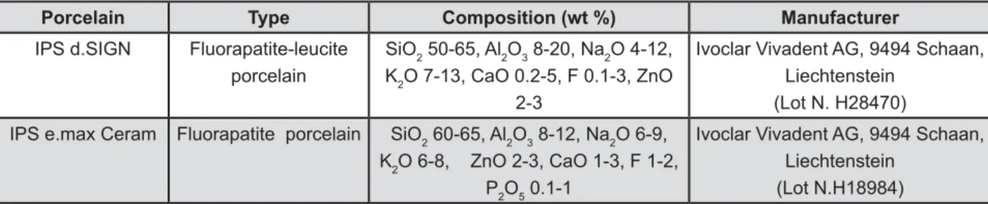
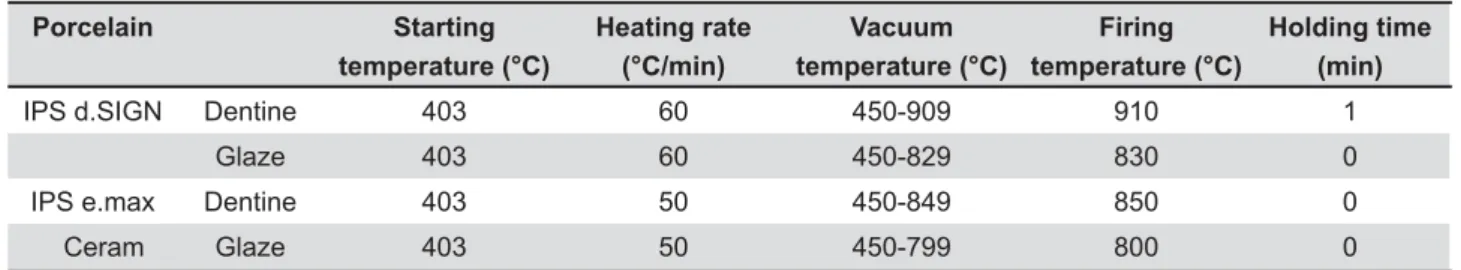
Two commercial dentin shade A3 porcelain powders were used: IPS d.SIGN and IPS e.max Ceram (Ivoclar Vivadent AG) (Figure 1). IPS d.SIGN and IPS e.max Ceram are indicated to be used as veneering porcelain for porcelain fused to metal and all ceramic restorations, respectively. One hundred and twenty bar specimens from each of the 2 porcelains were fabricated using the 26.0X6.0X3.0 mm silicone mold (Provil novo putty; Heraeus Kulzer GmbH, D-63450 Hanau, Germany). The porcelain }?> the silicone mold and condensed with a condenser (Ceramosonic II; Shofu Inc, Higashiyama-ku, ~? &# > according to the manufacturer's instructions (Table <&#> ? Phoenix 4000; Buehler GmbH, 40599 Düsseldorf, Germany) under running water using 600- and 1,200-grit silicon carbide paper (3M ESPE, St. Paul, MN, USA) to the dimensions of 25.0x5.0x2.0 mm, following the guidelines of the ISO 6872 standard13. Then, the specimens were ultrasonically cleaned in distilled water for 10 min, and subjected to self-glazing according to the manufacturer’s instructions (Table 1).
Erosive Agents Exposure
The porcelain bars were divided into 8 groups of 15 specimens each. Subsequently, the specimens were alternately immersed in 25 mL of an erosive agent for 30 min and in 25 mL of deionized water for 5 min for 7 cycles at 37°C. This amount of H & > to completely cover the specimen. In order to maintain the original pH level of the erosive agent, the agents were refreshed every cycle throughout the experiment. The same protocol was used with different types of 5 erosive solutions included in the study (citrate buffer solution, pineapple juice, green mango juice, cola soft drink and 4% acetic acid; see Figure 2) and deionized water (control). The specimens’ immersion protocol simulated an
Porcelain Type Composition (wt %) Manufacturer
IPS d.SIGN Fluorapatite-leucite
porcelain
SiO2 50-65, Al2O3 8-20, Na2O 4-12, K2O 7-13, CaO 0.2-5, F 0.1-3, ZnO
2-3
Ivoclar Vivadent AG, 9494 Schaan, Liechtenstein (Lot N. H28470)
IPS e.max Ceram Fluorapatite porcelain SiO2 60-65, Al2O3 8-12, Na2O 6-9, K2O 6-8, ZnO 2-3, CaO 1-3, F 1-2,
P2O5 0.1-1
Ivoclar Vivadent AG, 9494 Schaan, Liechtenstein (Lot N.H18984)
individual eating acidic food, sour fruits and drinks. Total immersion time was 245 min. Seventh group was continuously immersed in 4% acetic acid at 69;' <= > " =6R13) in order to examine the extensive effect which could occur. A 8th group was kept dry at 37°C for 245 min in order to compare the effect of moisture condition. After the immersion sequence was completed, the specimens were rinsed with deionized water, blotted #
Flexural Strength Measurements
universal testing machine (model LRX-plus; Ametek Lloyd Instruments, Farnborough, Hampshire, UK). Bar-shaped specimens were centered and placed on two steel spheres (1.6 mm in diameter) of a supporter part positioned 12 mm apart from each other. Three point bending tests were carried out using a 250 N load cell at crosshead speed 0.25 mm/min. The load at failure was recorded % MPa (3WL/2BD²; W=failure load, L=span length, B=specimen’s width, and D=specimen’s thickness).
Statistical Analysis
Two-way ANOVA was analyzed to measure > of erosive agents and the type of porcelains after being exposed to erosive agents. Tukey’s Honestly " > E D"E&
post hoc comparisons (D=0.05). The t-test was used
types of porcelain for each erosive agent (D=0.05).
RESULTS
porcelain were showed in Table 2. ANOVA results showed that the interaction between the two variables (type of porcelain and erosive agent) > 9#9&# Between the two dental porcelains, a statistically > 9#9<&? none was found among the types of erosive agents (p=0.46).
values between the porcelains for each group, the results of the t-test showed that all IPS e.max ' > U9#9H&!"#"$% groups.
Erosive agent Form Manufacturer
Citrate buffer solution Instant BDH Laboratory Supplies, Poole, England
pH 4.99±0.01
100% pineapple juice Instant Tipco Foods Co. Ltd., Prajuabkirikhan,
Thailand
pH 3.64±0.01
Green mango juice Prepared from fresh green mango _
pH 2.39±0.01
Cola soft drink Instant Hadthip Ltd, Songkhla, Thailand
pH 2.41±0.06
4% acetic acid Diluted from 100% acetic acid Merck KGaA, Darmstadt, Germany
pH 2.47±0.01
Figure 2- Erosive agents used in the present study
Porcelain Starting
temperature (°C)
Heating rate (°C/min)
Vacuum temperature (°C)
Firing temperature (°C)
Holding time (min)
IPS d.SIGN Dentine 403 60 450-909 910 1
Glaze 403 60 450-829 830 0
IPS e.max Dentine 403 50 450-849 850 0
Ceram Glaze 403 50 450-799 800 0
DISCUSSION
The results of this study support acceptance of ? porcelains was not affected by the erosive agents. found in the dry condition group (stored at 37°C) of both porcelains and decreased in all groups when the porcelains were immersed in erosive agents as well as in water. The possible explanation for these results could be the effect of glazing in determining #
! ? ? or defects on their surface and internal body. # D? }? } } #J> > a self-glaze layer. This layer may increase the strength of the porcelain restoration from two possible mechanisms8. Firstly, when the restoration ?}> ? # This should increase strength because, for given porcelains, strength increases with decreasing #"? based porcelains, the self-glaze layer has a lower > rich interior. This places the outer surface in compression when cooled. The compressive stress state diminishes the local tensile stress produced ? propagation from the external surface.
The IPS e.max Ceram had higher flexural strength than the IPS d.SIGN in all groups. A possible explanation for this result could be the microstructure of these porcelains11. The IPS d.SIGN, feldspathic-based porcelain, is unique and distinct from other porcelains since its microstructure
to having leucite particles in a feldspathic glassy matrix10, while the IPS e.max Ceram consists of glassy matrix28. In feldspathic-based porcelains, the leucite particles contract more than the surrounding glass upon cooling. Above a critical particle size, the stresses created during cooling can induce microcracks circumferential to the leucite particles22. Previous studies have documented that the size of leucite particles in feldspathic porcelain increases during heat treatment within the normal > 5,21. This can increase the probability of microcracking22. It is possible that microcracking occurred during the self-glaze # ? @ chemical durability11.
The erosive agents used in this present study, pineapple juice and green mango juice, are favorite sour fruit juices in many Asia countries. They consist of citric acid and other organic acid12,14,16, which give an acidic pH. However, in the present study, these juices did not affect the flexural strength of the tested porcelain after immersion, > studies that showed an impact of acidic agents on porcelains17-19,24. This study was a short-term experiment and could be the reason to explain > acidic agents and this aspect should be explored. So, a long-term evaluation of the effect of erosive agents on porcelains is required.
It must be noted that there are some limitations to this present study. This study did not consider the different conditions found in the oral environment. For example, the presence of water, temperature change, the pH level and the role of saliva25 in the of restorations. In addition, the present study
3 *"! #$& 4<= > ?@
IPS d.SIGN IPS e.max Ceram
Deionized water (control) 48.41±7.04a 71.07±12.16a
Citrate buffer solution 51.48±10.82b 67.69±12.73b
Pineapple juice 51.06±9.57c 64.28±14.98c
Green mango juice 47.78±13.18d 77.05±9.58d
Cola soft drink 53.25±9.43e 63.59±10.66e
4% Acetic acid 52.37±9.49f 65.95±12.17f
4% Acetic acid, 16 h 50.59±7.82g 66.54±11.14g
Dry condition 55.3±12.63h 78.60±11.97h
Table 2- !
porcelains. Further studies are required to investigate the effect on other porcelains.
CONCLUSIONS
? & > different. For both types of porcelain, dry storage at QR;' ? > #
> #
ACKNOWLEDGEMENTS
This study is supported in part by a grant from the Graduate School, Prince of Songkla University.
REFERENCES
1- Anusavice KJ. Degradability of dental ceramics. Adv Dent Res. 1992;6:82-9.
2- Clayton JA, Green E. Roughness of pontic materials and dental plaque. J Prosthet Dent. 1970;23(4):407-11.
?QWX[\]WX!^& # # on porcelain strength. Dent Mater. 2000;16(6):381-8.
`Q {&X { | |# "# ## # on overglazed and autoglazed porcelain surfaces. Int J Prosthodont. 1992;5(5):434-40.
5- Fairhurst CW, Anusavice KJ, Hashinger DT, Ringle RD, Twiggs SW. Thermal expansion of dental alloys and porcelains. J Biomed Mater Res. 1980;14(4):435-46.
~Q[#;X#X|# # strength of veneer ceramics. J Dent Res. 2003;82(12):972-5.
7- Goodacre CJ, Bernal G, Rungcharassaeng K, Kan JY. Clinical #"#$"#W?< ?Q` QW]X&"WX] #W|#\ Q glaze treatment on porcelain strength. J Dent Res. 1996;75(6):1414-7. 9- Haselton DR, Diaz-Arnold AM, Hillis SL. Clinical assessment of high-strength all-ceramic crowns. J Prosthet Dent. 2000;83(4):396-401. 10- Höland W, Rheinberger V, Apel E, Van 't Hoen C, Höland M, Dommann A, et al. Clinical applications of glass-ceramics in dentistry. J Mater Sci Mater Med. 2006;17(11):1037-42.
11- Höland W, Rheinberger V, Wegner S, Frank M. Needle-like apatite-leucite glass-ceramic as a base material for the veneering of metal restorations in dentistry. J Mater Sci Mater Med. 2000;11(1):11-7. Q& $X#$#'| W Sci. 1996;104(2):151-5.
13- International Organization for Standartization. ISO 6872: Dental ceramic. Geneva: The Organization; 1995.
14- Jaeggi T, Lussi A. Prevalence, incidence and distribution of erosion. Monogr Oral Sci. 2006;20:44-65.
15- Kelly R, Giordano R, Pober R, Cima MJ. Fracture surface analysis of dental ceramics: clinically failed restorations. Int J Prosthodont. 1990;3(5):430-40.
16- Khan F, Young WG, Law V, Priest J, Daley TJ. Cupped lesions of early onset dental erosion in young southeast Queensland adults. Aust Dent J. 2001;46(2):100-7.
17- Kukiattrakoon B, Hengtrakool C, Kedjarune-Leggat U. Chemical durability and microhardness of dental ceramics immersed in acidic agents. Acta Odontol Scand. 2010;68(1):1-10.
18- Kukiattrakoon B, Hengtrakool C, Kedjarune-Leggat U. The effect of acidic agents on surface ion leaching and surface characteristics of dental porcelains. J Prosthet Dent. 2010;103(3):148-62.
19- Kukiattrakoon B, Junpoom P, Hengtrakool C. Vicker's microhardness *"Q** "Q # " ceramics cyclically immersed in acidic agents. J Oral Sci. 2009;51(3):443-50. Q#!X^#$X!]; X elastic modulus and surface integrity after beverage contact. Braz Dent J. 2008;19(1):68-72.
21- Mackert JR Jr, Evans AL. Effect of cooling rate on leucite volume fraction in dental porcelains. J Dent Res. 1991;70(2):137-9.
22- Mackert JR Jr, Rueggeberg FA, Lockwood PE, Evans AL, Thompson WO. Isothermal anneal effect on microcrack density around leucite particles in dental porcelain. J Dent Res. 1994;73(6):1221-7.
23- Magalhães, AC, Wiegand A, Rios D, Honório HM, Buzalaf MA. Insights into preventive measures for dental erosion. J Appl Oral Sci. 2009;17(2):75-86.
24- Milleding P, Wennerberg A, Alaeddin S, Karlsson S, Simon E. Surface corrosion of dental ceramics in vitro. Biomaterials. 1999;20(8):733-46. 25- Piangprach T, Hengtrakool C, Kukiattrakoon B, Kedjarune-Leggat U. The effect of salivary factors on dental erosion in various age groups and tooth surfaces. J Am Dent Assoc. 2009;140(9):1137-43.