DOI: http://dx.doi.org/10.5007/1980-0037.2014v16n3p287
original article
Licence Creative Commom CCRBCDH
1 Centro Universitário Augusto Motta. Programa de Pós-Gradua-ção em Ciências da ReabilitaPós-Gradua-ção, Rio de Janeiro, RJ, Brasil.
2 Centro Universitário Augusto Motta. Graduação em Fisioterapia, Rio de Janeiro, RJ, Brasil.
3 Centro Universitário Augusto Motta. Graduação em Fisioterapia, Rio de Janeiro, RJ, Brasil.
Received: 20 September 2012 Accepted: 18 September 2013
Lower limb joint alignment and postural
control in elderly women
Alinhamento articular de membros inferiores e controle
postural em idosas
Míriam Raquel Meira Mainenti1
Erika de Carvalho Rodrigues1
Raíssa Christina Mendes de Sousa2
Dalila Terrinha Ribeiro da Silva3
Arthur de Sá Ferreira1
Abstract– he aim of this study was to test whether quiet stance body sway is associated with ankle and knee joint angles in elderly women. Joint angles were measured using a manual goniometer and body sway was assessed using a force platform and four pos-tural tasks with a combination of feet positions and eye condition. he sample (N = 58) showed the following angle values: 102 (100-104) for the tibiotarsal joint, 176 (174-180) for the subtalar joint, 184 (181-187) for knee lexion-extension, and 13 (10-15) for the Q-angle. Q-angle was signiicantly correlated (p < 0.05) with center of foot pressure (CP) displacement area (r = 0.36), anteroposterior (SDy, r = 0.34) and lateral (SDx, r = 0.31) CP standard deviation, and anteroposterior CP range (r = 0.38) during the closed base, eyes opened trial (CBEO). he valgus group showed statistically higher values than the normal and varus groups for SDy (0.56 vs. 0.52 and 0.46 mm; p = 0.02), SDx (0.55 vs. 0.49 and 0.36 mm; p = 0.02) and anteroposterior range (3.32 vs. 2.78 and 2.38 mm; p = 0.01), CBEO. he displacement velocity of the CP was signiicantly higher for the asymmetric than the symmetric Q-angle group (8.0 vs. 5.3 mm/s – closed base, eyes closed trial). Knee alignment was correlated with measures of body sway in elderly women, but ankle alignment showed no correlation. Knee morphology should be considered an associated factor for quiet stance postural control.
INTRODUCTION
Posture control depends on the integration of sensory information from various senses so that appropriate motor adjustments are selected during a speciic postural task1,2. he contribution of the visual, vestibular, and somatosensory systems in quiet standing is well described in the literature3,4 and the objective method most oten used to evaluate body stability is
plat-form stabilometry, also known as posturography1,2. his technique
meas-ures the displacement of the center of pressure (CP), which is the spatial coordinate of the vertical ground reaction force. hus, the CP is considered
the neuromuscular response to imbalances in the body’s center of mass2.
he aged population is increasing worldwide. he percentage of people over 65 years of age is expected to reach 21.6% in Europe, 20% in North America, and 11.9% in Latin America by 20305. In Rio de Janeiro, Brazil, 12.8% of the population is already considered elderly6. Accordingly, health care professionals dedicate more of their clinical practice to body stability issues. Postural instability can result from impairments in sensory, motor
and other central processing systems7. he somatosensory system provides
information from muscle spindles, Golgi tendon organs, and joint and skin receptors7, all of which can be inluenced by lower limb morphology. Because somatosensory perturbations during quiet stance increase body oscillation8 and joint impairments lead to augmented values of CP
displace-ment9, it can be argued that lower limb morphology and joint alignment
are linked to upright posture control, particularly in the elderly, who show an age-related deterioration of the sensory and neuromuscular control mechanisms10, as well as structural deformities related to the degeneration of joint cartilage.
he relationship between lower limb and balance characteristics in young adults has been studied by Chiari et al.11. hose authors showed that some biomechanical factors (e.g., maximum foot width, base-of-support area and foot opening angle) signiicantly inluenced stabilometric vari-ables. Another group of researchers12 evaluated 166 older people and found that ankle lexibility and toe plantarlexor strength were associated with participant performance on balance tests. In another study13, young subjects (26.9 ± 5.2 years old) with genu varus (subjective assessment) presented a lower oscillation velocity than those with genu valgus in protocols where the feet were together. However, none of those studies assessed the rela-tionship between objective measures of lower limb joint alignments and standing postural control.
present study was to determine whether quantitative parameters of quiet standing body sway are associated with ankle and knee joint angles in elderly Brazilian women.
METHODOLOGICAL PROCEDURES
Subjects
his cross-sectional study enrolled 58 women from the Open University for the Elderly (UNATI) program at Bonsucesso, Rio de Janeiro, Brazil. he subjects volunteered ater a lecture explaining the study protocol. Women over 60 years of age that agreed to participate in the study were included. Exclusion criteria were: (1) the presence of any musculoskeletal impairment or pain that could afect the ability to maintain the orthostatic posture; (2) diagnosed neurological diseases or any clinical manifestation of neurological impairment; (3) acute dizziness; and (4) alcohol intake in the previous 24h. Written informed consent was obtained from all volunteers before they participated in the study and the protocol was approved by the local ethics committee (number 003/10). No sample size calculation was performed because all women from the UNATI institutional program were invited and subsequently screened for eligibility criteria.
Ankle and knee angle measurements
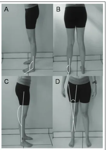
To characterize the ankle and knee alignment of the participants, four angles (tibiotarsal, subtalar, knee lexion-extension and Q-angle) were measured bi-laterally while the participant was in a bipedal quiet standing position without shoes and with no joint replacement allowance, as described elsewhere14,15,16 (Figure 1). To reduce measurement errors, all subjects were measured with the same goniometer with values rounded to the nearest two degrees (EMG Systems do Brasil, São Paulo, Brazil) and by the same expert examiner.
Platform stabilometry
Figure 1. Goniometer positions for the measurement of the: (A) tibiotarsal angle (ankle); (B) subtalar angle (ankle); (C) lexion-extension angle (knee); and (D) Q-angle (knee).
Anthropometric and body composition measurements
Subject weight and height were measured with an analog balance scale with a stadiometer applied (R110; Welmy - Santa Bárbara d’Oeste, São Paulo, Brazil). he body mass index (BMI) was calculated as the Quetelet
index (BMI = Weight/Height2) and the WHO classiication was used for
group characterization17. Waist circumference was measured at the
nar-rowest point between the lower costal border and the iliac crest18. A lexible steel tape (Terrazul; Cambuci, São Paulo, Brazil) was used to measure this
girth and the WHO classiication was used for group characterization17.
Body composition analysis was performed with a bioelectrical imped-ance analyzer (BIA 310e; Biodynamics, Seattle, Washington, USA). he test current used was 800 μA at 50 kHz. he equation chosen to predict fat free mass19 was previously validated in an elderly Brazilian sample20. he Deurenbert et al.21 classiication for obesity (body fat percentage > 35%) was used.
Statistical data analysis
as the median (irst quartile - third quartile). Frequency distributions for categorical variables were analyzed with a chi-square test. Box plots il-lustrate median, irst and third quartiles, and minimum and maximum values. he angle value was calculated as the mean of the right and let angles, except for symmetric analysis, as explained below.
A Spearman correlation coeicient was used to quantify the association between stabilometric parameters and lower limb joint angles at the p < 0.05 signiicance level. Subjects also were divided into three Q-angle groups: physiological valgus or ‘normal’ (Q-angle from 10º to 14º), varus (Q-angle < 10º), or valgus (Q-angle > 14º)22,23. A Kruskal-Wallis test was applied to detect diferences between those groups (at the p < 0.05 signiicance level). A Mann-Whitney test was used for each pair of groups to identify where the diferences were found when the Bonferroni correction was considered (0.05/3 = 0.017).
Additional data analysis was performed for the Q-angle bilateral sym-metry: each angle value (right and let body sides) was considered and clas-siied (valgus, varus, or normal) separately, and then all individuals were categorized as symmetric or asymmetric. he symmetric group comprised those elderly women with both knees categorized as the same classiication
(valgus, varus or normal). he asymmetric group had one knee
classiica-tion diferent from the other. For this comparison, a Mann-Whitney test was used at the p < 0.05 signiicance level. he SPSS statistical sotware program (version 13.0 for Windows; SPSS, Chicago, IL, USA) was used for all statistical analyses.
RESULTS
he characteristics of the studied sample are presented in Table 1 (data from all subjects and from each Q-angle group) and Table 2 (data from symmetric and asymmetric Q-angle groups). hese data show high values of adiposity for the entire sample, as assessed by the BMI, fat percentage, and waist circumference.
he investigation into the relationship between knee and ankle angles and posture control variables was performed by using three approaches. First, a Spearman analysis showed statistically signiicant (p < 0.05) cor-relations between the Q-angle and the SDx (CBEO, r = 0.31), SDy (OBEO, r = 0.28; CBEO, r = 0.34; and CBEC, r = 0.31), RANGEy (OBEO, r = 0.39; and CBEO, r = 0.38) and Area (CBEO, r = 0.36). he lexion-extension knee angle showed a few weakly signiicant correlations with the SDy (r = -0.36), RANGEy (r = -0.28) and Area (r = -0.29), all of them during the OBEC trial. In contrast, the ankle angles did not show signiicant correla-tions with stabilometric variables.
SDy (CBEO), RANGEy (CBEO), and Area (CBEC), speciically between the extreme – varus and valgus – groups (p < 0.017; for each pair mentioned comparison the Bonferroni correction was applied: 0.05/3 = 0.017; Figure 2).
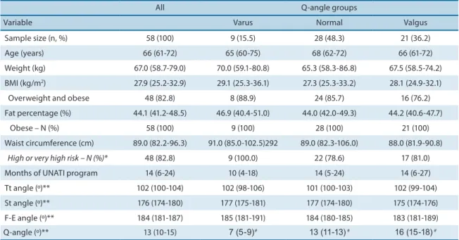
Table 1. Characteristics of the elderly women studied (N = 58)
All Q-angle groups
Variable Varus Normal Valgus
Sample size (n, %) 58 (100) 9 (15.5) 28 (48.3) 21 (36.2)
Age (years) 66 (61-72) 65 (60-75) 68 (62-72) 66 (61-72)
Weight (kg) 67.0 (58.7-79.0) 70.0 (59.1-80.8) 65.3 (58.3-86.8) 67.5 (58.5-74.2)
BMI (kg/m2) 27.9 (25.2-32.9) 29.1 (25.3-36.1) 27.3 (25.3-33.2) 28.1 (24.9-32.1)
Overweight and obese 48 (82.8) 8 (88.9) 24 (85.7) 16 (76.2)
Fat percentage (%) 44.1 (41.2-48.5) 46.9 (40.4-51.0) 44.0 (42.0-49.3) 44.2 (40.6-47.7)
Obese – N (%) 58 (100) 9 (100) 28 (100) 21 (100)
Waist circumference (cm) 89.0 (82.2-96.3) 91.0 (85.0-102.5)292 89.0 (82.3-106.0) 88.0 (81.9-90.8)
High or very high risk– N (%)* 48 (82.8) 9 (100.0) 22 (78.6) 17 (81.0)
Months of UNATI program 14 (6-24) 10 (4-18) 14 (5-24) 14 (6-27)
Tt angle (º)** 102 (100-104) 102 (98-106) 101 (100-103) 102 (99-104)
St angle (º)** 176 (174-180) 177 (175-181) 177 (174-180) 175 (174-176)
F-E angle (º)** 184 (181-187) 185 (181-191) 184 (180-185) 183 (181-189)
Q-angle (º)** 13 (10-15) 7 (5-9)# 13 (11-13)# 16 (15-18)#
Values are expressed as the median (1st-3rd quartile) for numerical variables and absolute number (percentage) for categorical variables. BMI = body mass index; F-E = lexion-extension angle; St = subtalar angle; Tt = tibiotarsal angle. *Risk for obesity-associated metabolic complications. **Mean value of the left and right sides # p < 0.001 when comparing Q-angle groups (Kruskal-Wallis test)
Table 2. Characteristics of Q-angle bilateral symmetry groups
Q-angle bilateral symmetry
Variable Symmetric Asymmetric
Sample size (n, %) 30 (51.7) 28 (48.3)
Age (years) 66 (61-72) 66 (61-72)
Weight (kg) 67.3 (58.8-80.3) 66.3 (58.2-74.4)
BMI (kg/m2) 28.8 (25.4-33.2) 27.3 (25.1-32.7)
Overweight and Obese 26 (86.7) 22 (78.6)
Fat percentage (%) 45.0 (41.6-48.6) 43.9 (40.5-48.0)
Obese – N (%) 30 (100) 28 (100)
Waist circumference (cm) 89.0 (82.9-96.8) 84.0 (80.5-96.5)
High of very high risk – N (%)* 27 (90.0) 21 (75.0)
Months of UNATI program 13 (3-23) 15 (9-24)
Tt angle (º)** 102 (100-104) 102 (99-104)
St angle (º)** 176 (175-181) 175 (173-178)
F-E angle (º)** 184 (181-188) 183 (180-186)
Q-angle (º)** 13 (11-17) 13 (10-15)
Figure 2. Box plots (median, 1st and 3rd quartiles, minimum and maximum) showing the body sway of elderly women classiied as varus, normal, and valgus (knee Q-angle) during CBEO (2.A, 2.B, and 2.C) and CBEC trials (2.D). (A) Standard deviation of the CP values in the lateral direction (SDx); (B) Standard deviation of the CP values in the anteroposterior direction (SDy); (C) Range of the CP displacement in the anteroposterior direction (RANGEy); (D) Area of the CP displacement (Area). * p < 0.017 comparing varus versus valgus (Mann-Whitney test after Kruskal-Wallis).
he analysis of the Q-angle symmetry (third approach) revealed that the CP mean displacement velocity was higher for the asymmetric group (n = 28) than the symmetric group (n = 30) for both closed base trials (Figure 3). No signiicant diference was found for the open base trials.
DISCUSSION
he present results showed that high knee Q-angles are correlated with
increased body sway. Subjects with genu valgus showed higher
stabilo-metric variable values than those with genu varus. Furthermore, upon
analyzing both knees of each participant, an asymmetric knee alignment was also associated with a greater oscillation. hese indings highlight the importance of regular assessments of lower limb alignment (particularly of knee angles), especially for elderly people, who frequently sufer from instability complaints and falls. Health professionals should concentrate their preventive approaches on Q-angle and joint alignment to minimize balance disorder manifestations.
he lexion-extension angle and Q-angle of the elderly women showed a large number of correlations with the analyzed stabilometric parameters. he lexion-extension angle has not been studied as oten as the Q-angle. Previous studies14,24,25 revealed the mean lexion-extension angle values that classiied the knees of the participants as genu recurvatum, as in the present study. he correlations observed between the lexion-extension angle and the stabilometric variables were weak, therefore it is not likely that the knee alignment in the sagittal plane inluence body sway. he Q-angle has already been extensively explored in the literature15,16,26.Normal values for this angle range from 10º to 14º 22,23, but research has not focused on the relationship between this angle and body sway. he present study focused on the elderly because they usually show standing instability. he relationship between joint alignment and body balance is of greater importance to this population, so every efort should be made to screen those who are more unstable and prone to falling. It is important to notice that the presented coeicients do not characterize strong correlations for
Q-Angle analysis. hey were mainly weak and regular ones27. In fact, as
posture control depends on a great number of factors, a simple bivariate correlation would rarely present a strong or very strong correlation. his motivated the other approaches performed in this research into Q-Angle.
When the entire sample was divided into varus, valgus, and normal
groups, the varus group showed the lowest number of participants. he
younger sample studied by Ferreira et al.13 also had the lowest prevalence
(22.6%) in the varus group. hese researchers revealed that the varus
knee group showed a lower CP mean velocity than the neutral and valgus
rigid control must be exerted by the postural system. his helps explain why signiicant diferences were observed only for closed base trials.
Livingston and Mandigo26 reported that almost half of the individuals studied by them demonstrated a diference of at least 4º between the right and let Q-angles. he present results show that asymmetry resulted in greater mean velocity values during both closed base conditions. No other study has, to the authors’ knowledge, investigated the relationship between Q-angle asymmetry and stabilometric variables during the quiet stance.
In the present sample, the tibiotarsal and subtalar angles showed no rel-evant correlation with the CP variables for most conditions. Ferreira et al.13, analyzing young adults, also found no association between stabilometric variables and subtalar alignment. In another study exploring the relation-ship between foot features and stabilometric variables11, it was found that maximum foot width, base-of-support area and foot opening angle were relevant biomechanical factors that inluenced lateral direction stabilometric variables. Menz et al.12 observed that ankle lexibility, plantar tactile sensi-tivity, and toe plantarlexor strength were associated with body sway. he same group of researchers, comparing fallers and nonfallers, found similar values for foot posture index, arch index, and navicular height, but not for ankle lexibility, presence of hallux valgus deformity, tactile sensitivity, toe plantar lexor strength and foot pain. herefore, while ankle angle measures do not appear to be an important factor in risk for falls, the evaluation of anthropometric and morphological characteristics in elderly people should still be considered when assessing possible risk factors for falls.
he correlations and diferences found in the present study corroborate the inluence of somatosensory information on postural control and suggest the possible efects of structural morphological changes due to joint aging. Horlings et al.29 state that it is generally assumed that lower limbs proprio-ception provides the main contribution to posture control. Ankle and knee morphological diferences will provide diferent inputs to the central nervous systems, so it is reasonable that motor adjustments to a good posture control will also be diferent, relecting CP variables. If we extrapolate the present results about the relationship between the Q-angle and posture control to a health professional’s practice, it is expected that the higher the Q-angle, the higher the body sway when an assessment is performed with only one angle value. If one assessment is performed for the two limbs showing asymmetry, it also seems that this fact will inluence body sway.
CONCLUSIONS
In the present study, knee alignment was associated with posture control in elderly Brazilian women. his was principally evident by the higher
stabilometric variable values obtained for the valgus and asymmetric
Q-angle groups. Conversely, ankle alignment showed no correlation with stabilometric variables.
Acknowledgments
he authors are grateful to all the elderly women who participated in the present study. We would also like to thank UNATI manager Rose Cristina Soares, and physiotherapist Maria Madalena da Costa Glinardello, for their contribution inviting volunteers.
REFERENCES
1. Mainenti MRM, Oliveira LF, Lima MAMT, Nadal J. Stabilometric signal analysis in tests with sound stimuli. Exp Brain Res. 2007; 181(2): 229-36.
2. Duarte M, Freitas SMSF. Revision of posturography based on force plate for balance evaluation. Rev Bras Fisioter 2010; 14(3): 183-192.
3. Lemos LFC, Teixeira CS, Mota CB. Lombalgia e o equilíbrio corporal de atletas da seleção brasileira feminina de canoagem velocidade. Rev Bras Cineantropom Desempenho Hum. 2010; 12(6):457-463.
4. Melzer I, Damry E, Landau A, Yagev R. he inluence of an auditory–memory attention-demanding task on postural control in blind persons. Clin Biomech. 2011; 26(4): 358-362.
5. Ferrucci L, Giallauria F, Guralnik JM. Epidemiology of Aging. Radiol Clin North Am. 2008; 46(4): 643–652.
6. IBGE – Instituto Brasileiro de Geograia e Estatística. Peril dos Idosos Respon-sáveis pelos Domicílios no Brasil. Estudos & Pesquisas – Informação Demográica e Socioeconômica. 9, 2002. Available at: <http://www.ibge.gov.br/home/estatistica /populacao/perilidoso/peridosos2000.pdf> Access: 25/11/2011.
7. Sturnieks DL, ST George R, Lord SR. Balance disorders in the elderly. Neurophysiol Clin. 2008; 38(6): 467-478.
8. Ramstrand N, huesen AH, Nielsen DB, Rusaw D. Efects of an unstable shoe construction on balance in women aged over 50 years. Clin Biomech. 2010; 25(5): 455-460.
9. Negahban H, Hadian MR, Salavati M, Mazaheri M, Talebian S, Jafari AH et al. he efects of dual-tasking on postural control in people with unilateral anterior cruciate ligament injury. Gait Posture. 2009; 30(4): 477-481.
10. Laughton CA, Slavin M, Katdare K, Nolan L, Bean JF, Kerrigan DC et al. Aging, muscle activity, and balance control: physiologic changes associated with balance impairment. Gait Posture. 2003; 18(2): 101-108.
11. Chiari L, Rocchi L, Cappello A. Stabilometric parameters are afected by anthro-pometry and foot placement. Clin Biomech. 2002; 17(9): 666-677.
12. Menz HB, Morris ME, Lord SR. Foot and Ankle Characteristics Associated With Impaired Balance and Functional Ability in Older People. J Gerontol. 2005; 60A(12): 1546-1552.
13. Ferreira AS, Gave NS, Abrahão F, Silva JG. Inluence of feet and knees morphology on balance during bipodal stance. Fisioter Mov. 2010; 23(2): 193-200.
Corresponding author
Míriam Raquel Meira Mainenti Programa de Pós-Graduação em Ciências da Reabilitação Praça das Nações, 34, 3º andar – Bonsucesso – Rio de Janeiro – Brazil – 21.041-021
E-mail: [email protected]
15. Merchant AC, Arendt EA, Dye SF, Fredericson M, Grelsamer RP, Leadbetter WB et al. he Female Knee - Anatomic Variations and the Female-speciic Total Knee Design. Clin Orthop Relat Res. 2008; 466(12): 3059-3065.
16. Omolulu BB, Ogunlade OS, Gopadalsani VK. Normal Q-angle in an Adult Nigerian Population. Clin Orthop Relat Res. 2009; 467(8): 2073-2076.
17. WHO - World Health Organization. Obesity – Preventing and Managing the Global Epidemic. World Health Organ Tech Rep Ser. 2000; 894(i-xii): 1-253.
18. ISAK - International Society for the Advancement of Kinanthropometry. Interna-tional Standards for Anthropometric Assessment. Australia: ISAK, 2001.
19. Kyle UG, Genton L, Karsegard L, Slosman DO, Pichard C. Single prediction equa-tion for bioelectrical impedance analysis in adults aged 20–94 years. Nutriequa-tion. 2001; 17(3): 248-253.
20. Reach CR, Cordeiro BA, Petroski EL, Vasconcelos FAG. Validation of Bioelectrical Impedance for the Prediction of Fat-free Mass in Brazilian Elderly Subjects. Arq Bras Endocrinol Metabol. 2008; 52(7): 1163-1171.
21. Deurenberg P, Andreoli A, Borg P, Kukkonen-Harjula K, De Lorenzo A, Lichtenbelt VMWD et al. he validity of predicted body fat percentage from body mass index and from impedance in samples of ive European populations. Eur J Clin Nutr. 2001; 55(11), 973-979.
22. Ali F. Clinical examination of the knee. Orthopaedics and Trauma. 2013; 27(1): 50-55.
23. Ribeiro DC, Loss JF, Cañeiro JPT, Lima CS, Martinez FG. Electromyographical analysis of the quadriceps during knee extension at diferent speeds. Acta Ortop Bras. 2005; 13(4): 189-193.
24. Siqueira CM, Moya GBL, Cafaro RR, Fu C, Kohn AF, Amorim CF et al. Misalign-ment of the knees: Does it afect human stance stability. J Bodyw Mov her. 2011; 15(2): 235-241.
25. Souza JA, Pasinato F, Basso D, Corrêa ECR, Silva AMT. Biofotogrametria: conia-bilidade das medidas do protocolo do sotware para avaliação postural (SAPO). Rev Bras Cineantropom Desempenho Hum. 2011; 13(4): 299-305.
26. Livingston LA, Mandigo JL. Bilateral Q angle asymmetry and anterior knee pain syndrome. Clin Biomech. 1999; 14(1): 7-13.
27. Malgady RG, Krebs DE. Understanding correlation coeicients and regression. Phys her. 1968; 66: 110-120.
28. Melzer I, Kurz I, Oddsson LIE. A retrospective analysis of balance control param-eters in elderly fallers and non-fallers. Clin Biomech. 2010; 25(10): 984-988.