Licence Creative Commom CC
RBCDH
1 Federal University of Pampa. Research Group in Applied neuro-mechanics. Uruguaiana. RS. Brazil. 2 Federal University of Rio Grande do Sul. Exercise Research Labora-tory. Porto Alegre. RS. Brazil.
Received: 20 June 2013 Accepted: 01 December 2013
Isometric muscle force, rate of force
development and knee extensor
neuromuscular eiciency asymmetries
at diferent age groups
Assimetrias na força muscular isométrica, taxa de
desenvolvimento de força e na eiciência neuromuscular
de extensores do joelho em diferentes faixas de idade
Helen L Schimidt1
Álvaro S Machado1
Marco A Vaz2
Felipe P Carpes1
Abstract– he aim of this study was to evaluate force, rate of force development and knee extensor neuromuscular eiciency asymmetries in children, adults and elderly. Each subject performed maximal isometric voluntary contractions (MIVC) and submaximal trials (15% and 30% MIVC). Maximal force, rate of force development (RFD) and neuro-muscular eiciency were evaluated and compared between groups and between preferred and non-preferred lower limb. Children (mean age 8.4, SD 0.7 yrs), female adults (mean age 23.2, SD 3.5 yrs) and elderly (mean age 65.9 SD 7.0 yrs) were evaluated. RFD was higher in young adults, and similar between children and elderly. Neuromuscular eiciency decreased signiicantly with aging (P<0.05). Inter-limb asymmetries were observed for force and RFD in favor of the preferred lower limb in the elderly (P<0.05). Force and RFD asymmetries in the elderly are supported by the right hemisphere-aging model contribut-ing to increased motor asymmetries. It was suggested that both physical assessment and training in the elderly should consider asymmetries that apparently are inherent to the aging process. A simple protocol for maximal and submaximal force assessment may be useful for delineating impairments in force and power in the elderly.
Key words: Aging; Functional lateralization; Isometric contraction; Lower limbs.
Knee extensor neuromuscular asymmetry in diferent ages Schmidt et al.
INTRODUCTION
In the next decades, elderly will represent up to 20-25% of the population in Belgium, 6-20% in South America and 10-18% in the United States1.
Mobility impairments are among the factors inluencing health and quality of life in the elderly, as well as the need for further assistance from health services and professionals. he impact of aging on motor control is in gen-eral inluenced by deicits in proprioception2, cognition3, brain structure
and function4, and neuromuscular impairments. he latter plays a major
role in falling events. It has been demonstrated that subjects older than 65 years may fall at least once a year, which can happen again for up to 50% of cases5. Falls potentiate the occurrence of bone fractures and the post-fall
syndrome, with higher risk for subjects with osteoporosis, which negatively decreases mobility and increases rigidity, afecting independence, quality of life and engagement in regular physical activity programs5.
he reduced use of skeletal muscle accelerates sarcopenia or atrophy in the elderly6, contributing to reduce knee extensor power7. Rate of force
development (RFD) and power8, muscle activation9, and bilateral
asym-metries7,10 have been described as potential tools when screening for risk of
falls in the elderly. Indeed, loss of power predicts functional performance in older adults, especially those with mobility-related impairments11.
Perry et al.7 reported knee extension force asymmetries around 10%
for young subjects and 14% in the elderly, without signiicant diferences between fallers and non-fallers. Portegijs et al.10 reported high leg extension
power asymmetries associated to lower gait speed and impaired postural control in the elderly. Assessments of performance asymmetries in children, young adults and elderly could be a satisfactory strategy to understand if asymmetries are related to the aging process.
Here, asymmetries were evaluated in the maximal voluntary force production, RFD and neuromuscular eiciency (NME) between chil-dren, young adults and elderly. he EMG/force ratio was used to quantify
NME12,13. Our primary hypothesis was that aging would negatively impact
force production, RFD and NME, and leg asymmetries would be found among older adults. he hypothesis of asymmetries in the elderly is consist-ent with the right hemisphere-aging model, related to greater age-related decline in the right cerebral hemisphere contributing to increased motor asymmetries14.
METHODOLOGICAL PROCEDURES
Experimental Design
Each subject was assessed in a single session at home, school or recreation center using a portable setup. Leg preference was veriied using the Water-loo inventory15. To assess neuromuscular function, subjects performed (a)
tested irst, but the irst leg to perform the MIVC was alternated between subjects. Subjects warmed-up by walking during 5 min at a comfortable self-selected speed on a treadmill. Ater warm-up exercise, subjects were prepared for the tests and instructed to exert maximal efort to extend the knee against a load cell at the verbal signal from the researcher.
A three-minute interval was observed between each contraction, and a ive-minute interval was observed between trials. All measurements were conducted with subjects comfortable seated on a chair, with trunk, hip and thigh stabilized, and hip, knee and ankle joints positioned at 90° of lexion. According to instructions from the researcher, subjects should fully contract their quadriceps to extend the knee unilaterally. Foot position was guided by the researcher to ensure that the knee was slightly extended against the cable at the beginning of the contraction with a pre-tension that was kept at minimum level (less than 7 N) in order to eliminate any slack from the load cell cable. his pre-tension level was controlled in the acquisition sot-ware to ensure that the subject was not applying higher tension to the cable. he contractions lasted 5 seconds. For each leg, a familiarization trial was performed before data collection. All subjects signed the informed consent form approved by the local ethics committee (protocol number 2007791). For children, parents or legal guardians were requested to sign the form.
Subjects
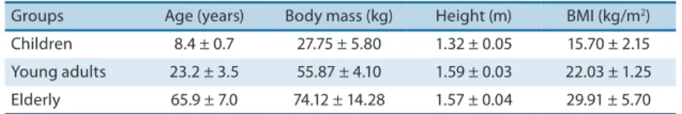
hirty-four female subjects recruited from the local community volun-teered for this study. Subjects were distributed into the following groups: children (n=8), young adults (n=11) or elderly (n=15). Subjects should not be involved in any regular physical activity program in the last 3 months, should be free from orthopedic problems afecting posture, should not have any previous fall, and should not have any balance or other health problems that could afect gait or upright position. All subjects had right footedness. Physical characteristics of the subjects are described in table 1.
Table 1. Physical characteristics of subjects presented as mean ± standard deviation for each group. BMI: body mass index.
Groups Age (years) Body mass (kg) Height (m) BMI (kg/m2)
Children 8.4 ± 0.7 27.75 ± 5.80 1.32 ± 0.05 15.70 ± 2.15
Young adults 23.2 ± 3.5 55.87 ± 4.10 1.59 ± 0.03 22.03 ± 1.25
Elderly 65.9 ± 7.0 74.12 ± 14.28 1.57 ± 0.04 29.91 ± 5.70
Force assessment
Knee extensor neuromuscular asymmetry in diferent ages Schmidt et al.
Biomedical Inc., Porto Alegre, Brazil). Of-line force signal analyses were conducted with a custom-written Matlab low-pass ilter (Butterworth, cut-of frequency of 10 Hz) routine (MATLAB 7.0, Mathworks Inc.). he MIVC of the highest force was used to determine submaximal trials as 15% and 30% of MIVC force. he MIVC force was analyzed considering a time domain approach with maximal force being determined by the highest value achieved during the plateau of the force-time curve during MIVC. he rate of force development (RFD) was determined by the highest force value achieved within the irst 200 ms ater MIVC starts, similar to procedure described in Amaral et al.12. For all subjects, the beginning of
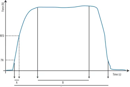
contraction was considered as the moment when the force produced was higher than 7 N. In the submaximal trials, visual feedback was provided to guide the subjects while producing 15% and 30% of MIVC. Force was normalized to the individual’s body weight. Signals were recorded in the same way for all trials and subjects, with central window of 3 seconds of contraction considered for further analysis (Figure 1).
Figure 1. Schematic representations of the MIVC force contraction and ENM computation. A represents the interval of 200ms considered to compute the rate of force development. B represents the central 3- second window considered for computation of the neuromuscular eiciency and C represent the total trial duration, considering the moment when force was higher than 7N as the beginning of contraction and the moment when force was lower than 7N as the end of contraction.
Muscle activation analysis
he electrical activation of the vastus lateralis (VL) and vastus medialis (VM) muscles from the preferred and non-preferred lower limb was moni-tored using surface electromyography synchronized to force signals. Ag/ AgCl electrode pairs (bipolar coniguration; diameter of 22 mm; Kendall Meditrace Inc., Canada) were positioned on the skin ater careful shaving and cleaning of the area with abrasive cleaner and alcohol swabs to reduce skin impedance16. A reference electrode was placed over the acromion skin
between legs and between groups.
Muscle activity was monitored throughout all trials, diferentially am-pliied and sampled at 2000 Hz with 14-bit resolution, common rejection mode of 120 dB and impedance input of 1TΩ (Miograph system, Miotec Biomedical Inc, Porto Alegre, Brazil). he raw EMG signals were smoothed with a 4th order band-pass Butterworth digital ilter at 10-500 Hz. From the
full-wave rectiied signals, the mean was calculated from the individual time series. Ater full-wave rectiication and ofset correction, the onset and ofset of the EMG activity were determined by two standard-deviations above the baseline value recorded at rest before EMG burst18. Of-line signal
analyses were developed with custom-written Matlab routines (MATLAB 7.0, Mathworks Inc) considering a time domain approach. he average root mean square (RMS) values were calculated from a 5-second window during maximal and submaximal contractions. From this time window, irst and last seconds were excluded and then the average RMS was used as an indicator of muscle activation magnitude19.
Neuromuscular eiciency
Neuromuscular eiciency was computed for each MIVC and submaximal trials. For each trial, NME was computed considering the ratio between the average muscle activation and force output during a time window of 3 seconds. he neuromuscular eiciency (NME) obtained from each trial was therefore averaged for MIVC, 15% submaximal and 30% submaximal contractions to infer on the muscle activation magnitude required to pro-duce a given level of force as described elsewhere20.
Statistical Analyses
Data were averaged for group mean and standard-deviation. Shapiro-Wilk, Mauckly and Levene tests were used to verify data normality, sphericity, and equality of variances, respectively. Data regarding force, NME and RFD were compared by analysis of variance (3 groups x 2 legs) with Bonferroni’s correction. Where interactions were found, groups were compared using one-way Anova; when leg efects were found, within group comparisons were accomplished using independent t-tests. Statistical power higher than 70% was ensured for all comparisons. he signiicance level was set at 0.05 for all data analysis using a commercial statistical package.
RESULTS
Knee extensor neuromuscular asymmetry in diferent ages Schmidt et al.
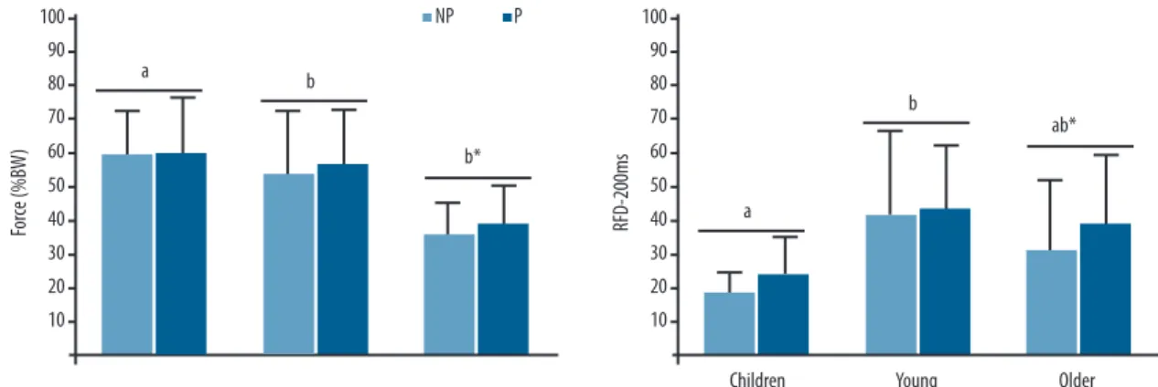
RFD data presented main efect for group. RFD was lower in children compared to young adults [F(2)=3.95; P=0.04], but no diferences were ob-served between young adults and older adults (igure 2). RFD was asym-metric for older adults [t(14)=2.45; P=0.03]. In this case, the preferred leg presented greater RFD.
here was a main efect for group in the NME (igure 3) measured in the MIVC [F(2)=28.44; P=0.01], at 15% MIVC [F(2)=10.829; P<0.01] and at 30% MIVC [F(2)=17.464; P<0.01]. NME from VM was similar to VL and lower in both muscles for children and elderly compared to young adults. here was no NME asymmetry for VM and VL, despite the lower NME in the elderly.
Figure 2. At the top, knee extensor force during MIVC normalized by body mass (%BW) for preferred (P) and non-preferred (NP) limb of children, young adults and elderly subjects. At the bottom, rate of force development (RFD-200ms) obtained during MIVC for P and NP limb in children, young adults and
elderly subjects. Diferent letters indicate statistical signiicant diference between groups (P<0.05); * indicates signiicant asymmetry between P and NP
limb within each group (P<0.05).
he knee extensor function was evaluated in subjects of diferent ages. Maximal and submaximal knee extensor voluntary force, RFD and NME were assessed for preferred and non-preferred limb in all age groups. he novelty of our study was the assessment of these variables in subjects from childhood to elderly subjects. he lower force production observed in the elderly group was anticipated due to the expected aging efects on the neuromuscular system. his force loss was accompanied by lower NME and RFD compared to children and young adults. Maximal force and RFD asymmetries were observed only in elderly subjects, and always in favor of the preferred limb. Overall, these results suggest a general neuromus-cular loss with signiicant knee extensor asymmetry in the elderly group. While for children and young adults, asymmetry indexes might not be relevant for neuromuscular function assessment, it becomes important when assessing older subjects. Additionally, the results are supported by the right hemisphere-aging model which contributes to increased motor asymmetries.
Muscle weakness observed in the lower limbs, especially ater 50 years of age21, inluences pain and/or osteoarthritis, mobility and movement
control6,22. Lower RFD in the elderly may result from morphological and
mechanical tendon changes. Decreased tendon stifness reduces force transmission from the muscle to the bone insertion23 and consequently
decreases power output24. Although still a speculation, RFD and force
asymmetries observed in favor of the preferred limb in the elderly might suggest some speciic adaptation between limbs, which may inluence force transmission during isometric contractions in diferent ways for each limb and may cause speciic imbalances during daily life activities.
While elderly subjects presented a well-deined asymmetry pattern in the knee extension force and RFD, children and young adults did not present signiicant asymmetry. A plausible explanation for the symmetry observed in the children and young adults is their higher daily involvement in bilateral actions, which contribute to similar ability between limbs25.
In the case of the elderly, asymmetries could be explained by the right hemisphere-aging model, for which the right cerebral hemisphere shows greater age-related decline than the let hemisphere14. From this
perspec-tive, an overall magnification of motor asymmetries would be expected with aging14, which is in agreement with our results. he dependence on
the right hemisphere-aging model is still an inference in our study as we were not able to measure cerebral activity during experiments. It could be possible that due to the decreased activity of the right hemisphere, lower muscle activation and consequent force production could be decreased in the let limb, therefore eliciting asymmetries.
Our data are also in agreement with a previous study, which suggested similar NME between vastii muscles26. he lower NME observed in the
Knee extensor neuromuscular asymmetry in diferent ages Schmidt et al.
as the biceps femoris27. Additionally, decreased number and iring rate of
active motor units in the elderly negatively afects the ability to fully activate the muscle, which also inluences NME28. Mau-Moeller et al.29 reported
age-related decreased voluntary muscle activation in maximal isometric voluntary torque. However, if the changes in the number and iring rate of active motor units manifest in a speciic way for each limb remains unclear.
he physical condition level in the elderly might not be inluenced by the magnitude of force asymmetries11. No leg preference efects were observed
for NME. Considering the force and RFD asymmetries observed in the elderly, further studies on neuromuscular eiciency should consider factors such as antagonist co-activation27, and degenerative articular diseases such
as osteoarthritis30, which have not been addressed in literature. Among the
limitations of this study, (1) assessment of only two muscles in females and (2) assessment of a single knee joint position, which is known to inluence the activation magnitude between the muscles evaluated stand out. he application of these results to male subjects may have some limitations since aging has some diferences between sexes and older women`s muscle iber distribution plays an important role in muscle force, RFD and fatigue.
CONCLUSION
In the present study, it was shown that leg preference may signiicantly afect knee extensor neuromuscular performance in the elderly. he decline in neu-romuscular function observed in the elderly is also accompanied by signiicant leg asymmetries. Force and RFD asymmetries in the elderly are supported by the right hemisphere-aging model contributing to increased motor asym-metries. he implication of these asymmetries for long term joint function should be considered in rehabilitation programs and sports physiotherapy.
REFERENCES
1. United Nations. he Millennium Development Goals Report. New York; 2011.
2. Patel M, Magnusson M, Kristinsdottir E, Fransson PA. he contribution of mecha-noreceptive sensation on stability and adaptation in the young and elderly. Eur J Appl Physiol 2009;105(2):167-73.
3. Holtzer R, Friedman R, Lipton RB, Katz M, Xue X, Verghese J. he relation-ship between speciic cognitive functions and falls in aging. Neuropsychology 2007;21(5):540-8.
4. Sullivan EV, Rose J, Rohling T, Pfeferbaum A. Postural sway reduction in aging men and women: relation to brain structure, cognitive status, and stabilizing fac-tors. Neurobiol Aging 2009;30(5):793-807.
5. Nancye MP. Epidemiology of Falls in Older People. Can J Aging 2011;30(1):7-19.
6. Scott D, Blizzard L, Fell J, Jones G. Prospective study of self-reported pain, ra-diographic osteoarthritis, sarcopenia progression, and falls risk in community-dwelling older adults. Arthritis Care Res 2012;64(1):30-7.
7. Perry MC, Carville SF, Smith IC, Rutherford OM, Newham DJ. Strength, power output and symmetry of leg muscles: efect of age and history of falling. Eur J Appl Physiol 2007;100(5):553-61.
Corresponding author
Felipe P Carpes, Ph.D Federal University of Pampa - Laboratory of Neuromechanics. BR 472 km 592 - Po box 118 - ZIP 97500-970, Uruguaiana, RS, Brazil Phone oice: +55 55 3413 4321 Fax: +55 55 3414 1484 E-mail: [email protected]
of recurrent falls. Gait Posture 2011;34(1):60-4.
10. Dain PL, Summer BC, Krzysztof M. Strength asymmetry increases gait asymmetry and variability in older women. Med Sci Sports Exerc 2012;44(11):2172-81.
11. Carabello RJ, Reid KF, Clark DJ, Phillips EM, Fielding RA. Lower extremity strength and power asymmetry assessment in healthy and mobility-limited popu-lations: reliability and association with physical functioning. Aging Clin Exp Res 2010;22(4):324-9.
12. Amaral JF, Castro EA, Mancini M, Doimo LA, Novo Jr JM. Rate of force develop-ment of upper and lower limbs in elderly women. Motri 2012; 8(2): 454-461.
13. David P, Mora I, Perot C. Neuromuscular eiciency of the rectus abdominis difers with gender and sport practice. J Strength Cond Res 2008;22(6):1855-61.
14. Raw RK, Wilkie RM, Culmer PR, Mon-Williams M. Reduced motor asymmetry in older adults when manually tracing paths. Exp Brain Res 2012;217(1):35-41.
15. Elias LJ, Bryden MP, Bulman-Fleming MB. Footedness is a better predictor than is handedness of emotional lateralization. Neuropsychologia 1998;36(1):37-43.
16. De Luca CJ, Sabbahi MA, Roy SH. Median frequency of the myoelectric signal. Efects of hand dominance. Eur J Appl Physiol Occup Physiol 1986;55(5):457-64.
17. Hermens HJ, Freriks B, Disselhorst-Klug C, Rau G. Development of recommenda-tions for SEMG sensors and sensor placement procedures. J Electromyogr Kinesiol 2000;10(5):361-74.
18. Brennan JT, Eric DR, Trent JH, Pablo BC, Ashley AW, Eric JS, et al. Consistency of rapid muscle force characteristics: Inluence of muscle contraction onset detection methodology. J Electromyogr Kinesiol 2012;(22):893–900.
19. Moritani T, Muro M, Nagata A. Intramuscular and surface electromyogram changes during muscle fatigue. J Appl Physiol 1986;60(4):1179-85.
20. Mills KR, Willison RG. Quantiication of EMG on volition. Electroenceph Clin Neurophysiol 1987;39:27-32.
21. Lynch NA, Metter EJ, Lindle RS, Fozard JL, Tobin JD, Roy TA, et al. Muscle quality. I. Age-associated diferences between arm and leg muscle groups. J Appl Physiol 1999;86(1):188-94.
22. Segal NA, Glass NA Is quadriceps muscle weakness a risk factor for incident or progressive knee osteoarthritis? Phys Sportsmed 2011;39(4):44-50.
23. Moerch L, Pingel J, Boesen M, Kjaer M, Langberg H. he efect of acute exercise on collagen turnover in human tendons: inluence of prior immobilization. Euro J Appl Physiol 2013;(113)449-55.
24. Lichtwark GA, Barclay CJ. he inluence of tendon compliance on muscle power output and eiciency during cyclic contractions. J Exp Biol 2010;213(5):707-14.
25. Teixeira LA, de Oliveira DL, Romano RG, Correa SC. Leg preference and interlateral asymmetry of balance stability in soccer players. Res Q Exerc Sport 2011;82(1):21-7.
26. Ebersole KT, Malek DM. Fatigue and the electromechanical eiciency of the vastus medialis and vastus lateralis muscles. J Athl Train 2008;43(2):152-6.
27. Pereira MP, Goncalves M. Muscular coactivation (CA) around the knee reduces power production in elderly women. Arch Gerontol Geriatr 2011;52(3):317-21.
28. Klass M, Baudry S, Duchateau J. Age-related decline in rate of torque development is accompanied by lower maximal motor unit discharge frequency during fast contractions. J Appl Physiol 2008;104(3):739-46.
29. Mau-Moeller A, Behrens M, Lindner T, Bader R, Bruhn S. Age-related changes in neuromuscular function of the quadriceps muscle in physically active adults. J Electromyogr Kinesiol 2013;23(3):640-8.