The role of magnetic resonance cholangiography
in the evaluation of biliary anatomy in living liver
donors*
O papel da colangiografia por ressonância magnética na avaliação da anatomia biliar em doadores de transplante hepático intervivos
Elaine Cristina de Moraes Arruda1, Julio Cezar Uili Coelho2, Jorge Massayuki Yokochi3, Jorge Eduardo Fouto Matias4
OBJECTIVE: The present study was aimed at evaluating the accuracy of magnetic resonance cholangiography in the assessment of the biliary anatomy in living liver donors in correlation with surgical findings. MATERIALS AND METHODS: Fifty living liver donors were retrospectively evaluated at Hospital de Clínicas da Universidade Federal do Paraná, Curitiba, PR, Brazil. Cholangiographic images were analyzed and results were compared with intraoperative findings. Only anatomical alterations that affected the surgical strategy and had not been previously observed at magnetic resonance cholangiography were considered as being in disagreement. RESULTS: Anatomical variations were found in 7 donors at magnetic resonance cholangiography, and in 14 during surgery. Agreement between imaging and surgical findings was observed in 41 of the 50 patients, and disagreement in 9. Magnetic resonance cholangiography sensitivity, specificity, positive and negative predictive values, and accuracy were respectively 43%, 97%, 86%, 81% and 81.6%. CONCLUSION: Magnetic resonance imaging is a safe and noninvase method for preoperative evaluation of the biliary tract in living liver donors. However some anatomical abnormalities are not detected by magnetic resonance cholangiography.
Keywords: Liver transplant; Living donors; Magnetic resonance imaging; Biliary tract.
OBJETIVO: Avaliar a acurácia da colangiografia por ressonância magnética no estudo da anatomia biliar de doadores de fígado em correlação com achados operatórios. MATERIAIS E MÉTODOS: Estudo retrospectivo de 50 doadores submetidos a transplante hepático intervivos no Hospital de Clínicas da Universidade Fede-ral do Paraná, Curitiba, PR. As colangiografias foram analisadas e os resultados dos exames foram compa-rados com os achados intra-operatórios. Apenas alterações anatômicas que promoveram mudança de estra-tégia cirúrgica, não-evidenciadas previamente pela colangiografia por ressonância magnética, foram consi-deradas como discordantes. RESULTADOS: Foram encontradas variações pela colangiografia por ressonân-cia magnética em 7 doadores e em 14 durante a cirurgia. Do total de pacientes, 41 resultados foram concor-dantes e 9 foram discorconcor-dantes. A sensibilidade, a especificidade, o valor preditivo positivo, o valor preditivo negativo e a acurácia da colangiografia por ressonância magnética foram, respectivamente, de 43%, 97%, 86%, 81% e 81,6%. CONCLUSÃO: Conclui-se que a ressonância magnética é um método de imagem se-guro e não-invasivo para avaliação pré-operatória das vias biliares de doadores e que algumas anomalias não são detectadas pela colangiografia por ressonância magnética.
Unitermos: Transplante de fígado; Doadores vivos; Imagem por ressonância magnética; Vias biliares.
Abstract
Resumo
* Study developed at Hospital de Clínicas da Universidade Federal do Paraná (UFPR), Curitiba, PR, Brazil.
1. Master, MD, Pediatric Radiologist at Instituto de Radiodiag-nóstico – Ultra-X, São José do Rio Preto, SP, Brazil.
2. Post-doctoral Fellow, Full Professor, Department of Surgery – Universidade Federal do Paraná (UFPR), Curitiba, PR, Brazil.
3. MD, Radiologist at Hospital Vita, Curitiba, PR, Brazil. 4. PhD, Associate Professor, Department of Surgery – Univer-sidade Federal do Paraná (UFPR), Curitiba, PR, Brazil.
Mailing address: Dra. Elaine Cristina de Moraes Arruda. Rua Boa Vista, 1026, ap. 103, Bairro Boa Vista. São José do Rio Preto, SP, Brazil, 15025-010. E-mail: [email protected]
Received December 16, 2007. Accepted after revision May 27, 2008.
should be dedicated to the planning for management of the biliary ducts during liver lobe resection and implantation, con-sidering that the high variability of the bil-iary anatomy in the population and the pat-tern of second order biliary branches could change the surgical technique or even rep-resent a contraindication for liver dona-tion(3). Anatomic variants have been
de-scribed in as many as 19.7%(4) to 43%(5) of
individuals. Studies have evidenced the Arruda ECM, Coelho JCU, Yokochi JM, Matias JEF. The role of magnetic resonance cholangiography in the evaluation of biliary anatomy in living liver donors. Radiol Bras. 2008;41(6):361–365.
INTRODUCTION
Liver transplant represents the sole treatment possibility for many patients af-fected by irreversible hepatic diseases, but the scarce offering of organs has been one of the limiting factors in the survival of waiting-list patients with hepatic failure(1).
utilization of magnetic resonance imaging as the method of choice for preoperative evaluation of the biliary anatomy(2,6–8) and
also as a sole method for preoperative evaluation in these cases, considering its accuracy in the detection of hepatic paren-chyma abnormalities hepatic and lobar volume, besides the depiction of the por-tal artery, and of the venous and biliary systems anatomy(9–11). However, the
utili-zation of this method for this purpose still remains to be more deeply evaluated.
The present study was aimed at retro-spectively evaluating the accuracy of mag-netic resonance cholangiography (MR cho-langiography) for depicting the biliary tract in living liver donors in correlation with intraoperative findings.
MATERIALS AND METHODS
MR cholangiography images and records of all living liver donors were re-viewed in the period between November 1998 and May 2006. The present casuistic included 50 donors. All the living donors for liver transplant were included. The in-dividuals with significant vascular or bil-iary anomalies resulting in contraindication for liver donation were excluded because of the increased risk for postoperative com-plications. Also, one donor whose records had not been found was excluded. The present study was approved by the Com-mittee for Ethics in Research in Humans of the Institution (Register No. CEP/HC 759.178/2003-11).
The studies were performed in a Gyro-scan ACS15 model system (Philips Medi-cal Systems; Best, The Nederlands), with 1.5 T magnetic field and body coil.
The examination protocol included the following sequences:
– Axial, coronal and sagittal, turbo field echo, T1-weighted sequences, adopting the following parameters: 11/4 msec repetition time (TR)/echo time (TE); flip angle = 25°. – Axial turbo spin eco (TSE) T2-weighted sequence for evaluating the liver (TR/TE: 1,800/160 msec; thickness: 8 mm; gap: 0.8; matrix: 258 × 205 reconstructed
for 512 × 410; field of view (FOV):
rang-ing between 300 mm and 380 mm, accord-ing to the patient; number of signal aver-aged (NSA): 4; number of sections: 24).
– Coronal, with overcontinuous slices, 1.5 mm overlapping, with inversion recov-ery technique for fat suppression, T2-weighted STIR (TSE) for evaluating the biliary tract (TR/TE: 1,800/500 msec; FOV: 230 mm; inversion time: 160 msec; matrix: 256 × 179; NSA: 2; thickness: 3 mm; number of sections: 65 to 80).
The sequences were acquired with res-piratory gating mode, and the total exami-nation time ranged between 30 and 50 min-utes, depending on the patient´s respiration regularity. Immediately after acquisition, the images were reconstructed and trans-mitted to the console for processing. The biliary tract anatomy was reviewed on the images acquired and based on maximum-intensity-projection reconstruction. The images were reviewed both in workstations and hardcopies. All the images were re-viewed by a same specialized radiologist, with about ten years experience in the in-terpretation of abdominal images.
The analysis included an evaluation of any variations, particularly those involving the presence of segmental ducts of the right and left lobes, or even the presence of com-mon hepatic duct trifurcation, and the evaluation of the choledocal duct and its main branches.
The extrahepatic biliary tract was con-sidered as normal in the presence of only one right hepatic duct and one left hepatic duct joining to form the common hepatic
duct, and the cystic duct joining the com-mon hepatic duct at right (Figure 1). The intraoperative findings described by the surgeon were considered as a reference pattern. Only those anatomical alterations that affected the surgical strategy and had not been previously observed at magnetic resonance cholangiography were consid-ered as being in disagreement.
RESULTS
Six of the 50 cases involved pediatric liver transplants (recipients with < 18 years of age) and 44, adult liver transplants. The donors ranged in age from 18 to 60 years (mean = 32.4 years). Thirty-one patients (62%) were male, with mean age of 30.8 years. Female patients were 19 (38%), with mean age of 35.2 years. Three types of grafts were utilized as follows: Couinaud liver segments II and III (lateral left mentectomy) in two cases; Couinaud seg-ments II, III and IV (left lobectomy) in one case; and Couinaud segments V, VI, VII and VIII (right lobectomy) in 47 cases. Anatomical alterations which affected the surgical strategy were found in only two right-lobe donors.
Normal hepatic anatomy could be found by MR cholangiography in 43 patients, and anatomical alterations were found in seven (14%). Three donors (6%) presented junc-tion of the right anterior and posterior
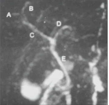
patic ducts with the left hepatic duct (com-mon hepatic duct trifurcation) (Figure 2). In one case (2%), the right posterior hepatic duct was tributary of the left hepatic duct (Figure 3). In one case (2%) the right pos-terior and anpos-terior hepatic ducts drained separately into the left hepatic duct. In one case (2%) physiological narrowing at the level of the hepatic ducts junction. Finally, in one case (2%) the bile duct of the seg-ment IV drained to the left hepatic segseg-ment, near the junction with the right hepatic duct.
Intraoperative findings corresponding to biliary tract alterations were described in 14 donors (28%). Three patients (6%) pre-sented common hepatic duct trifurcation; three (6%) presented right hepatic duct duplication; and two patients (4%), right hepatic triplication, one of these patients with also an accessory duct. In one donor (2%), a fine accessory bile duct originating from the right posterior duct was observed. Five donors (10%) presented an accessory right hepatic duct, one of them with the accessory duct tributary of the left hepatic duct, another with the accessory duct drain-ing into the choledocal duct, and another with two cystic ducts besides the accessory hepatic duct.
In the analysis of the 50 donors, 34 pre-sented normal anatomy at the surgery, in agreement with the findings at MR cholan-giography. Anatomical variations were
found at MR cholangiography in seven donors (14%), and in 14 donors (28%) during surgery. These variations were found in two cases with right hepatic ducts, three with triplication of right hepatic duct, one case of common hepatic duct trifurca-tion, and four cases with accessory hepatic duct. In one of these donors, the accessory duct was tributary of the choledocal duct, in another, of the left hepatic duct, and another with the presence of two cystic ducts. In one donor who had a very small accessory duct that was linked during the surgery, this finding was not considered as being in disagreement. In two patients (4%) anatomical variations were found at MR cholangiography, but intraoperative find-ings demonstrated a normal anatomy. In one donor, the right anterior and posterior ducts drained separately into the left he-patic duct, and in another case, non relevant from the surgical point of view, a physi-ological narrowing was described at the level of the hepatic ducts junction (also this case was not considered as being in dis-agreement).
Among the seven studies with biliary tract abnormalities, five presented results in agreement with the intraoperative find-ings, two of them with common hepatic duct trifurcation, and one with the right posterior hepatic ducts draining into the left hepatic ducts (the latter demonstrating an accessory duct draining to the left hepatic
duct at surgery, interpreted as being in agreement, considering its non-relevance from the surgical point of view). In two donors, the MR cholangiography demon-strated anatomic alterations which al-though not properly corresponding to the intraoperative findings, neither resulted in change of the surgical strategy nor in the planned anastomotic changes. In one of these donors, MR cholangiography demon-strated trifurcation, but two right hepatic ducts were present, and in the other with an accessory hepatic duct, it was interpreted as a biliary duct of the segment IV. The comparison between MR cholangiography and intraoperative findings is shown on Chart 1.
Therefore, 41/50 donors (82%) pre-sented correspondence between imaging and intraoperative findings, and 9/50 (18%) did not. The MR cholangiography sensitivity was 43%, specificity, 97%, posi-tive predicposi-tive value, 86%, negaposi-tive predic-tive value, 81%, and accuracy, 81.6%.
DISCUSSION
Living-donor liver transplant is a defi-nite method of treatment for patients with irreversible hepatic diseases, particularly in countries where cadaver liver donors are scarce or even non-existent(12). In Brazil,
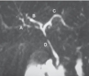
the number of living liver donors increased 10% in 2005, and in 2006 remained Figure 2. MR cholangiography demonstrating common hepatic duct
trifurca-tion.A, right anterior hepatic duct; B, right posterior hepatic duct; C, left he-patic duct; D, common hehe-patic duct.
stable(13). The safety of the donors is
ex-tremely important, considering that they are healthy individuals submitted to an exten-sive surgical procedure. So, the detection of the biliary tract anatomy is crucial for allowing the surgical planning and avoid-ing unnecessary surgery in donors with anatomical variations, besides preventing possible postoperative complications both for liver donors and recipients(7).
Accord-ing to Liu et al.(14), biliary complications
remain as the most noticeable weakness in living-donor liver transplant, playing a sig-nificant role in the occurrence of postop-erative morbidities occasionally caused by graft loss. According to Marcos et al.(15),
biliary reconstruction corresponds to the most challenging part of the surgery in the liver recipient, considering that double or triple anastomosis certainly represents a risk factor for biliary complications(16).
Biliary anatomical variations which lack pathological meaning in the general population, assume a greater relevance in cases of right lobe donation. These varia-tions include common hepatic duct trifur-cation, accessory right hepatic duct and drainage from the right anterior or poste-rior segmental duct directly into the right hepatic duct. Although such variations do not contraindicate liver donation, the pre-operative identification prevents that these ducts are inadvertently connected, result-ing in atrophy of the involved portions of the liver(6).
Retrograde endoscopic cholangiogra-phy or percutaneous transhepatic cholan-giography are considered as
“golden-stan-dard” methods for evaluating the biliary tract, but are not routinely performed be-cause of their invasiveness and association with high risks for complications(8,14,17).
The rate of complications from retrograde endoscopic cholangiography ranges be-tween 0.5% and 5%, and from percutane-ous transhepatic cholangiography, 3.4%(17).
Recently, some studies reported mag-netic resonance imaging as the sole preop-erative method for evaluating living liver donor candidates, demonstrating good re-sults in the evaluation of the biliary tract(9,11,18). This assumption is based on the
fact that conventional MR cholangiogra-phy T2-weighted sequences demonstrate high signal intensity from static fluid struc-tures while the background signal is sup-pressed(19). On the other hand, there is a
difficulty in the evaluation of a non-dilated biliary tract(3,19). Innovations such as the
utilization of a biliary contrast agent (mangafodipir trisodium) have allowed the acquisition of higher resolution images, with good results because of the better en-hancement of the biliary tract and higher differentiation from the hepatic paren-chyma and from the vascular system(7,8, 20,21). Ayuso et al. have observed a
sensitiv-ity of 93.7% and specificsensitiv-ity of 100%(22).
However, high cost, limited contrast agent availability, possibility of allergic reactions and increased images acquisition time con-stitute limiting factor for the utilization of this method(21).
Other authors have compared contrast-enhanced (gadobenate dimeglumine – Gd-DTPA) MR cholangiography T1-weighted
sequences with findings at conventional MR cholangiography T2-weighted se-quences and there was a preponderant pref-erence for contrast-enhanced MR cholan-giography in the evaluation of the biliary tract(23,24). This contrast agent combines the
properties of a gadolinium-based extracel-lular contrast agent as a hepatocyte-direct excreted at about 2%–4% through the bil-iary tract. An et al(25) have described an
accuracy of 75% with MR cholangiogra-phy T2-weighted sequences, 79% with paramagnetic contrast-enhanced T1-weighted sequences, and an increase to 92% in accuracy with the evaluation by means of a combination of both methods. The results of the present study, like-wise those previously reported by Lee et al.(9) reflect a diligent analysis in relation to
the consistency of MR cholangiography for appropriately visualizing the biliary anatomy in liver donors. The present study could demonstrate that MR cholangiogra-phy presents a good reproducibility in re-lation to the surgical findings. However, the low sensitivity of the method and the fail-ure in detecting anatomical variations in nine cases (18%), among them, the pres-ence of common hepatic duct trifurcation, duplicated or triplicated right hepatic ducts and accessory hepatic ducts, inspires pru-dence and demonstrates that the segmen-tal ducts definition is not clear. As previ-ously reported, the utilization of MR cho-langiography with specific contrast agents for studying the biliary tract, as well as further investigation about the utilization of paramagnetic contras agents such as Chart 1 Comparison between MR cholangiography and intraoperative findings.
No. of patients
2 (9 and 44)
2 (15 and 48)
2 (21 and 34)
1 (23)
1 (29)
2 (36 and 47)
1 (38)
2 (40 and 42)
1 (43)
1 (45)
1 (46)
Findings at MR cholangiography
Normal
Normal
Normal
Right anterior hepatic duct and posterior hepatic duct draining separately to the left hepatic duct
Physiological narrowing at the level of the hepatic ducts junction
Trifurcation of the common hepatic duct
Right posterior hepatic duct draining to the left hepatic duct
Normal
Trifurcation of the common hepatic duct
Normal
Segment IV bile duct draining to the left hepatic duct
Findings at surgery
Two right hepatic ducts
Three right hepatic ducts
Right accessory hepatic duct
Normal
Normal
Trifurcation of the common hepatic duct
Right accessory hepatic duct draining to the left hepatic duct
Right accessory hepatic duct draining to the choledocal duct and another coursing with two cystic ducts
Two right hepatic ducts
Trifurcation of the common hepatic duct
gadobenate dimeglumine have contributed for improvement of the method. Recently, single-shot fast spin echo sequences were adopted as a standard method for MR cho-langiography(26), and new sequences such
as half Fourier RARE have demonstrated technical advances, allowing imaging of the whole biliary tract during a single 18-second breath-hold(27). Maybe the
utiliza-tion of the described sequences, if avail-able, could result in better images as those already reported by some authors(9,11).
Fur-ther studies are required to evaluate tech-nological developments such as new se-quences and utilization of paramagnetic and biliary contrast agents.
Considering the retrospective character o the present study, the authors could not determine the number of patients excluded as well as the identified anatomical varia-tions.
It can be concluded that MR cholang-iography performed with T2-weighted se-quences presented a high accuracy, high specificity and low sensitivity. MRI is a safe and non-invasive method with poten-tial capacity and applicability in the preop-erative evaluation of the biliary tract in liv-ing liver donors. Continued technical inno-vations will certainly allow an expansion of the utilization of this method in a near future.
REFERENCES
1. Emond JC, Renz JF, Ferrel LD, et al. Functional analysis of grafts from living donors. Ann Surg. 1996;224:544–54.
2. Bassignani MJ, Fulcher AS, Szucs RA, et al. Use of imaging for living donor liver transplantation. Radiographics. 2001;21:39–52.
3. Yeh BM, Breiman RS, Taouli B, et al. Biliary tract depiction in living potential liver donors: com-parison of conventional MR, mangafodipir triso-dium-enhanced excretory MR, and multi-detec-tor row CT cholangiography – initial experience.
Radiology. 2004;230:645–51.
4. Chisuwa H, Hashikura Y, Mita A, et al. Living liver donation: preoperative assessment, anatomic considerations, and long-term outcome. Trans-plantation. 2003;75:1670–6.
5. Couinaud C. Le foie: études anatomiques et chi-rurgicales. Paris: Masson; 1957.
6. Fulcher AS, Szucs RA, Bassignani MJ, et al. Right lobe living donor liver transplantation: pre-operative evaluation of the donor with MR imag-ing. AJR Am J Roentgenol. 2001;176:1483–91. 7. Kapoor V, Peterson MS, Baron RL, et al. Intra-hepatic biliary anatomy of living adult liver do-nors: correlation of mangafodipir trisodium-en-hanced MR cholangiography and intraoperative cholangiography. AJR Am J Roentgenol. 2002; 179:1281–6.
8. Kim RD, Sakamoto S, Haider MA, et al. Role of magnetic resonance cholangiography in assess-ing biliary anatomy in right lobe livassess-ing donors. Transplantation. 2005;79:1417–21.
9. Lee VS, Morgan GR, Teperman LW, et al. MR imaging as the sole preoperative imaging modal-ity for right hepatectomy: a prospective study of living adult-to-adult liver donor candidates. AJR Am J Roentgenol. 2001;176:1475–82. 10. Goyen M, Barkhausen J, Debatin JF, et al.
Right-lobe living related liver transplantation: evalua-tion of a comprehensive magnetic resonance im-aging protocol for assessing potential donors. Liver Transpl. 2002;8:241–50.
11. Cheng YF, Chen CL, Huang TL, et al. Single im-aging modality evaluation of living donors in liver transplantation: magnetic resonance imaging. Transplantation. 2001;72:1527–33.
12. Hashikura Y, Makuuchi M, Kawasaki S, et al. Successful living-related partial liver transplan-tation to an adult patient. Lancet. 1994;343:1233– 4.
13. Associação Brasileira de Transplante de Órgãos. Área para profissionais. Gráficos 2005/2006. [acessado em: 9/10/2007]. Disponível em: http:// www.abto.org.br/profissionais.asp
14. Liu CL, Lo CM, Chan SC, et al. The right may not be always right: biliary anatomy contraindi-cates right lobe live donor liver transplantation. Liver Transpl. 2004;10:811–2.
15. Marcos A, Ham JM, Fisher RA, et al. Surgical management of anatomical variation of the right lobe in living donor liver transplantation. Ann Surg. 2000;231:824–31.
16. Fan ST, Lo CM, Liu CL, et al. Biliary reconstruc-tion and complicareconstruc-tions of right lobe live donor
liver transplantation. Ann Surg. 2002;236:676– 83.
17. Caoili EM, Paulson EK, Heyneman LE, et al. Helical CT cholangiography with three-dimen-sional volume rendering using an oral biliary contrast agent: feasibility of a novel technique. AJR Am J Roentgenol. 2000;174:487–92. 18. Sahani D, D’souza R, Kadavigere R, et al.
Evalu-ation of living liver transplant donors: method for precise anatomic defining by using a dedicated contrast-enhanced MR imaging protocol. Radio-graphics. 2004;24:957–67.
19. Song GW, Lee SG, Hwang S, et al. Preoperative evaluation of biliary anatomy of donor in living donor liver transplantation by conventional nonenhanced magnetic resonance cholangiogra-phy. Transpl Int. 2007;20:167–73.
20. Lee VS, Rofsky NM, Morgan GR, et al. Volumet-ric mangafodipir trisodium-enhanced cholangiog-raphy to define intrahepatic biliary anatomy. AJR Am J Roentgenol. 2001;176:906–8.
21. Lee VS, Krinsky GA, Nazarro CA, et al. Defin-ing intrahepatic biliary anatomy in livDefin-ing liver transplant donor candidates at mangafodipir trissodium-enhanced MR cholangiography ver-sus conventional T2-weighted MR cholangiogra-phy. Radiology. 2004;233:659–66.
22. Ayuso JR, Ayuso C, Bombuy E, et al. Preopera-tive evaluation of biliary anatomy in adult live liver donors with volumetric mangafodipir triso-dium enhanced magnetic resonance cholangiog-raphy. Liver Transpl. 2004;10:1391–7. 23. Lim JS, Kim MJ, Kim JH, et al. Preoperative
MRI of potential living-donor-related liver trans-plantation using a single dose of gadobenate dimeglumine. AJR Am J Roentgenol. 2005;185: 424–31.
24. Papanikolaou N, Prassopoulos P, Eracleous E, et al. Contrast-enhanced magnetic resonance cho-langiography versus heavily T2-weighted mag-netic resonance cholangiography. Invest Radiol. 2001;36:682–6.
25. An, SK, Lee JM, Suh KS, et al. Gadobenate dimeglumine-enhanced liver MRI as the sole pre-operative imaging technique: a prospective study of living liver donors. AJR Am J Roentgenol. 2006;187:1223–33.
26. Irie H, Honda H, Kuroiwa T, et al. Pitfalls in MR cholangiopancratographic interpretation. Radio-graphics. 2001;21:23–37.