JSCS–4858 549.753.1–36 Original Scientific paper
Cerium-doped hydroxyapatite nanoparticles synthesized by the
co-precipitation method
CARMEN STELUTA CIOBANU1,2, CRISTINA LIANA POPA1 and DANIELA PREDOI1*
1National Institute of Materials Physics, P. O. Box MG 07, 07725, Magurele, Romania and 2University Politehnica of Bucharest, Faculty of Applied Chemistry and Materials Science,
Department of Science and Engineering of Oxide Materials and Nanomaterials, 1–7 Polizu Street, P. O. Box 12–134, 011061 Bucharest, Romania (Received 24 August, revised 20 December, accepted 25 December 2015) Abstract: The present work reports a simple adapted co-precipitation method for the synthesis of stable Ce-substituted Ca hydroxyapatite (HAp) nano-particles. The structural and morphological properties of the Ce-doped hydro-xyapatite (Ce:HAp) were characterized by X-ray diffraction (XRD), trans-mission electron microscopy (TEM),scanning electron microscopy (SEM) and energy dispersive X-ray analysis (EDAX). The optical properties of the Ce-doped hydroxyapatite were also investigated using Fourier transform infrared (FTIR) spectroscopy, Fourier transform Raman spectroscopy and photolumine-scence analysis. The results of the XRD studies revealed the progressive inc-rease in the a- and c-axes with increasing Ce concentration. In the FTIR studies of Ce:HAp powders, a structure similar to that of hydroxyapatite was observed. The IR and Raman wavenumbers and the peak strengths of the bands asso-ciated with the P–O and O–H bonds decreased progressively with increasing Ce concentration. All the emission maxima could be attributed to 5d–4f tran-sitions of the Ce ions. The displacements of the maximum emission bands with increasing Cerium in the samples were in agreement with the results obtained by XRD studies. The Ce:HAp samples with xCe = 0.03 and 0.05 exhibited significant antibacterial activity against Staphylococcus aureus and Escherichia coli bacterial strains compared to Ce:HAp samples with xCe = 0 (pure HAp) and 0.01.
Keywords: biomaterials; FTIR; FT Raman; photoluminescence; Staphylococ-cus aureus; Escherichia coli.
INTRODUCTION
Hydroxyapatite is one of the most studied biomaterial with the chemical for-mula Ca10(PO4)6(OH)2, (HAp) and represents the principal inorganic component
of bones and teeth. Its excellent biological properties (biocompatibility, osteo-conductivity, inductivity, etc.) have made HAp a suitable material for the
bio-medical field.1–6 Recent studies demonstrated that the structure of apatite allows
a large number of substitutions.7–9 These substitutions modify the physical,
che-mical and biological properties of HAp. Moreover, the crystallinity, solubility and thermal stability are also influenced by the substitution of Ca with different ions (Eu, Ag, Sm, Zn, Cu, etc.). The substitution of Ca with Ce is also possible
because their radius and electronegativity are very similar.
Studies conducted by Doat et al. showed that lanthanide-doped
hydroxy-apatite (e.g., europium-doped hydroxyapatite) nanoparticles could be used as
bio-active fluorescent carriers of biological molecules and drugs.10–12
It is well known that the morphology, structure and physicochemical pro-perties of HAp powders depend on the synthesis method. Thus, various tech-niques are used to synthesize hydroxyapatite nanopowders, such as precipitation, sol–gel, solid-state reactions, pyrolysis, microwave synthesis, a mechanochem-ical route.13–16
It was found that cerium (Ce) can act similarly to calcium in the human body and accumulate in bones. According to these results, cerium-doped hydroxy-apatite could improve/stimulate metabolism.17,18 On the other hand, cerium ions
also have antimicrobial activity and unique regenerative properties. Moreover, recent studies showed that the use of cerium in dentistry may help prevent cavities.19,20 These biological properties make cerium suitable for applications in
the biomedical field.21
A large number of studies were also conducted in order to discover new biomaterials with improved biological and physicochemical properties (espe-cially antibacterial and luminescent) in order to be successfully used in the bio-medical field.
As Ce cations possess luminescent properties (under UV excitation22,23) and
antimicrobial activity, their incorporation in the structure of HAp could lead to a biomaterial being obtained with promising applications in medicine (for imaging, drug delivery, etc.).24
The present structural, morphological and optical studies were conducted on Ca10–xCex(PO4)6(OH)2 (Ce:Hap) synthesized by the co-precipitation method.
Small concentrations of cerium (xCe = 0.01, 0.03 and 0.05) were incorporated in
various complementary characterization techniques for a general understanding of what occurs in the apatite lattice when Ca is replaced by Ce.
The aim of this work was also to study the influence of cerium on the physicochemical, photoluminescent and antimicrobial properties of hydroxy-apatite doped with small concentrations of cerium.
In addition, the results obtained in this study could be of importance for future studies concerning the influence of rare earths on various ceramic mat-erials.
EXPERIMENTAL Powders preparation
In this work, Ce-doped Ca10(PO4)6(OH)2 powders, (Ca10-xCex(PO4)6(OH)2; Ce:HAp) with different concentrations of cerium (xCe = 0.01, 0.03 and 0.05) were synthesized by the
co-precipitation method. In order to obtain Ce:HAp powders, appropriate amounts of cerium nitrate hexahydrate Ce(NO3)3·6H2O (99.999 %, Sigma–Aldrich) and calcium nitrate
tetra-hydrate Ca(NO3)2·4H2O (99.999 %, Sigma–Aldrich) were dissolved in deionised water,
resulting 350 mL of solution (solution 1). In a separate beaker, an appropriate amount of (NH4)2HPO4 (≥98 %,Sigma–Aldrich) was dissolved in deionised water, resulting 350 mL of
solution (solution 2). Both solutions were obtained under vigorous stirring (300 rpm, 150 °C, 1 h) on a magnetic stirrer (Velp Scientifica). Solution 2 was dropped into solution 1 under vigorous stirring (300 rpm) at 100 °C. The pH of the mixture was adjusted to 10 by the addition of NH4OH. The resulting precipitate was washed several times with deionised water
and then filtered. The powder was dried at 100 °C for 48 h in an air oven. Powders characterization
The powders were investigated by X-ray diffraction using a Bruker D8 Advance diffractometer, with nickel filtered CuKα (λ = 1.5418 Å) radiation and a high efficiency
one-dimensional detector (Lynx Eye type) operated in the integration mode. The diffraction pat-terns were collected in the 2θ range 20–60°, with a step of 0.02° and 34 s measuring time per step. Insightful analyses of the Ce:HAp structures were performed by Rietveld refinement analysis using the MAUD code.25,26 Transmission electron microscopy (TEM) studies were
realised using a JEOL 200 CX instrument. Scanning electron microscopy (SEM) measure-ments were performed using a Quanta Inspect F microscope from the FEI Company, equipped with an energy-dispersive X-ray spectrometer (EDAX). The FTIR studies of the powders were performed on a Spectrum BX Spectrometer using the KBr pellet technique. The spectra were registered in the spectral region 400–4000 cm-1 with a resolution 4 cm-1 and 128 scans.
An FT Raman Bruker RFS 100 spectrophotometer was used in order to obtain complementary information about optical properties of the Ce:HAp powders. The powders were excited using a laser wavelength of 1064 nm. The spectra were recorded with a resolution of 4 cm-1 and 100
scans. The fluorescence emission spectra were obtained on a HORIBA Scientific Fluorolog-3 spectrofluorometer (model FL3-2iHR320 with Hamamatsu R-928 PMT).
Antibacterial tests
from American Type Culture Collection. The Ce:HAp samples were inoculated on Mueller– Hinton agar plates followed by incubation at 37 °C for 24 h. To evaluate the antibacterial activity of the Ce:HAp samples, the inhibition areas were examined. The total diameter (in mm) of the inhibition zone was measured for each sample. The survival rates of the S. aureus and E. coli bacterial strains were evaluated after they had been exposed to the Ce:HAp and pure HAp (as reference) bioceramic powders. The tests for all Ce:HAp samples were per-formed in triplicate.
RESULTS AND DISCUSSIONS
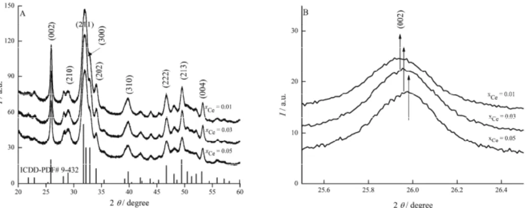
The X-ray diffraction patterns of cerium doped hydroxyapatite, Ca10–xCex(PO4)6(OH)2, with different concentrations of cerium (xCe = 0.01, 0.03
and 0.05) are depicted in Fig. 1A. The broad peaks in the X-ray diffraction patterns assigned to the characteristic (002), (210), (211), (300), (202), (310), (222), (123) and (004) planes of Ca10–xCex(PO4)6(OH)2 were in accordance with
the expected patterns for crystalline hydroxyapatite with a hexagonal structure (JCPDS No. 9-432). Separate phases for cerium oxide or other impurity phases were not observed in these diffraction patterns. Nevertheless, when some Ca sites were replaced by Ce, there was some distortion of the lattice cell parameters. For the peaks registered at lower degrees, a slight shift was observed. This behaviour seems to be the effect of substitutions that change the crystal structure (Fig. 1B), which according to Lim et al.28 suggests a significant incorporation of the
dopants into the apatite lattice.
Fig. 1. A) X-ray diffraction patterns of Ce:HAp with xCe = 0.01, 0.03 and 0.05; B) comparison
of the X-ray diffraction patterns for Ce:HAp with xCe = 0.01, 0.03 and 0.05.
The crystallite sizes of all the Ce:HAp samples decreased with increasing Ce concentration in the samples (Table I). In this study, both the a- and c-axes
These results obtained from XRD studies demonstrate that the Ce:HAp pow-ders synthesised by the adapted co-precipitation method revealed the charac-teristic structure of hydroxyapatite with good crystal structure without the pre-sence of any new phase or impurity.
TABLE I. Lattice cell parameters and the crystallite sizes of Ca10-xCex(PO4)6(OH)2 obtained from Rietveld refinement
xCe Cell parameters Crystallite size, nm
a / nm c / nm
(JCPDS 9-432) 0.9418 0.6884 –
0.01 0.9420±0.0001 0.6886±0.0001 25.45±0.1
0.03 0.9424±0.0002 0.6889±0.0003 22.86±0.1
0.05 0.9436±0.0002 0.6895±0.0004 18.95±0.1
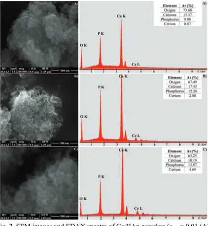
In order to examine the microstructure and elemental composition of Ce:HAp powders, scanning electron microscopy studies were performed. The SEM images and EDAX spectra of the Ce:HAp samples are presented in Fig. 2. From the
Fig. 2. SEM images and EDAX spectra of Ce:HAp powders (xCe = 0.01 (A),
SEM images, it could be observed that the increase in the Ce concentration slightly influenced the morphology of the samples. However, the ellipsoidal shape of the nanoparticles was preserved in all the samples. The SEM micro-graphs confirmed the nanometric dimensions of the particles but also their ten-dency to agglomerate. This tenten-dency to agglomerate is due most probably to the nanometric dimensions of the particles.
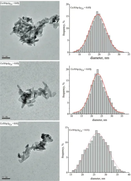
Fig. 3. TEM images and size distribution of Ce:HAp samples (xCe = 0.01 (A),
The EDAX spectra (Fig. 2b) of the studied powders confirmed the presence of all constituent elements of the Ce:HAp powders: Ce, Ca, P and O. In the tables inserted in the EDAX spectra, the calculated atomic ratios of the constituent elements for each powder are presented. These results suggest that cerium ions were incorporated in the hydroxyapatite structure. Moreover, these studies con-firmed the increase of the Ce concentrations in the samples.
The TEM images of the synthesized samples are presented in Fig. 3 (left). The TEM images clearly demonstrated the morphologies of Ce:HAp nanopar-ticles. The grain size of the Ce:HAp samples determined from TEM images are in full agreement with the results from the XRD studies. The mean particle size was determined from TEM images after counting approximately 500 nanopar-ticles Fig. 3 (right). Therefore, the average particle size obtained for Ce:HAp with xCe = 0.01 was 26.5±0.5 nm, for the Ce:HAp powder with xCe = 0.03, it was
23±0.1 nm and for Ce:HAp with xCe = 0.05, it was 19.5±0.1 nm.
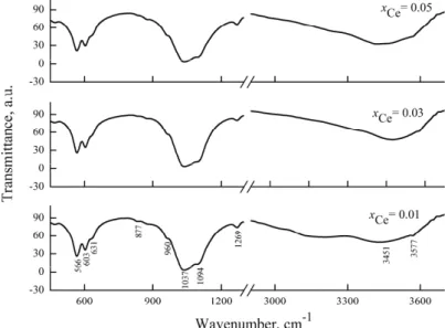
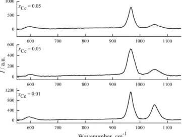
In the FTIR spectra of cerium doped hydroxyapatite (Fig. 4), characteristic vibrational bands related to phosphate, hydroxyl and adsorbed water were pre-sent. Moreover, an additional band attributed to the carbonate group (1269 cm–1)
was also presented.
Fig. 4. FTIR spectra of the cerium-doped hydroxyapatite powders.
The vibrational bands from the region 3100–3460 cm–1 were attributed to
adsorbed water. The bands at 3577 and 631 cm–1 were assigned to the stretching
bands at 566, 603 (assigned to the ν4 bending mode of PO43– groups) and 631
cm–1 indicate the formation of a well crystallized powder.
The phosphate ν1 stretching mode band was present at 960 cm–1 and the
PO43– ν3 stretching mode band at 1094 and 1037 cm–1. Furthermore, the band at
470 cm–1 was assigned to the ν2 bending mode band of the PO43– group. In
addition, the IR wavenumber and the peak strength of the P–O and O–H bonds decreased progressively with increasing Ce concentration.
Complementary information to those obtained by FTIR spectroscopy was obtained by Raman spectroscopy. The main vibrational bands observed in the Raman spectra (Fig. 5) were attributed to the stretching (ν3 and ν1) and bending
(ν4) modes of the phosphate group from the HAp structure. The bands from 594
and 610 cm–1 were assigned to the ν4 bending mode of the PO43– group. On the
other hand, the phosphate ν1 stretching mode band appeared at 965 cm–1 and
another stretching mode (ν3) was observed at 1053 cm–1.
Fig. 5. Raman spectra of cerium-doped hydroxyapatite powders.
In the Raman spectra we could also observe that the intensity of the vib-rational bands decreased with the increase of Cerium concentration in the pow-ders. This behaviour is due to the substitution of Ca ions with larger ions (Ce) in the HAp lattice which leads to a decrease of bonding strength of P–O.23 These
results obtained by Raman spectroscopy are in good agreement with the inform-ation derived from the FTIR studies presented above. On the other hand, the results obtained by FTIR and Raman spectroscopy confirmed the XRD analysis.
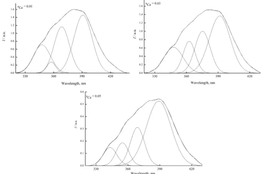
The emission spectra of Ce:HAp samples with xCe = 0.01; 0.03 and 0.05 are
at around 390 nm and a shoulder at about 350 nm that covered the wavelength range from 330 to 430 nm.
Fig. 6. Emission spectra of Ce:HAp powders (xCe = 0.01, 0.03 and
0.05);λexc = 250 nm.
As the spectra were asymmetric, they were deconvoluted into four Gaussian peaks with maxima at around 390, 375, 357 and 347 nm (Fig. 7). All these emission maxima could be attributed to 5d–4f transitions of the Ce ions.
In the structure of HAp two types of Ca site (positions) are present: Ca (I) and Ca (II). Here, the emission maximum around 360 nm is attributed to Ce ion which substituted the Ca(I) position in the hydroxyapatite structure.36 In
addi-tion, the emission band at around 390 nm occurred due to Ce ion that substituted the Ca(II) position. The results reported in this paper are in good agreement with data reported in the literature.36 Furthermore, it is obvious that the intensity of
the emissions bands increased with increasing cerium concentrations in the samples. Furthermore, the maximum emission bands were displaced with inc-reasing cerium in the samples. These results are in agreement with the results obtained by XRD studies.
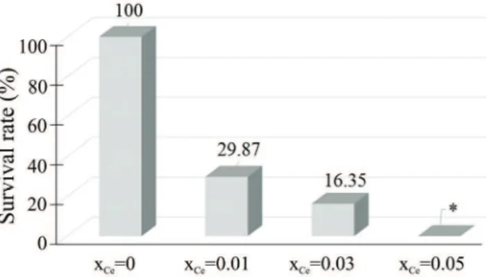
The antimicrobial activity of the cerium substituted hydroxyapatite against
E. coli 714 and S. aureus ATCC 6538 bacteria was studied after 24 h incubation
(Figs. 8 and 9, respectively). The antimicrobial activity of the cerium-substituted hydroxyapatite was compared with that of pure hydroxyapatite (xCe = 0), which
was considered as the reference. According to a previous study,37 pure HAp did
not exhibit any antibacterial effect against E. coli 714 and S. aureus ATCC 6538.
The results of this study suggest that the survival rate of E. coli 714 bacterial
strain decreased with increasing concentration of Ce in the hydroxyapatite. Moreover, for xCe = 0.05, the antibacterial effect against E. coli 714 was
sig-nificant (*p < 0.05, compared to pure HAp).
Fig. 8. The survival rate of E. coli 714 in dependence on the concentration of Ce in the analyzed samples; *p < 0.05.
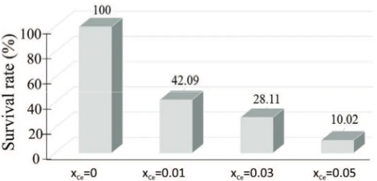
The survival rate of
S. aureus
in dependence on the concentration of
Ce in the analyzed samples is presented in Fig. 9. The survival rate of the
S. aureus
ATCC 6538 bacterial strain decreased with increasing
concentration of Ce in the hydroxyapatite. However, the survival rate of
S.
aureus
ATCC 6538 was still around 10 % when
x
Ce= 0.05. To
-negative bacteria than Gram-positive bacteria, future studies on the effect
against several strains of Gram-positive and Gram-negative are required.
Fig. 9. The survival rate of S. aureus ATCC 6538 in dependence on the concentration of Ce in the analyzed samples.
The results of this research demonstrated that cerium-doped hydroxyapatite may be prepared by the co-precipitation method at low temperatures. Ce3+ can
enter into the HAp structure by substituting calcium ions. After substituting Ca2+
in hydroxyapatite structure, the crystallite sizes of Ce:HAp decreased with inc-reasing cerium concentration. According to previous studies,38 the crystallinity
decreased gradually with increasing cerium concentration in the Ce:HAp samples. Many studies have been focused on the synthesis of various materials based on rare earth elements with antimicrobial activity and their applications in different biological fields was highlighted.39
The antimicrobial activity of the cerium-substituted hydroxyapatite with low concentrations of cerium was compared to that of pure HAp. The antimicrobial activities of pure HAp against E. coli 714 and S. aureus ATCC 6538 were
com-parable with previous results.8,9 On the other hand, the present study
demon-strated that the antimicrobial activities of hydroxyapatite doped with low concen-trations of cerium against E. coli 714 and S. aureus ATCC 6538 were more
pro-nounced than those reported in previous studies for copper-doped hydroxy-apatite.40 In addition, the present study also showed that the antimicrobial
acti-vity of cerium-substituted hydroxyapatite was more pronounced than the antimic-robial activity of both unsubstituted hydroxyapatite and fluorapatite.41,42 This
result could be explained by the interaction of cerium ions present in the hydro-xyapatite structure with the cell membrane that could lead to structural damage and death of the cells.43,44 This research demonstrated that cerium is a good
CONCLUSIONS
In this article, the synthesis of hydroxyapatite doped with small concen-tration of cerium is reported. The lattice parameters showed that Ce substituted Ca in the apatite structure. The crystal size decreased with increasing concen-tration of Ce. The results obtained in the XRD studies demonstrated that the Ce:HAp powders synthesized by an adapted co-precipitation method gave hydro-xyapatite with a good crystalline structure without any new phases or impurities. The nanometric dimensions of the particles were confirmed by SEM and TEM micrographs. Raman and FTIR studies confirmed the initial results obtained by XRD investigations. The deconvolution of the emission spectra of all Ce:HAp samples revealed four peaks that could be attributed to 5d–4f transitions of the Ce ions. This research demonstrated that cerium is a good candidate that can provide HAp an excellent antibacterial activity. In conclusion, the results pre-sented in this paper encourage further studies and research on the antimicrobial properties of cerium-doped hydroxyapatite aimed at possible applications as an antimicrobial agent.
Acknowledgements. The work was funded by the Sectoral Operational Programme Human Resources Development 2007–2013 of the Ministry of European Funds through the Financial Agreement POSDRU/159/1.5/S/134398. We thank HORIBA Scientific and espe-cially Dr. Reynald Hurteaux and Dr. Patrick Chapon from Horiba Jobin Yvon SAS France for the access to their specific instrumentation.
И З В О Д
ЦЕРИЈУМОМДОПИРАНЕНАНОЧЕСТИЦЕХИДРОКСИАПАТИТАСИНТЕТИСАНЕ
МЕТОДОМКОПРЕЦИПИТАЦИЈЕ
CARMEN STELUTA CIOBANU1,2, CRISTINA LIANA POPA1и DANIELA PREDOI1
1
National Institute of Materials Physics, P.O. Box MG 07, 07725, Magurele, Romania и2
University Politehnica of Bucharest, Faculty of Applied Chemistry and Materials Science, Department of Science and
Engineering of Oxide Materials and Nanomaterials, 1–7 Polizu Street, P.O. Box 12–134, 011061 Bucharest, Romania
Урадујеприказанједноставнимодификованикопреципитационипоступакзасин
-тезу стабилних Cе-допираних наночестица Cа-хидроксиапатита (HAp). Структурна и
морфолошка својства синтетисаних прахова Cе:HAp су испитивана применом ренд
-генске дифракционе анализе (XRD), трансмисионе електронске микроскопије (TEM),
скенирајућеелектронскемикроскопије (SEM) иенергетскедисперзивнеспектроскопије
(EDS). Оптичка својства Cе-допирараних хидроксиапатита су испитивана применом
инфрацрвене спектроскопије са Фуријеовим трансформацијама (FTIR), Раман спек
-троскопијесаФуријеовимтрансформацијамаифотолуминисцентноманализом. Резул
-тати XRD анализесу показалида долазидо континуалногповећањааиc параметара
јединичнећелијесаповећањемконцентрацијецеријума. FTIR анализапрахова Cе:HAp
је показала сличну структуру добијених прахова. IR и Раман таласни бројеви, као и интензитетитракакојеодговарајувезама P–ОиО–H опадајуконтинуалносаповећањем
концентрације Cе. Свиемисионимаксимумимогусеприписати 5d–4f прелазу Cе-јона.
сагласности сарезултатима XRD анализе. Cе:HAp узорцисаxCе = 0,03 и 0,05 показују
значајно већу антибактеријску активност према бактеријским врстама Staphylococcus aureus ATCC 6538иЕscherichia coli 714 уодносунаузорак Cе:HAp саxCе = 0 (чист HAp) и
xCе = 0,01.
(Примљено 24. августа, ревидирано 20. децембра, прихваћено 25. децембра 2015)
REFERENCES
1. R. Gorcia, R. H. Doremus, J. Mater. Sci. – Mater. Med. 3 (1992) 154 2. C. K. Hsu, Mater. Chem. Phys. 9470 (2002) 1
3. T. Kokubo, Bioceramics and their Clinical Applications, CRC Press, Boca Raton, FL, 2008
4. J. Park, Bioceramics: Properties, Characterizations and Applications, Springer, New York, USA, 2008, p. 359
5. S. V. Dorozhkin, Calcium orthophosphates: applications in nature, biology and medi-cine, Pan Stanford, Singapore, 2012, p. 850
6. S. V. Dorozhkin, J. Mater. Sci. – Mater. Med. 24 (2013) 1335 7. C. S. Ciobanu, C. L. Popa, D. Predoi, J. Nanomat.2014 (2014) 1
8. C. S. Ciobanu, F. Massuyeau, L. V. Constantin, D. Predoi, Nanoscale Res. Lett. 6 (2011) 613
9. C. S. Ciobanu, S. L. Iconaru, F. Massuyeau, L. V. Constantin, A. Costescu, D. Predoi, J. Nanomater. 2012 (2012) 1
10. S. P. Mondejar, A. Kovtun, M. Epple, J. Mater. Chem. 17 (2007) 4153 11. P. Yang, Z. Quan, C. Li, X. Kang, H. Lian, J. Lin, Biomaterials29 (2008) 4341 12. A. Doat, F. Pellé, N. Gardant, A. Lebugle, J. Solid State Chem. 177 (2004) 1179 13. S. Koutsopoulos, J. Biomed. Mater. Res. 62 (2002) 600
14. S. V. Dorozhkin, Int. J. Chem. Mater. Sci. 1 (2013) 105
15. M. S. Djošić, M. Mitrić, V. B. Mišković-Stanković, J. Serb. Chem. Soc. 80 (2015) 237 16. A. Janković, S. Eraković, A. Dindune, D. Veljović, T. Stevanović, D. Janaćković, V.
Mišković-Stanković, J. Serb. Chem. Soc.77 (2012) 1609
17. J. Emsley, Nature's Building Blocks: An A–Z Guide to the Elements, Oxford University Press, Oxford, 2011
18. M. A. Jakupec, P. Unfried, B. K. Keppler, Rev. Physiol. Biochem. Pharmacol. 153 (2005) 101
19. J. Kiss, J. Bánóczy, E. Fehérváry, Z. Gintner, M. Albrecht, Acta Morphol Hung. 38
(1990) 61
20. X. F. Liu, M. J. Tu, Modern Chemical Industry 25 (2005) 145 21. Y. Lin, Z. Yang, J. Cheng, J. Rare Earths. 25 (2007) 452
22. Z. Feng, Y. Liao, M. Ye, J. Mater. Sci. - Mater. Med. 16 (2005) 417 23. L. Yingguang, Y. Zhuoru, C. Jiang, J. Rare Earth. 25 (2007) 452
24. Ž. Radovanović, Đ. Veljović, B. Jokić, S. Dimitrijević, G. Bogdanović, V. Kojić, R. Petrović, D. Janaćković, J. Serb. Chem. Soc.77 (2012) 1787
25. L. Lutterotti, Nucl. Instr. Meth. Phys. Res. 268 (2010) 334 26. N. C. Popa, J. Appl. Crystallogr. 31 (1998) 176
27. A. W. Bauer, W. M. Kirby, J. C. Sherris, M. Truck, Am. J. Clin. Pathol. 45 (1966) 493 28. P. N. Lim, B. Y. Tay, C. M. Chan, E. S. Thian, J. Biomed. Mater. Res., B 100 (2012) 285 29. C. Ergun, T. J. Webster, R. Bizios, R. H. J. Biomed. Mater Res. 59 (2002) 169
32. O. Kaygili, S. V. Dorozhkin, S. Keser, Mat. Sci. Eng., C 42 (2014) 78
33. I. V. Berezovskaya, N. P. Efryushina, G. B. Stryganyuk, A. S. Voloshinovskii, E. V. Zubar, V. P. Dotsenko, Func. Mater. 15 (2008) 164
34. A. Groza, A. Surmeian, J. Nanomater. 2015 (2015) 1 35. A. Groza, Rom. Rep. Phys. 64 (2012) 1227
36. I. V. Berezovskaya, N. P. Efryushina, E. V. Zubar, V. P. Dotsenko, in Proceedings of the International Conference Nanomaterials: Applications and Properties, Sumy State University, Ukraine, 2012, Abstract No. 01PCN24
37. G. Dai, A. Yu, X. Cai, Q. Shi, Y. Ouyang, S. Tan, J. Rare Earth 30 (2012) 820 38. L. Yingguang, Y. Zhuoru, C. Jiang, J. Rare Earth 25 (2007) 452
39. T. S. H. Perera, Y. Han, X. Lu, X. Wang, H. Dai, S. Li, J. Nanomater. 2015 (2015) 1 40. S. Shanmugamn, B. Gopal, Ceram. Int. 40 (2014) 15655
41. M. Turkoza, A. Atillab, Z. Evis, Ceram. Int. 39 (2013) 8925
42. V. Stanic, S. Dimitrijevic, D. G. Antonovic, B. M. Jokic, S. P. Zeca, S. T. Tanaskovic, S. Raicevi, Appl. Surf. Sci. 290 (2014) 346