https://doi.org/10.1590/0004-282X20180008 ARTICLE
The importance of central auditory evaluation
in Friedreich’s ataxia
A importância da avaliação auditiva central na ataxia de Friedreich
Bianca Simone Zeigelboim1, Hélio A. G. Teive2, Michèlli Rodrigues da Rosa1, Jéssica Spricigo Malisky1, Vinicius Ribas Fonseca1, Jair Mendes Marques1, Paulo Breno Liberalesso3
Hereditary ataxias take up about 10% of genetic
dis-eases affecting the nervous system. Their classification is made according to their etiopathogenesis. Among them, Friedreich’s ataxia, which was initially described by Nicholas Friedreich, stands out. It is a progressive neurodegenerative disease with recessive autosomal inheritance and early onset in most cases1,2,3.
The mutation responsible for this disease is found
in chromosome 9, where the GAA triplet repeat expan
-sion in the FXN gene occurs4. The affected gene encodes
the mitochondrial protein, frataxin, involved in iron
metabolism2,3,4. The deficit of that protein causes iron
accumulation within the mitochondria, thus impairing the respiratory chain2,3,4,5.
The first symptoms are usually observed in childhood or
early adolescence. However, in some cases, it can be diag
-nosed before two or after 20 years of age. The main charac
-teristics of this disease are: ataxia (impaired coordination), initially in the lower limbs and subsequently in the upper limbs; absence of tendon reflexes and weakness of lower limbs; dysarthria; loss of deep distal sensitivity; and bilateral Babinski signs. Studies on neural conduction have shown
sensitive axonal neuropathy4,5,6. Other features may be asso
-ciated with the main symptoms, such as: nystagmus, optical
1Univesidade Tuiuti do Paraná ,Departament of Otoneurology, Curitiba – Paraná State, Brazil;
2Universidade Federal do Paraná, Hospital de Clínicas, Neurology Service, Curitiba – Paraná State, Brazil; 3Hospital Pequeno Príncipe, Departament of Neurology, Curitiba – Paraná State, Brasil.
Correspondence: Bianca Simone Zeigelboim; Departamento de Otoneurologia da Universidade Tuiuti do Paraná, Rua Sydnei Antonio Rangel Santos, 238; 82010-330 Curitiba PR, Brasil; E-mail: [email protected]
Conflict of interest: There is no conflict of interest to declare.
Received 19 September 2017; Received in final form: 26 November 2017; Accepted: 14 December 2017. ABSTRACT
Objective: To assess central auditory function in Friedreich’s ataxia. Methods: A cross-sectional, retrospective study was carried out. Thirty patients underwent the anamnesis, otorhinolaryngology examination, pure tone audiometry, acoustic immittance measures and brainstem auditory evoked potential (BAEP) assessments. Results: The observed alterations were: 43.3% in the pure tone audiometry, bilateral in 36.7%; 56.6% in the BAEP test, bilateral in 50%; and 46.6% in the acoustic immittance test. There was a significant difference (p < 0.05) in the comparison between the tests performed. Conclusion: In the audiological screening, there was a prevalence of the descending audiometric configuration at the frequency of 4kHz, and absence of the acoustic reflex at the same frequency. In the BAEP test, there was a prevalence of an increase of the latencies in waves I, III and V, and in the intervals of interpeaks I-III, I-V and III-V. In 13.3% of the patients, wave V was absent, and all waves were absent in 3.3% of patients.
Keywords: spinocerebellar degenerations; hearing disorders; Friedreich ataxia; evoked potentials, auditory; ataxic gait.
RESUMO
Objetivo: Avaliar a função auditiva central na ataxia de Friedreich (AFRD). Métodos: Foi realizado um estudo retrospectivo de corte transversal. 30 pacientes realizaram anamnese, avaliações otorrinolaringológica, audiológica, imitanciométrica e do potencial evocado auditivo de tronco encefálico (PEATE). Resultados: As alterações observadas foram: 43,3% no exame audiométrico sendo 36,7% dos casos, bilateralmente; 56,6% na avaliação do PEATE com 50% dos casos, bilateralmente e 46,6% no exame imitanciométrico. Houve diferença significativa (p < 0,05) na comparação entre os exames realizados. Conclusão: No exame audiológico, ocorreu uma preponderância maior da configuração audiométrica descendente a partir da freqüência de 4kHz e ausência do reflexo acústico na mesma frequência. No exame do PEATE, houve prevalência do aumento das latências nas ondas I, III e V, e nos intervalos dos interpicos I-III, I-V e III-V. Em 13,3% dos casos, a onda V estava ausente, e em 3,3% dos casos, todas as ondas estavam ausentes.
atrophy, hearing loss (may be present), hand atrophy and distal atrophy in the lower limbs, scoliosis, pes cavus and claw-toe deformity2,3,4,5,6. Diabetes may be present in 10%
of the patients, and cardiomyopathy may occur in about
two-thirds of the patients, whichis the main cause of mor
-tality6,7. There are significant differences in the lifespan of
affected individuals, which tends to be around four decades
from the disease onset until death4,5,8.
Screening of the peripheral and central auditory system is carried out by means of behavioral, acoustic-electric and electrophysiological assessment methods.
The brain is responsible for speech processing, beginning in the cochlea, where mechanical activity turns into nerve
impulses. From the neurophysiological point of view, hear
-ing entails a complex system, compris-ing a peripheral and a central part (cortical and subcortical structures). Whenever there is a physical dysfunction, a deficit in speech recognition skills occurs. Sound perception is performed by the central activity, and sound sensation is generated by the peripheral activities. While the peripheral auditory system receives and analyzes environmental auditory stimuli, the brain analyzes
inner representations of acoustic stimuli9.
The effects caused by degenerative processes may involve the central auditory system, and disorders occur due to changes that directly affect brain mechanisms that process
auditory information10.
In the past decades, a growing number of research stud
-ies, involving auditory function in neurodegenerative dis
-eases, has been reported. Biacabe et al.11 state that the most
evidenced auditory dysfunctions in neurodegenerative dis
-eases have been observed in the brainstem auditory evoked potential (BAEP) testing, and usually occur in the inferior colliculus, lateral lemniscus and cochlear nuclei.
The current study assessed the auditory function in patients suffering from Friedreich’s ataxia.
METHODS
This study was approved by the Institutional Ethics Board,
Brazil Platform, opinion no. 832.502/2014, and was authorized
by patients after signing the Free Informed Consent Form. A cross-sectional, retrospective study was carried out. Thirty (30) patients (10 females and 20 males), diagnosed with Friedreich’s ataxia, were referred by the Department
of Internal Medicine of the Hospital de Clinicas for assess
-ment in the Depart-ment of Otoneurology of a teaching insti
-tution in the samecity. An ataxia diagnosis was reached by means of genetic testing using the polymerase chain reaction technique12,13. In order to measure the severity of
the cerebellar ataxia in an easier and more practical way,
Schmitz-Hübsch et al.14 propose a scale for the assessment
and rating of ataxia – SARA- translated and validated in
Brazilian Portuguese by Braga-Neto et al.15. SARA has eight
items that yield a total score of 0 (no ataxia) to 40 (most severe ataxia); 1: gait (score 0 to 8), 2: stance (score 0 to 6), 3: sitting (score 0 to 4), 4: speech disturbance (score 0 to 6), 5: finger chase (score 0 to 4), 6: nose-finger test (score 0 to 4), 7: fast alternating hand movements (score 0 to 4), 8: heel-shin slide (score 0 to 4). Limb-kinetic functions (items 5 to 8) are rated independently for both sides, and the arithmetic mean
of both sides is included in the SARA total score15. This scale
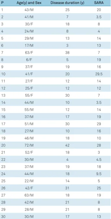
has proven to be valid and reliable in patients with ataxia. The patients’ ages ranged from six to 72 years, with a mean age of 38.7 years, and standard deviation of 17.7 years. Disease duration was between three and 42 years, with a mean duration of 14.7 years and standard deviation of 9.4 years (Table 1).
Table 1. Summary of patient demographics.
P Age(y) and Sex Disease duration (y) SARA
1 43/M 25 20
2 41/M 7 3.5
3 30/F 18 8
4 24/M 8 4
5 29/M 13 14
6 17/M 3 13
7 63/F 38 7
8 6/F 5 19
9 37/F 19 16
10 41/F 20 29.5
11 27/F 12 14
12 25/F 12 12
13 55/F 30 7
14 44/M 10 3.5
15 55/M 12 14
16 37/M 17 19
17 51/M 30 29
18 27/M 10 16
19 46/M 18 10
20 72/M 42 28
21 52/F 18 3
22 30/M 4 4.5
23 37/M 19 18
24 44/M 18 9.5
25 22/M 14 5
26 42/F 31 25
27 63/M 18 19
28 42/M 21 8
29 28/M 21 8
30 30/M 17 13
Patients excluded from the research had otologic dis
-orders or other abnormalities that prevented them from undergoing the examinations. Seven (7) patients were excluded from the study (three of them died and four
refused to participate in the research). Audiological assess
-ments were carried out in a single session, which lasted an average of 50 minutes.
The patients were submitted to the following procedures:
Anamnesis: Patients answered a questionnaire with empha
-sis on otoneurological signs and symptoms.
Otorhinolaryngology examination: Performed with the objec
-tive of excluding any other disorders that could interfere with the examination.
Pure tone audiometry: Patients were submitted to pure tone air conduction threshold audiometry at frequencies from
250Hz to 8kHz; pure tone bone conduction threshold audi
-ometry at frequencies from 500Hz to 4kHz, speech recogni
-tion threshold and speech discrimina-tion tests. For those tests, the Madsen Itera audiometer – GN Optometrics, with TDH-39 headphones from GN-ReSound, was used and hearing level (HL) thresholds were measured indB. The
equipment was calibrated according to ISO 8253. The lev
-els and types of hearing loss were analyzed according to
Davis et Silverman16.
Brainstem auditory evoked potential: This test used two
channels with a click stimulus at 90dBHL, alternate polar
-ity with a presentation frequency of 21.1 cycles per second, window of 15ms, 30 Hz to 3kHz filter and at least 2,000 stimuli, and two rounds of repetition. Kendall Med trace
2000 electrodes were placed on the right and left mas
-toids, as well as on the Fz position (10–20 system), and ground electrodes on the forehead. Clicks were presented via 3A insert earphones. Wave latencies I, III and V and
interpeak intervals I-III, III-V, I-V were analyzed accord
-ing to Hall’s criteria17. These waves represent structures
of the auditory pathway, having the following generator sites: wave I – distal portion of the cochlear nerve; wave
II – proximal portion of the cochlear nerve; wave III – gen
-erated in the cochlear nucleus; wave IV – superior olivary complex; wave V – lateral lemniscus; wave VI – inferior colliculus; and wave VII: medial geniculate body. In this study, latency values of waves I, III and V were used, as well as their interpeak latencies I-III, III-V and I-V. The choice ofthose three waves was because they feature greater amplitude and stability. In patients with hearing loss, it was necessary to increase the intensity of the click stimulus to 100 dBHL. The equipment used was Bio-logic’s Evoked Potential System.
Acoustic immittance evaluation: This procedure was done to assess the integrity of the middle ear according to Jerger’s
criteria18. The immittance equipment used was the Madsen
OTOflex tympanometer, with TDH 39P earphones by GN-ReSound.
Statistical analysis
Pearson’s correlation coefficient was used to correlate
age, disease duration and the SARA scale; the two-propor
-tion z-test was used to determinedifferences in propor-tion for symptoms analysis; and Fisher’s exact test was carried out to compare the results of the audiological examinations,
the BAEP and the measure of acoustic immittance (analyz
-ing normal and abnormal results). Statistica 13.1 software was used, and the null hypothesis was rejected at 0.05 or 5%.
RESULTS
By correlating age, disease durationand the result of the
SARA scale, Pearson’s correlation coefficient was signifi
-cant in the correlation between disease duration and age (p = *0.0000), and in the correlation betweendisease duration and the SARA scale (p = *0.0022) (Table 2).
The most reported complaints in the anamnesis were: uncoordinated movement (66.7%), gait imbalance (56.7%), and dizziness (50%). Hearing loss occurred in 10% of the patients (Table 3).
Table 2. Correlation between age, disease duration and SARA.
Correlation Pearson’s coefficient
correlation (r) p
Age and disease
duration 0.7171 *0.0000
Age and SARA 0.1911 0.0312
Disease duration
and SARA 0.4169 *0.0022
SARA: scale for the assessment and rating of ataxia. *Significant comparison; p-values for Pearson’s coefficient correlation shown.
Table 3.Symptoms in 30 patients withFriedreich’s ataxia. Symptoms N. patients Frequency (%)
Incoordination of movement 20 66.7
Gaitimbalance 17 56.7
Dizziness 15 50.0
Dysarthria 14 46.7
Headache 10 33.4
Dysphagia 9 30.0
Diplopia 9 30.0
Falling 8 26.7
Tremor 8 26.7
Depression 8 26.7
Fatigue 7 23.4
Anxiety 7 23.4
Pain, difficulty in neck movement 6 20.0
Tingling in extremities 4 13.4
Insomnia 3 10.0
Hearingloss 3 10.0
Olfactory alteration 2 6.7
Gustatory alteration 2 6.7
In the two-proportion z-test, in orderto determinediffer
-ence in proportions, there were significant differ-ences for the symptoms of uncoordinated movement (p < *0.0010), gait imbalance (p < *0.0370) and dizziness (p < *0.0330) in relation to the other reported symptoms.
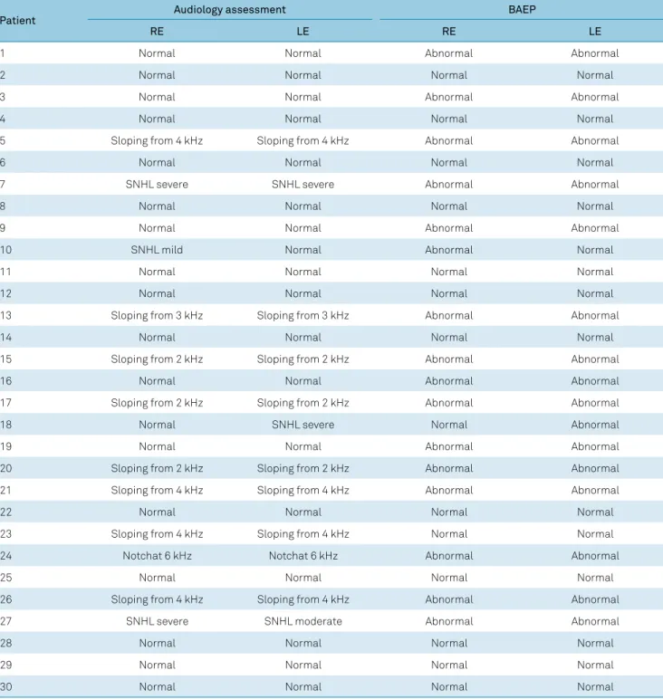
In the audiological assessment, 13/30 patients (43.3%) hadhearing alterations:3.3% in the right ear only, 3.3% in the left ear only, and 36.7% bilaterally (Table 4).
Results for the speech recognition threshold and speech recognition percentage index were comparable with the pure tone thresholds.
In the BAEP assessment, 17/30 patients (56.6%) featured alterations: 3.3% in the right ear only, 3.3% in the left ear only, and 50% bilaterally, as shown in Table 4.
In the acoustic immittance evaluation,14/30 patients (46.6%) showed alterations in the acoustic reflex, all of which were bilateral (Table 5).
The results of the audiological assessments, BAEP and acoustic immittance evaluation, regarding the proportions for normal and alterations, are shown in Table 6.
Fisher’sexact test showed a significant difference between the audiological evaluation and BAEP (p = *0.0007), between
Table 4. Summary of each patient’s audiological and BAEP results.
Patient Audiology assessment BAEP
RE LE RE LE
1 Normal Normal Abnormal Abnormal
2 Normal Normal Normal Normal
3 Normal Normal Abnormal Abnormal
4 Normal Normal Normal Normal
5 Sloping from 4 kHz Sloping from 4 kHz Abnormal Abnormal
6 Normal Normal Normal Normal
7 SNHL severe SNHL severe Abnormal Abnormal
8 Normal Normal Normal Normal
9 Normal Normal Abnormal Abnormal
10 SNHL mild Normal Abnormal Normal
11 Normal Normal Normal Normal
12 Normal Normal Normal Normal
13 Sloping from 3 kHz Sloping from 3 kHz Abnormal Abnormal
14 Normal Normal Normal Normal
15 Sloping from 2 kHz Sloping from 2 kHz Abnormal Abnormal
16 Normal Normal Abnormal Abnormal
17 Sloping from 2 kHz Sloping from 2 kHz Abnormal Abnormal
18 Normal SNHL severe Normal Abnormal
19 Normal Normal Abnormal Abnormal
20 Sloping from 2 kHz Sloping from 2 kHz Abnormal Abnormal
21 Sloping from 4 kHz Sloping from 4 kHz Abnormal Abnormal
22 Normal Normal Normal Normal
23 Sloping from 4 kHz Sloping from 4 kHz Normal Normal
24 Notchat 6 kHz Notchat 6 kHz Abnormal Abnormal
25 Normal Normal Normal Normal
26 Sloping from 4 kHz Sloping from 4 kHz Abnormal Abnormal
27 SNHL severe SNHL moderate Abnormal Abnormal
28 Normal Normal Normal Normal
29 Normal Normal Normal Normal
30 Normal Normal Normal Normal
audiological evaluation and acoustic immittance evaluation (p = *0.0000), and between BAEP and acoustic immittance evaluation (p = *0.0002).
DISCUSSION
In the analysis of the disease duration and age variables,
it was observed that the older the patient, the longer the dis
-ease duration, as most patients suffered from the dis-ease since childhood/adolescence; and the longer the disease duration, the worse the score result on the SARA scale.
The anamnesis disclosed a prevalence of gait disorders, which were expected in spinocerebellar ataxias. Speech disorders, dizziness, dysphagia, dysphonia, and hearing loss are also described in several studies1,2,3. According to
Payne19, patients develop primary neurodegeneration in the
Table 5. Summary of each patient’s acoustic immittance results.
Patient
Acoustic immittance
Right ear Left ear
Tympanometry curve Acousticreflex Tympanometry curve Acousticreflex
1 Type A Absent at 3–4 kHz Type A Absent at 3–4 kHz
2 Type A Present Type A Present
3 Type A Absent at 4 kHz Type A Absent at 4 kHz
4 Type A Present Type A Present
5 Type A Absent at 4 kHz Type A Absent at 4 kHz
6 Type A Present Type A Present
7 Type A Absent Type A Absent
8 Type A Present Type A Present
9 Type A Absent at 4 kHz Type A Absent at 4 kHz
10 Type A Present Type A Present
11 Type A Present Type A Present
12 Type A Present Type A Present
13 Type A Absent at 3–4 kHz Type A Absent at 3–4 kHz
14 Type A Present Type A Present
15 Type A Absent at 3–4 kHz Type A Absent at 3–4 kHz
16 Type A Present Type A Present
17 Type A Absent at 2–4 kHz Type A Absent at 2–4 kHz
18 Type A Absent at 3–4 kHz Type As Absent at 3–4 kHz
19 Type A Present Type A Present
20 Type A Absent at 3–4 kHz Type A Absent at 3–4 kHz
21 Type A Absent at 4 kHz Type A Absent at 4 kHz
22 Type A Present Type A Present
23 Type A Absent at 4 kHz Type A Absent at 4 kHz
24 Type A Present Type A Present
25 Type A Present Type A Present
26 Type A Absent at 4 kHz Type A Absent at 4 kHz
27 Type A Absent Type A Absent
28 Type A Present Type A Present
29 Type A Present Type A Present
30 Type A Present Type A Present
Statistical findings are reported in the results text.
Table 6. Distributions of audiological, BAEP and acoustic immittance test results in Friedreich’s ataxia.
Variables Normal (n) Abnormal (n) Total (n) P
Audiology findings BAEP
Normal 12 5 17
*0.0007
Abnormal 1 12 13
Total 13 17 30
Acoustic immittance
Normal 14 3 17
*0.0000
Abnormal - 13 13
Total 14 16 30
BAEP findings, N Acoustic immittance
Normal 12 1 13
*0.0002
Abnormal 4 13 17
Total 16 14 30
dorsal root ganglia, which explains the loss of propriocep
-tion and coordina-tion.
Abnormalities in the audiological evaluation were present
in 43.3% of the ataxic patients. Knezevic and Stewart-Wynne20
assessed 18 patients with spinocerebellar ataxia and observed normal hearing in all of them; however, five of the seven (71%) Friedreich’s ataxia patients had abnormal BAEP results, where
only wave I was identified, and 71% of the olivopontocerebel
-lar atrophy patients had abnormal BAEP results. The results
showed severe abnormalities in the brainstem auditory path
-ways in patients with spinocerebellar ataxia.
In the current study, 56.6% of the patients had altera
-tions in the BAEP test, with the occurrence of latency
increases in waves I, III and V, and in the interpeak inter
-vals I-III, I-V and III-V for 12/17 patients (40%); wave V was absent in 4/17 (13.3%) patients, and all waves were absent in one (3.3%) patient.
Yokoyamaet al.21assessed 30 patients with spinocerebel
-lar degeneration and verified increases in latency and inter
-peak intervals of waves I-III and I-V; wave V was absent in 30% of the patients, and 82.5% showed altered wave ranges. Rance et al.22 assessed two patients with audiological fol
-low-ups for a period of three years, BAEP and central audi
-tory processing examinations being performed periodically. The global severity of the disease in the initial assessment
was equivalent in both cases. The authors showed axonopa
-thy in the cochlear nerve in both cases. They observed hear
-ing loss (significant neural reduction) along the course of
the disease. Zeng et al.23 showed that diseases that affect the
integrity of the auditory nerve drastically impair hearing
perception. In another study, Rance et al.24 observed inter
-rupted neural activity in the BAEP in nine out of 14 patients with Friedreich’s ataxia. The same authors reported that an
impaired auditory pathway is a relatively common conse
-quence in this disease. Satya-Murti et al.25 showed a nor
-mal audiological evaluation, and alteration in all waves of the BAEP in four patients diagnosed with Friedreich’s
ataxia. They reported that these alterations could be attrib
-uted to the degeneration of the spiral ganglion neurons. A study by López-Diaz-de León et al.26 assessed two ado
-lescents with Friedreich’s ataxia, who showed abnormali
-ties in the BAEP with normal otoacoustic emissions, point
-ing to auditory neuropathy. Thus, auditory thresholds were normal in one patient, and the other was diagnosed with
a mild sensorineural hearing loss. Pelosiet al.27 assessed
15 patients with Friedreich’s ataxia and observed the pres
-ence of wave I, and abs-ence of wave V in all patients, irre
-spective of the symptom durationor the clinical severity of
the disease, raising the hypothesis that these alterations were related to primary axonal degeneration. Santarelli et
al.28 stated that the sensory neural hearing loss is one of the
clinical features of Friedreich’s ataxia, and most patients present auditory neuropathy. According to the authors, the neuropathy was explained by the presence of faulty nerve connections due to the loss of inner hair cells, causing the
interruption of the acoustic signal. Spinelli et al.29 reported
that auditory neuropathy was a dysfunction of the auditory
nerve, which caused a disconnection in the nerve conduc
-tion, probably related to an alteration in the myelination of those fibers, probably located in the inner hair cells and in their synapses. Auditory neuropathy has been observed in Friedreich’s ataxia, Guillan-Barré and Charcot-Marie-Tooth type II diseases.
The measurement of the acoustic immittance was altered in 46.6% of the Friedreich’s ataxia patients in our study, and
there was no reference to this finding in the literature to com
-pare with our results. It is known thatfibers leave the ante
-rior cochlear nuclei and go, via the trapezoidal body, to the
nuclei of the contralateral facial nerve, and on to the supe
-rior olivary complex, which in turn, make synapses with the nucleus of the facial nerve. The ipsilateral fibers from the anterior cochlear nuclei establish these connections and,
from the nuclei of the facial nerves, axons innervate the sta
-pes muscles. Thus, in neurodegenerative diseases, the ante
-rior cochlear muscles are impaired, with possible interfer
-ences in the mechanism of the acoustic reflex30.
In the current study, there was a higher prevalence of
alterations in the BAEP test, which showed significant altera
-tions in the integrity of the brainstem auditory pathway. This
finding corroborates the literature, where Yokoyama et al.21
reported alterations in several structures of the central audi
-tory pathway, showing a higher sensitivity of the BAEP test in
detecting alterations of the acoustic impulse along the cen
-tral auditory pathway.
In conclusion, the most-reported change in the audiologi
-cal assessment was the prevalence of the descending audio
-metric configuration at the frequency of 4kHz, and bilateral absence of the acoustic reflex at the frequency of 4kHz.
In the electrophysiological evaluation, 40% of the patients had alterations, mostly showing an increase of the latencies in waves I, III and V, and in the interval of interpeaks I-III, I-V and III-V. In 13.3% of the patients, wave V was absent and, in one patient (3.3%), all waves were absent.
Therefore, it is importantto study the central auditory sys
-tem using an electrophysiological assessment to detect abnor
-malities in the brainstem auditory pathway in this population.
References
1. Fogel BL, Perlman S. Clinical features and molecular genetics of autosomal recessive cerebellar ataxias. Lancet Neurol. 2007 Mar;6(3):245-57. https://doi.org/10.1016/S1474-4422(07)70054-6
3. Ell J, Prasher D, Rudge P. Neuro-otological abnormalities in Friedreich’s ataxia. J Neurol Neurosurg Psychiatry. 1984 Jan;47(1):26-32. https://doi.org/10.1136/jnnp.47.1.26
4. Albano LM, Zatz M, Kim CA, Bertola D, Sugayama SM, Marques-Dias MJ et al. Friedreich’s ataxia: clinical and molecular study of 25 Brazilian cases. Rev Hosp Clin Fac Med Sao Paulo. 2001 Sep-Oct;56(5):143-8. https://doi.org/10.1590/S0041-87812001000500003
5. Alper G, Narayanan V. Friedreich’s ataxia. Pediatr Neurol. 2003 May;28(5):335-41. https://doi.org/10.1016/S0887-8994(03)00004-3
6. Pandolfo M. Friedreich ataxia. Arch Neurol. 2008
Oct;65(10):1296-303. https://doi.org/10.1001/archneur.65.10.1296
7. Melo M, Fagulha A, Barros L, Guimarães J, Carrilho F, Carvalheiro M. [Friedreich ataxia and diabetes mellitus: family study]. Acta Med Port. 2005 Nov-Dec;18:479-83. Portuguese.
8. Baloh RW, Konrad HR, Honrubia V. Vestibulo-ocular function in patients with cerebellar atrophy. Neurology. 1975 Feb;25(2):160-8. https://doi.org/10.1212/WNL.25.2.160
9. Pereira LD, Schochat E. Processamento auditivo central: manual de avaliação. São Paulo: Lovise; 1997.
10. Aquino AMCM, organizer. Processamento auditivo: eletrofisiologia & psicoacústica. São Paulo: Lovise; 2002.
11. Biacabe B, Chevallier JM, Avan P, Bonfils P. Functional anatomy of auditory brainstem nuclei: application to the anatomical basis of brainstem auditory evoked potentials. Auris Nasus Larynx. 2001 Jan;28(1):85-94. https://doi.org/10.1016/S0385-8146(00)00080-8
12. Dueñas AM, Goold R, Giunti P. Molecular pathogenesis of spinocerebellar ataxias. Brain. 2006 Jun;129(Pt 6):1357-70. https://doi.org/10.1093/brain/awl081
13. Pearson CE, Nichol Edamura K, Cleary JD. Repeat instability: mechanisms of dynamic mutations. Nat Rev Genet. 2005 Oct;6(10):729-42. https://doi.org/10.1038/nrg1689
14. Schmitz-Hübsch T, Montcel ST, Baliko L, Berciano J, Boesch S, Depondt C et al. Scale for the assessment and rating of ataxia: development of a new clinical scale. Neurology. 2006 Jun;66(11):1717-20. https://doi.org/10.1212/01.wnl.0000219042.60538.92
15. Braga-Neto P, Godeiro-Júnior C, Dutra LA, Pedroso JL, Barsottini OG. Translation and validation into Brazilian version of the scale of the assessment and rating of ataxia (SARA). ArqNeuropsiquiatr. 2010 Apr;68(2):228-30. https://doi.org/10.1590/S0004-282X2010000200014
16. Davis H, Silverman RS. Hearing and deafness. 3rd ed. New York: Holt, Rinehart & Wilson; 1970.
17. Hall J. Handbook of auditory evoked responses. Boston: Allyn & Bacon; 1992.
18. Jerger J. Clinical experience with impedance audiometry. Arch Otolaryngol. 1970 Oct;92(4):311-24. https://doi.org/10.1001/archotol.1970.04310040005002
19. Payne RM. The heart in Friedereich’s ataxia: basic findings and clinical implications. Prog Pediatr Cardiol. 2011 May;31(2):103-9. https://doi.org/10.1016/j.ppedcard.2011.02.007
20. Knezevic W, Stewart-Wynne EG. Brainstem auditory evoked responses in hereditary spinocerebellar ataxias. Clin Exp Neurol. 1985;21:149-55.
21. Yokoyama J, Aoyagi M, Suzuki T, Kiren T, Koike Y. Three frequency component wave forms of auditory evoked brainstem response in spinocerebellar degeneration. Acta Otolaryngol Suppl. 1994;511 sup511:52-5. https://doi.org/10.3109/00016489409128301
22. Rance G, Corben LA, Delatycki MB. Auditory pathway changes mirror overall disease progress in individuals with Friedreich ataxia. J Neurol. 2012 Dec;259(12):2746-8. https://doi.org/10.1007/s00415-012-6679-z
23. Zeng FG, Kong YY, Michalewski HJ, Starr A. Perceptual consequences of disrupted auditory nerve activity. J Neurophysiol. 2005
Jun;93(6):3050-63. https://doi.org/10.1152/jn.00985.2004
24. Rance G, Corben L, Barker E, Carew P, Chisari D, Rogers M et al. Auditory perception in individuals with Friedreich’s ataxia. Audiol Neurootol. 2010;15(4):229-40. https://doi.org/10.1159/000255341
25. Satya-Murti S, Cacace A, Hanson P. Auditory dysfunction in Friedreich ataxia: result of spiral ganglion degeneration. Neurology. 1980 Oct;30(10):1047-53. https://doi.org/10.1212/WNL.30.10.1047
26. López-Díaz-de-León E, Silva-Rojas A, Ysunza A, Amavisca R, Rivera R. Auditoryneuropathy in Friedreich ataxia. A report of two cases. Int J Pediatr Otorhinolaryngol. 2003 Jun;67(6):641-8. https://doi.org/10.1016/S0165-5876(03)00036-3 27.
27. Pelosi L, Fels A, Petrillo A, Senatore R, Russo G, Lönegren K et al. Friedreich’s ataxia: clinical involvement and evoked potentials. Acta Neurol Scand. 1984 Nov;70(5):360-8. https://doi.org/10.1111/j.1600-0404.1984.tb00837.x
28. Santarelli R, Cama E, Pegoraro E, Scimemi P. Abnormal cochlear potentials in Friedreich’s ataxia point to disordered synchrony of auditory nerve fiber activity. Neurodegener Dis. 2015;15(2):114-20. https://doi.org/10.1159/000375307
29. Spinelli M, Fávero-Brewel MI, Silva CM. [Auditory neuropathy: clinical, diagnostic and therapeutic aspects]. Ver Bras Otorrinolaringol. 2001;67(6):863-7. Portuguese. https://doi.org/10.1590/S0034-72992001000600017