Contents lists available atScienceDirect
Redox Biology
journal homepage:www.elsevier.com/locate/redox
CO-mediated cytoprotection is dependent on cell metabolism modulation
Cláudia Figueiredo-Pereira
a,1, Daniela Dias-Pedroso
a,b,1, Nuno L. Soares
a,b,
Helena L.A. Vieira
a,b,c,∗aCEDOC, Faculdade de Ciência Médicas/NOVA Medical School, Universidade Nova de Lisboa, 1169-056, Lisboa, Portugal bUCIBIO, Faculdade de Ciências e Tecnologia, Universidade Nova de Lisboa, Portugal
cInstituto de Biologia Experimental e Tecnológica (iBET), Apartado 12, 2781-901, Oeiras, Portugal
A R T I C L E I N F O Keywords: Carbon monoxide Metabolism Mitochondrial biogenesis Oxidative phosphorylation Glycolysis
Pentose phosphate pathway ROS signaling
A B S T R A C T
Carbon monoxide (CO) is a gasotransmitter endogenously produced by the activity of heme oxygenase, which is a stress-response enzyme. Endogenous CO or low concentrations of exogenous CO have been described to present several cytoprotective functions: anti-apoptosis, anti-inflammatory, vasomodulation, maintenance of home-ostasis, stimulation of preconditioning and modulation of cell differentiation. The present review revises and discuss how CO regulates cell metabolism and how it is involved in the distinct cytoprotective roles of CO. The first found metabolic effect of CO was its increase on cellular ATP production, and since then much data have been generated. Mitochondria are the most described and studied cellular targets of CO. Mitochondria exposure to this gasotransmitter leads several consequences: ROS generation, stimulation of mitochondrial biogenesis, increased oxidative phosphorylation or mild uncoupling effect. Likewise, CO negatively regulates glycolysis and improves pentose phosphate pathway. More recently, CO has also been disclosed as a regulating molecule for metabolic diseases, such as obesity and diabetes with promising results.
1. Introduction
Carbon monoxide (CO) was first described to be endogenously produced in 1949, when it was found in exhaled human breath [1]. Only 20 years later, the enzyme responsible for CO production was identified. Tenhunen and colleagues have discovered that heme de-gradation by microsomal heme oxygenase (HO) originates CO, iron (Fe2+) and biliverdin [2]. In fact, HO catalyzes the first and limiting rate step of heme oxidative degradation by opening Fe-protoporphyrin-IX ring into free Fe2+, CO and biliverdin, whose reaction consumes O2 and NADPH as electrons donor [3]. HO activity is cytoprotective for two main reasons: (i) it eliminates free heme, which is potentially very toxic, and (ii) its products present beneficial properties. First, biliverdin is rapidly converted by biliverdin reductase into bilirubin, which is a potent anti-oxidant [4]. Second, iron binds to iron regulatory proteins (IRP) increasing ferritin expression, which promotes cytoprotection [5]. Finally, CO emerges as a gasotransmitter with several beneficial bio-logical functions revised in Refs. [6–10], namely maintenance of tissue homeostasis, anti-inflammation, cytoprotection, cardioprotection,
promotion of preconditioning and tolerance state, regulation of cell differentiation/proliferation and modulation of cell metabolism. The present review tackles how endogenous CO or low concentrations of exogenous CO modulate cell metabolism in different cells and tissues, and discusses CO's potential impact on human health.
2. Carbon monoxide releasing molecules (CORM)
The development of carbon monoxide releasing molecules (CORM) emerges due to the potential application of this gasotransmitter as a therapeutic agent in Medicine. The use of CO gas presents intrinsic limitations because of its great affinity to hemoglobin, forming car-boxyhemoglobin and hampering optimal oxygen delivery. Therefore, the development of strategies to deliver CO in a safer mode is needed. CORMs are metal or transition metal carbonyl complexes that function as a prodrug to deliver CO under temporal and spatial controlled con-ditions [11,12]. In fact, CORM design must take into account water solubility, biocompatibility, toxicity, absorption, distribution, metabo-lism and excretion properties [11]. Herein four different types of
https://doi.org/10.1016/j.redox.2020.101470
Received 28 January 2020; Received in revised form 11 February 2020; Accepted 17 February 2020
Abbreviations: CNS, central nervous system; CO, carbon monoxide; CORM, Carbon monoxide-releasing molecule; DMSO, dimethyl sulfoxide; GSH, reduced
glu-tathione; GSSG, oxidized gluglu-tathione; HO-1, Heme-oxygenase 1; Nrf, nuclear respiratory factor; PGC-1α, peroxisome proliferator-activated receptor gamma co-activator-1α; PPP, Pentose phosphate pathway; ROS, reactive oxygen species; TCA, Tricarboxilic acid cycle; TFAM, mitochondrial transcription factor
∗Corresponding author. UCIBIO Departamento de Química Faculdade de Ciências e Tecnologia Universidade Nova de Lisboa, 2829-516, Caparica, Portugal.
E-mail address:[email protected](H.L.A. Vieira). 1equally contributed.
Redox Biology 32 (2020) 101470
Available online 19 February 2020
2213-2317/ © 2020 The Authors. Published by Elsevier B.V. This is an open access article under the CC BY-NC-ND license (http://creativecommons.org/licenses/BY-NC-ND/4.0/).
CORMs are discussed as modulators of cell metabolism. CORM-2 and CORM-3 contain a ruthenium center and release CO with t1/2of 1 min [13,14]. While CORM-2 is soluble in DMSO, CORM-3 is water soluble and only slightly increases carboxyhemoglobin levels when injected in vivo, which is a great advantage [15]. CORM-A1 has boron in its center and releases CO in a pH- and temperature-dependent manner with t1/2 of 21 min, being a slow releaser [16]. Of note, CORM biological effect might be also dependent on CO release kinetics. Finally, CORM-401 presents a manganese center, is water soluble and provides at least 3 mol of CO per mole of compound [17]. Moreover, CORM-401 is re-latively stable in PBS buffer, and 1 mM of CORM-401 releases 0.33 mol equivalent of CO gas in 4 h [17]. In biological systems, CORM-3 and CORM-A1 are the most studied with well-established functions of va-sodilation, anti-inflammation and cytoprotection.
3. ATP assessment following CO treatment
The first clues about the CO's potential modulation of cell metabo-lism emerged in 2004 when Lavitrano and colleagues have found an improved cardiac metabolic status in CO-treated pigs before ischemia and reperfusion injury [18]. In fact, pre-treatment with CO gas (250 ppm) increased heart levels of ATP and phosphocreatine, which is a high-energy phosphate cellular reserve. Likewise, in hepatocytes, endogenous CO derived from heme-oxygenase activity or exogenous CO exposure increases ATP production that, in turn, activates p38 MAPK signaling [19]. In vivo, CO gas exposure also increased ATP levels in liver and in in vitro primary cultures of hepatocytes. These higher concentrations of ATP are associated with higher viability of hepato-cytes and mice in response to TNF-α treatment and fulminant hepatitis, respectively [20]. The CO improvement of cellular energy metabolism is dependent on soluble guanylyl cyclase [20]. Nevertheless, in both models (heart and liver), CO-induced higher ATP levels can also be a consequence of increased cell viability rather than an improvement of cellular metabolism. Only later, more accurate studies properly re-vealed the role of CO in cell metabolism regulation, which is discussed below.
4. CO acts via mitochondrial ROS signaling
Several studies have demonstrated that many CO's biological ben-eficial effects are dependent on mitochondrial ROS generation, re-viewed in Refs. [10,21,22]. In fact, CO-induced low levels of mi-tochondrial ROS act as signaling molecules, as a preconditioning-like effect, which in turn, promote different biological responses: cytopro-tection, anti-inflammatory, modulation of cell metabolism or cellular differentiation. In this section, the molecular mechanisms of mi-tochondrial ROS generation are discussed. It is worth of note that for ROS to signal and to exert beneficial properties, they must be at low concentrations.
Despite all the described biological functions of CO, its pathways and in particular, its molecular targets in biological systems are still a matter of great debate. CO is a quite chemically inert molecule and it binds to transition metals present in proteins [23,24]. In biological
systems, the typical target of CO are heme-containing proteins, namely soluble guanylate cyclase, cytochromes, hemoglobin and myoglobin. Notably, CO can only bind to reduced Fe2+, limiting the potential target proteins, in contrast to NO that donates electrons to Fe3+and accepts electrons from Fe2+[24].
High levels of CO are toxic and lead to tissue hypoxia at systemic level and to mitochondrial damage at cellular level. In fact, cytotoxic effects of exogenous CO involve cytochrome c oxidase inhibition and defects on mitochondrial metabolism and energy production. This has been demonstrated in isolated mitochondria from human muscle by the direct analysis of the activity of respiratory chain complexes [25]. Likewise in HEK293 cells, it was also demonstrated that HO-1 over-expression and endogenous CO production also moderately decreased cellular respiration along with partial inhibition of cytochrome c oxi-dase [26]. In fact, CO binds to cytochrome c oxidase and slows down the rate of electron transport, leading to electrons accumulation, in particular at complex III. Thus, the lifetime of the ubisemiquinone state of coenzyme Q is prolonged, increasing the propensity to reduce O2into superoxide (O2−), which is enzymatically converted to other ROS, in particular hydrogen peroxide (H2O2) that can then act as signaling molecules [21,27]. Likewise, CO-mediated inhibition of cytochrome c oxidase can be partial or transitory, which enables low levels of ROS production without further damaging mitochondria [28] (Fig. 1).
Zuckerbraun and colleagues have demonstrated that CO generates mitochondrial ROS and promotes anti-inflammatory effects in macro-phages [27]. Low concentrations of exogenous CO partially inhibited cytochrome c activity, generating mitochondrial ROS, which in turn limited LPS-induced inflammation by promoting p38 MAPK phos-phorylation and decreasing TNF-α levels. The presence of antimycin A or in respiration deficiency ρ° cells CO's anti-inflammatory effect is re-verted, indicating its dependency on mitochondrial ROS signaling [27]. Likewise, CO is anti-inflammatory in macrophages via PPARβγ SU-MOlation and p38 activation, whose upstream effectors are mitochon-drial ROS. CO-induced generation of mitochonmitochon-drial ROS depends on a mild uncoupling effect via targeting the uncoupling protein 2 (UCP2), since whenever Ucp2 gene is knocked out, CO anti-inflammatory ca-pacity is lost along with a decrease on mitochondrial ROS production [29]. In airway smooth muscle cells (ASMC), CO modulates cell pro-liferation by generating mitochondrial ROS [30]. Finally, in primary culture of cerebellar neurons, CO gas prevented apoptosis mediated by excitotoxicity and by oxidative stress in a ROS dependent manner [31]. Co-treatment with the anti-oxidant butyl-hydroxytoluene and CO re-verted the anti-apoptotic role of this gasotransmitter [31]. In summary, low concentrations of exogenous CO or endogenous CO target mi-tochondria, generating low amounts of ROS, which act as signaling molecules (Fig. 1). Nevertheless, one cannot disregard that high CO levels inhibit mitochondrial respiratory chain and generate toxic amounts of ROS and oxidative stress. Subcellular localization of CO can also regulate ROS production, for instance Taillé and colleagues have demonstrated that CORM-2 treatment inhibited the activity of NAD(P) H oxidase cytochrome b558, which decreases plasmatic membrane ROS production, while there is an increase on mitochondrial ROS generation by targeting cytochrome c oxidase [30]. Therefore, exogenous CO
Fig. 1. CO promotes mitochondrial ROS generation. CO can bind and inhibit
cyto-chrome c oxidase, which accumulates elec-trons at the mitochondrial electron trans-port chain. Accumulation of electrons enhances the probability of O2 reduction into anion superoxide, which is quickly converted into other ROS, namely hydrogen peroxide. Because low amounts of CO par-tially and transitorily binds to cytochrome c oxidase low levels of ROS are generated that act as cell signaling molecules.
(delivered by CORMs) and endogenously produced CO may present different subcellular localizations, with potential different effects on ROS generation and signaling.
5. CO stimulates mitochondrial biogenesis
The consequence of CO targeting mitochondria is not limited to ROS production, since this gasotransmitter also activates mitochondrial biogenesis (Fig. 2). CO has been demonstrated to promote cytoprotec-tion via activacytoprotec-tion of mitochondrial biogenesis in several different cel-lular models with distinct damage triggering factors. In 2007, it was found by Suliman and colleagues that CO stimulates mitochondrial biogenesis in cardiomyocytes using in vivo and cell culture models [32,33]. Transient elevations of intracellular CO levels increased number of mitochondrial DNA and the protein content of complexes I–V. Likewise, CO upregulated the expression of nuclear respiratory factor-1 and -2 (Nrf-1, Nrf-2), peroxisome proliferator-activated re-ceptor gamma co-activator-1α (PGC-1α) and mitochondrial transcrip-tion factor (TFAM), which are key transcriptranscrip-tion factors controlling mitochondrial biogenesis. Moreover, the CO-induced mitochondrial biogenesis was dependent on hydrogen peroxide generation and in-dependent on nitric oxide synthase activation [32]. Still, CO stimula-tion of mitochondrial biogenesis was cardioprotective by limiting doxorubicin-induced apoptosis [33]. Likewise, in a mice sepsis model induced by peritonitis, CORM-3 treatment improved cardiac mi-tochondrial function via stimulation of mimi-tochondrial biogenesis in a PGC-1α and ROS dependent manner [34]. Interestingly, HO-1 over-expression and CO production activated the mitochondrial biogenesis Nrf2 transcription factor, along with upregulation of the anti-in-flammatory proteins IL-10 and the IL-1 receptor antagonist (IL-1Ra) [35]. These data couples mitochondrial biogenesis with the CO anti-inflammatory role in LPS-induced inflammation cellular model and in mice challenged with Escherichia coli sepsis [35]. Furthermore, activa-tion of mitochondrial biogenesis by HO-1 rescued mice from lethal Staphylococcus aureus sepsis, via redox-regulated NF-E2–related factor-2 signaling [36]. In another model, HO-1 activity, along with CO pro-duction, protects macrophages against endoplasmic reticulum stress via mitochondrial biogenesis and in a protein kinase RNA-like endoplasmic reticulum kinase (PERK)-dependent manner [37]. The naturally
occurring flavonoid Quercetin presents several beneficial biological effects including promotion of mitochondrial biogenesis in a HO-1/CO axis dependent manner in HepG2 cells [38]. Finally, CO gas protected astrocytes against apoptosis by increasing mitochondrial population in a Bcl-2 dependent manner [28]. Likewise, Kim and colleagues demon-strated that CORM-2 activation of L-type Ca2+channel mediates PGC-1α/ERRα axis, which leads to mitochondrial biogenesis and main-tenance of astrocyte function in astrocytes [39]. The same authors have recently demonstrated that CORM-2 increases HIF-1α stabilization, which is necessary for ERRα-mediated mitochondrial biogenesis [40]. This data can be controversial since HIF-1α stabilization is usually as-sociated with an increase of glucose uptake and glycolytic metabolism [41]. Semenza and colleagues have demonstrated in renal carcinoma cells that HIF-1 negatively regulates mitochondrial biogenesis by pre-venting C-MYC activity, which in turn reduces PGC-1β expression and mitochondrial biogenesis [42]. This controversy can be partially ex-plained by the different PGC-1 family members: PGC-1α and PGC-1β [41]. Moreover, the same researchers also shown that under hypoxia, HIF-1 modulates the expression of cytochrome c oxidase subunit 4 (COX4), by decreasing COX4-1 and increasing COX4-2, which promotes COX activity, oxygen consumption and ATP production [43]. Thus the same group demonstrated two different effects of HIF on glycolysis and mitochondrial metabolism. Of note, Semenza group work has been developed in cancer cell lines, while Kim's research has been done in primary cultures of astrocytes, which may also influence the potential different responses to HIF stabilization. Taken all together, in different cell types and models, CO-induced mitochondrial biogenesis is essential for its cytoprotective and anti-inflammatory roles (Fig. 2). Data are summarized inTable 1.
6. CO and mitochondrial quality control
CO-mediated mitochondrial function also implicates modulation of mitochondrial quality control. Mitochondrial quality control can be defined as the balance between mitochondrial biogenesis and elimina-tion of dysfuncelimina-tional mitochondria via autophagy (mitophagy) [44,45]. In hepatocytes, CO activates protein kinase R (PKR)-like en-doplasmic reticulum (ER) kinase (PERK), which in turn promotes nu-clear translocation of transcription factor EB (TFEB) [46]. TFEB
Fig. 2. CO stimulates mitochondrial biogenesis.
CO exposure upregulates the expression of peroxi-some proliferator-activated receptor gamma co-acti-vator-1α (PGC-1α) and the nuclear respiratory factor-1 and 2 (Nrf-1/2), which regulate the expres-sion of nuclear encoding mitochondrial proteins. PGC-1α interaction with Nrf-1/2 also coordinates the upregulation of the mitochondrial transcription factor (TFAM) that modulates transcription of mi-tochondrial DNA. These factors are all involved in CO-induced mitochondrial biogenesis.
Table 1
CO and mitochondrial function.
Biological function: mitochondrial biogenesis
Experimental Model Assessment of mitochondrial biogenesis Biological outcome of CO Ref.
Cardiomyocytes (in vivo and cell culture) Increased content of mtDNA, mitochondrial proteins (complexes I-IV)
Upregulation of Nrf1, Nrf2, TFAM and PGC-1α
Mitochondrial biogenesis in a ROS dependent manner [32] In vivo mouse model for doxorubicin-induced
heart failure Increased content of mtDNA, mitochondrial proteinsUpregulation of TFAM Cardioprotection against doxorubicin-induced apoptosis viamitochondrial biogenesis [33] In mice peritonitis-induced sepsis model Increased content of mtDNA
Upregulation of PGC-1α Cardioprotection in sepsis context via mitochondrialbiogenesis Reduces mortality
[34] In mice Escherichia coli-induced sepsis Upregulation of Nrf2 Anti-inflammatory effect
Upregulation of IL-10 and IL-1Ra [35]
In mice Staphylococcus aureus-induced sepsis Increased content of mtDNA, mitochondrial protein (citrate synthase)
Upregulation of Nrf1, Nrf2, TFAM and PGC-1α
Reduction of mortality
Anti-inflammatory effect via upregulation of NF-E2–related factor-2
[36]
Macrophages Increased content of mtDNA
Upregulation of Nrf1, TFAM and PGC-1α Cytoprotection against thapsigargin-induced endoplasmicreticulum stress [37] Hepatocytes (HepG2 cell line) Increased content of mtDNA, mitochondrial proteins
(complex IV)
Upregulation of Nrf1, TFAM and PGC-1α
Mitochondrial biogenesis promoted by Quercetin via CO
production [38]
Primary culture of mouse astrocytes Increased content of mtDNA Cytoprotection against oxidative stress-induced apoptosis [28] Primary culture of mouse astrocytes Increased content of mitochondrial proteins
(cytochrome c and cytochrome c oxidase) Upregulation of PGC-1α
Cytoprotection/maintenance of astrocytic function [39] Mouse ischemic brain injury model Upregulation of PGC-1α Improvement of astrocytic mitochondrial function [40]
Biological function: mitochondrial metabolism
Experimental Model Effects and Mechanisms of CO Biological outcome of CO Ref.
Diabetic kidney Upregulation of carnitine, citrate, deoxynucleotide, dicarboxylate and ADP/ATP carriers
Increase of cytochrome c oxidase activity
Improvement of mitochondrial metabolism
Reversion of diabetes-induced lower mitochondrial transport carriers
[49] Isolated mouse liver mitochondria Inhibition of mitochondria membrane depolarization
induced by oxidative stress
Increased activity of cytochrome c oxidase
Anti-apoptotic effect [50]
Primary culture of astrocytes Isolated mouse cortex non-synaptic mitochondria
Increased specific activity of cytochrome c oxidase and O2consumption
Increased content of mtDNA Bcl-2 dependent mechanism
Oxidative metabolism improvement
Increased ATP production [28]
Primary culture of astrocytes Isolated rat cortex non-synaptic mitochondria
Inhibition of mitochondria membrane depolarization induced by oxidative stress
ROS dependent mechanism Protein glutathionylation signaling
Anti-apoptotic effect [51]
Primary culture of astrocytes and neurons Increased ATP production
Purinergic signaling Astrocytic-neuronal communication for preventing neuronalcell death induced by oxidative stress [53] Microglia challenged with LPS Increased ATP production
Decreased glycolytic metabolism Maintenance of ATP production
Anti-neuroinflammatory effect [54]
Human teratocarcinoma NT2 cell line Enhanced mitochondrial population Higher expression of pyruvate and lactate dehydrogenase
Higher levels of tricarboxylic acid cycle metabolites
Improvement of neuronal differentiation [58]
Embryonic stem cell differentiation into
cardiomyocytes Improvement of mitochondrial functionIncreased mtDNA Differentiation and maturation of cardiomyocytes [59] Human prostate cancer cells Shifts from glycolytic into oxidative metabolism Anti-Warburg effect
Sensitizes cancer cells to respond to chemotherapy by cell metabolism regulation
Promotes apoptosis
[60]
Biological function: mitochondrial uncoupling & CO
Experimental Model Effects and Mechanisms of CO Biological outcome of CO Ref.
Isolated rat renal mitochondria (in vivo
treatment) Decreased levels of state 3 respirationDecreased respiratory control index Uncoupling mitochondrial respiration [65] Isolated rat heart mitochondria Increased levels of state 2 respiration
Modulation by adenine nucleotide translocator (ANT) and uncoupling proteins (UCP)
Uncoupling mitochondrial respiration [66] Isolated rat heart mitochondria Decreased hydrogen peroxide production at complex I
level
Dependent on phosphate carrier and dicarboxylate carrier
Uncoupling mitochondrial respiration [67]
Human endothelial EA.hy926 cells Increased oxygen consumption Increased proton leak Increased state 2 respiration
Uncoupling mitochondrial respiration [68] (continued on next page)
translocation is involved in lysosomal and mitochondrial biogenesis. In TFEB deficient cells, CO failed to promote Parkin translocation into mitochondria and to induce mitophagy. Likewise, knockdown expres-sion of TFEB also decrease expresexpres-sion of lysosomal genes (Lamp1, ca-thepsin B and TPP1), along with a decrease on mitochondrial DNA, mitochondrial biogenesis markers (PGC-1α, Nrf1 and TFAM) and mi-tochondrial proteins, such as COX II, COX IV and cytochrome c [46]. In summary, TFEB emerges as a key regulator of CO-dependent cytopro-tection in hepatocytes, being associated with modulation of mitophagy and mitochondrial biogenesis [46].
The term mitochondrial quality control can be also applied for the maintenance of mitochondrial function via regulation of fusion/fission processes, which promote dynamic changes in mitochondrial mor-phology in response to environment, nutritional conditions and po-tential stress. In fact, CO modulation of mitochondrial quality control also involves fusion and fission processes. Actually, increased expres-sion of HO-1, caused by hemin exposure, protects astroglial C6 cell line against manganese (Mn2+) induced oxidative stress and cell death via regulation of mitochondrial quality control [47]. Hemin treatment limited mitochondrial fragmentation and mitophagy and improved mitochondrial interconnectivity, along with a decrease on hydrogen peroxide and anion superoxide levels [47]. Nevertheless, it is not clear which are the downstream pathways related to mitochondrial fusion/ fission and mitophagy, and which are the involved HO-1 products. In addition, Hull and colleagues have described how mice overexpressing HO-1 are protected against DOX-induced cardiotoxicity via modulation of mitochondrial quality control [48]. Overexpression of HO-1 led to mitochondrial biogenesis activation assessed by increased expression of TFAM, PGC-1α and Nrf1, and presence of mitochondrial DNA. More-over, HO-1 overexpression also decreased fission and increased fusion levels, measured by morphology and expression of mitochondrial fis-sion 1 (Fis1) and mitofusin 1 and 2 (Mfn1 and Mfn2). Finally, HO-1 improved basal levels of mitophagy immediately after DOX treatment, while at 14 days later HO-1 prevented mitophagy progression since it is much higher at WT animals [48].
In summary, a tight-controlled crosstalk between the mitochondrial processes: biogenesis, fusion, fission and mitophagy is necessary to promote cellular homeostasis. The exact role of CO in this tight control needs further clarification, in particular how CO regulates mitophagy, which are the molecular players and its crosstalk to biogenesis and fission/fusion processes.
7. CO modulation of mitochondrial metabolism
The direct effect of CO on mitochondrial metabolism has also been described, namely the activity of mitochondrial respiratory chain complexes, oxygen consumption or mitochondrial membrane permea-bilization.
It is well established that in diabetes, the kidney presents lower levels of mitochondrial transport carriers. This effect is reverted by CO exposure. Overexpression of HO-1 or its up-regulation by treatment with cobalt protoporphyrin (CoPP) leaded to a robust increase in car-nitine, citrate, deoxynucleotide, dicarboxylate and ADP/ATP carriers, along with an increase on cytochrome c oxidase activity [49], sug-gesting an improvement on mitochondrial metabolism. In isolated liver mitochondria, low concentrations of CO gas (CO-saturated PBS solu-tions) improved ADP/ATP translocase activity and inhibited mi-tochondrial membrane permeabilization (MMP), which is a key step in apoptosis control. CO protection against MMP was assessed by
inhibition of mitochondrial swelling, mitochondrial depolarization, inner membrane permeabilization and cytochrome c release, as well as by modulation of cytochrome c oxidase activity [50]. CO treatment limited MMP in a ROS dependent manner, since whenever ROS are scavenged by β-carotene CO did not inhibit MMP. In addition, CO transiently decreased specific activity of cytochrome c oxidase during the first 10 min of exposition and at 30 min there was an increase of its activity [50]. This CO-induced two-step response in cytochrome c ac-tivity was also observed in non-synaptic mitochondria isolated from rat cortex [28]. In this study, CO prevented astrocytic cell death by re-inforcing mitochondrial metabolism, namely CO increased ATP pro-duction and oxygen consumption. CO pushed the glycolysis/oxidative phosphorylation (OXPHOS) balance towards OXPHOS and increased specific activity of cytochrome c oxidase, along with an enhancement of mitochondrial population [28]. Likewise, in astrocytic primary culture and enriched non-synaptic mitochondria from rat cortex, low con-centrations of CO gas also prevented MMP. In fact, CO improved ADP/ ATP mitochondrial inner membrane translocation and maintained mi-tochondrial metabolism in a ROS dependent manner via mimi-tochondrial protein glutathionylation signaling [51]. Therefore, one may speculate that CO generates small amounts of ROS by partially and transitorily inhibiting cytochrome c oxidase. These ROS act as signaling molecules that, in turn, improve mitochondrial metabolism and function [28,50,51]; including cytochrome c oxidase activity [28,50]. Never-theless, the molecular mechanisms underlying this dual effect of CO on cytochrome c oxidase activity is not yet clearly disclosed. The hypoth-esis about CO modulation of mitochondrial metabolism in a ROS de-pendent manner is deeper discussed in Refs. [10,22,52]. Finally, in the brain context, CO-increased astrocytic production of ATP and pur-inergic signaling is also important for glia cell-neuron communication and the maintenance of neuronal function and survival in response to oxidative stress [53].
Inflammatory response in macrophages and microglia is accom-panied by cellular metabolic changes, favoring glycolysis over oxidative phosphorylation. In microglia, CO presents anti-neuroinflammatory properties, which are associated with CO-induced cell metabolism modulation [54]. Actually, low concentrations of CORM-401 decreased glycolytic metabolism, maintaining ATP production and mitochondrial respiration in microglia challenged with lipopolysaccharide (LPS) [54]. The CO-increased respiration levels is associated with an uncoupling activity. While higher concentrations of CORM-401 or prolonged per-iods of CORM-401 exposure also inhibited mitochondrial respiration and decreased ATP production, suggesting a biphasic effect of CO on microglial metabolism.
During neuronal differentiation there is a metabolic switch: from mainly glycolytic metabolism (cell proliferation phase) into oxidative metabolism and increased mitochondrial population (cell differentia-tion phase) [55–57]. Likewise, CORM-A1 supplementation of entiation medium containing retinoic acid improved neuronal differ-entiation of human embryonic teratocarcinoma NT2 cell line by favoring mitochondrial metabolism [58]. CORM-A1 supplementation increased neuronal yield along with enhanced mitochondrial popula-tion, higher expression of pyruvate and lactate dehydrogenase and in-creased the percentage of13C incorporation from13C-glucose into the tricarboxylic acid cycle metabolites. Whenever NT2 cells were differ-entiated under 5% of oxygen levels, which promotes glycolytic meta-bolism, there was a reversion on CO-improved neuronal differentiation [58]. Likewise, in another cell type, HO-1/CO axis is essential for stem cell differentiation into mature and functional cardiomyocytes in a
Table 1 (continued)
Microglia challenged with LPS Higher levels of oxygen consumption and ATP generation
Dependent on mild uncoupling and ROS production
Mild uncoupling effect
manner that is dependent on mitochondrial biogenesis [59].
Cancer cells are known to present Warburg effect, which consists in having mainly glycolytic metabolism, even in the presence of normal levels of oxygen. In human prostate cancer cells, HO-1 is mainly present in the nucleus with low enzymatic activity and correlates with worse clinical outcomes. Overexpression of HO-1 or exogenous administration of CO improved prostate cancer cell response to chemotherapy in-creasing apoptosis levels and mitotic catastrophe. Likewise, CO limited growth of human prostate cancer xenografts. This sensitization is con-sequence of an increased mitochondrial metabolism induced by CO. CO increased oxygen consumption and pushed cell metabolism towards mitochondria, leading to cell metabolic exhaustion [60] and an anti-Warburg effect mode of action. Furthermore, CO act in a ROS depen-dent manner, since whenever ROS are depleted using a cocktail of pe-gylated catalase and superoxide dismutase, CO loses its effect on mi-tochondrial metabolism [60].
In summary, CO emerges as a molecule able to reinforce oxidative mitochondrial metabolism in several different models, kidney brain, heart, cancer cells leading to homeostasis, cell survival, cell differ-entiation and anti-tumorigenesis effects (Fig. 3,Table 1).
8. CO decreases glycolysis
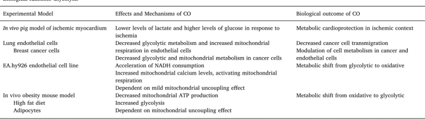
Along and in accordance with CO-mediated effect on oxidative metabolism, this gasotransmitter also decreases glycolysis in three distinct models (Fig. 3). In a model of ischemic myocardium, CO gas inhalation during 200 min and reaching 5% of COHb levels, improved metabolic conditions by decreasing glycolysis. Levels of lactate in the myocardium were 50% lower in pig pre-conditioned with CO gas than in control pigs. Accordingly, CO-treated animals presented higher glu-cose levels in myocardium than control animals [61]. In a research work about breast cancer cell adhesion and transmigration across lung endothelial cells, CORM-401 was found to decrease transmigration by cell metabolism modulation [62]. CORM-401 differently controls cell metabolism: in lung endothelial cells CO decreased glycolytic metabo-lism (assessed by extracellular acidification rate) and activated mi-tochondrial respiration (assessed via oxygen consumption rate). While in breast cancer cells CO decreased both mitochondrial respiration and glycolysis [62]. Cell adhesion and transmigration are highly bioener-getic demanding cellular processes, being cell metabolism a promising target for therapy. Finally, in EA.hy926 endothelial cell line CORM-401 treatment also shifts metabolism from glycolysis to oxidative
phosphorylation for ATP production. This effect is accompanied by a slight mitochondrial depolarization and mild uncoupling affect; which in turn activates mitochondrial respiration via mitochondrial Ca2+ signaling [63]. Moreover, CORM-401 accelerates mitochondrial NADH consumption and increases mitochondrial Ca2+ content, which is in accordance with the fact that Ca2+activates pyruvate dehydrogenase (and other hydrogenases) to feed NADH into electron transport chain, in particular complexes I and II [63]. Nevertheless, in obese mice fed with high fat diet, oral administration of CORM-401 reduces body weight and accelerates glucose metabolism in adipocytes by improving glycolysis [64]. Therefore CO action on metabolism might depend on tissue type and on pathophysiological conditions. One can speculate that CO pushes the biological system to an homeostatic status. For in-stance, in tumor context and cancer cell lines, CO accelerates oxidative phosphorylation pushing glycolysis down [62,63], in obesity models CO improves glucose homeostasis [64] or in ischemic conditions CO optimizes the use of oxygen [61]. Data and references are summarized in Table 2. Finally, it can also be considered that CO-induced mi-tochondrial uncoupling might increase mimi-tochondrial respiration as a compensatory effect, and this is further discussed and explored in the next section.
9. CO promotes a mild uncoupling effect
In apparent controversy, hemin-activation of HO-1 or treatment with CORMs (CORM-2, CORM-3 and CORM-A1) decreased state 3 oxygen consumption in isolated mitochondria [65]. State 3 represents oxygen consumption for oxidative phosphorylation that is associated with ATP production. Other studies using isolated mitochondria also point to a mild uncoupling effect of CO. In isolated mitochondria from rat hearts, low concentrations of CORM-3 promoted an augmentation of state 2 respiration (oxygen consumption in the absence of ADP, meaning an increase on oxygen consumption uncoupled of mitochon-drial potential and ATP production [66], while CORM-3 decreases state 3 respiration. This CO-induced mild uncoupling effect is mediated by adenine nucleotide translocator (ANT) and uncoupling proteins (UCP), since their pharmacological inhibition attenuated the CO effect on state 2 respiration [66]. Guanosine 5′-diphosphate (GDP) is general inhibitor for all UCP family proteins, nevertheless UCP2 and UCP3 are both ex-pressed in cardiac mitochondria. Furthermore, CORM-3-mediated un-coupling effect is also associated with modulation of mitochondrial ROS generation since CORM-3 decreases hydrogen peroxidase production at
Fig. 3. Main cellular metabolic pathways modulated by CO: mitochondrial meta-bolism, glycolysis and pentose phos-phate pathway. Low levels of CO enhance
TCA, improve mitochondrial OXPHOS, in-crease oxygen consumption and production of ATP. Accordingly, in most of the tested models (cell type and pathophysiological conditions) CO decreases glycolysis. Finally CO modulates redox processes: ROS appear as signaling molecules for CO mode of ac-tion and one of the cytoprotective proper-ties of CO is anti-oxidant. Thus it is not surprising that CO also stimulates PPP, a critical pathway responsible to maintaining the reducing capacity of the cell, as well as the cellular nucleotides pool.
complex I level. In addition, this uncoupling effect of CORM-3 in iso-lated mitochondria was also found to be dependent on phosphate car-rier [67]. Inhibition of phosphate carrier with N-ethylmaleimide re-verted CORM-3 effect on reducing mitochondrial potential. Likewise, higher levels of mitochondrial phosphate were found following treat-ment with CORM-3, along with mitochondrial swelling [67]. One could speculate that since these effects were found in isolated mitochondria, a cell-free system, could not be physiologically representative. Never-theless, in intact in human endothelial EA.hy926 cells, CORM-401 in-creased oxygen consumption rate simultaneously with an increase of proton leak and decrease of mitochondrial reserve capacity, indicating an uncoupling effect. This effect is dependent on the activation of mi-tochondrial large-conductance calcium-regulated potassium channel (mitoBKCa) [68]. Furthermore, low concentrations of CORM-401 are anti-inflammatory via cell metabolism modulation in microglia [54]. In this system, LPS-triggered inflammation promoted lower levels of oxygen consumption, mitochondrial function and cellular ATP gen-eration, while CORM-401 reverted all these effects. Still, CORM-401 promoted a mild uncoupling outcome without affecting cellular ATP concentration, but decreasing mitochondrial membrane potential and generation of ROS [54]. In conclusion, one can speculate that CO-mediated mild and transitory uncoupling (Fig. 4) promotes mitochon-drial homeostasis and anti-oxidant effect, which may in turn maintain mitochondrial function and cellular metabolic status. In summary, CO-induced mild uncoupling effect improves mitochondrial function via a compensatory mechanism and ROS signaling, these data are summar-ized inTable 1.
10. CO stimulates pentose phosphate pathway (PPP)
Mitochondria are the main source of cellular ROS generation under physiological and pathological conditions. Moreover, reduced glu-tathione (GSH) is the major anti-oxidant component of the cell, and its
recycling depends on NADPH as source of electrons. NADPH is mostly generated at the pentose phosphate pathway (PPP). Thus, it is not surprising that CO may also direct or indirectly modulate PPP. In fact, the first evidence of CO regulation o PPP was demonstrated in 2014 by two different groups and in different biological systems. CO-treatment of whole blood increased levels of reduced GSH in the cytosol of red blood cells. CO promoted stimulation of PPP, and whenever PPP was inhibited by 2-deoxyglucose, reduced, GSH levels are partially de-creased [69]. Nevertheless, other mechanisms in red blood cells were also found to be involved in CO-increased GSH levels. CO promoted deglutathionylation of hemoglobin at Cys93 and Cys112, which is a significant source of reduced GSH. Finally, CO also increased glu-tathione reductase activity without promoting glycolysis [69]. The axis HO-1/CO protects cancer cells against oxidative stress, in particular in leukemia. Yamamoto and colleagues have found that CO promotes a shift from glycolysis to PPP, supplying NADPH to reduce glutathione and limit oxidative stress [70]. CO inhibited the heme containing en-zyme cystathionine β-synthase (CBS) that, in turn, reduced methylation of PFKFB3, which decreases its activity and fructose 2,6-biphosphate (F-2,6-BP) production. Since F-2,6-BP is an allosteric activator of the rate limiting enzyme of glycolysis, phosphofructokinase-1, there was an acceleration of PPP with the consequent increased cellular resistance to oxidative damage by presenting higher levels of reduced glutathione [70].
In the human endothelial cell line EA.hy926, CORM-401 promoted NO production and triggered Ca2+release from the endoplasmic re-ticulum, increasing Ca2+intracellular levels. Inhibition of NO synthase by L-NAME limited NO production and prevents Ca2+ signaling, in-dicating that CO increase of Ca2+ is dependent on NO generation. Furthermore, CO-induced NO production was prevented by 6-amino-nicotinamide, which is an inhibitor of PPP. PPP is the major enzymatic source of NADPH, a molecule required for NO synthase [71]. Therefore, reinforcement of PPP by CORM-401 is also important for NADPH
Table 2
CO and glycolysis. Biological function: Glycolysis
Experimental Model Effects and Mechanisms of CO Biological outcome of CO Ref.
In vivo pig model of ischemic myocardium Lower levels of lactate and higher levels of glucose in response to
ischemia Metabolic cardioprotection in ischemic context [61]
Lung endothelial cells
Breast cancer cells Decreased glycolytic metabolism and increased mitochondrialrespiration in endothelial cells Decreased glycolytic and mitochondrial metabolism in cancer cells
Decreased cancer cell transmigration Modulation of cell metabolism in cancer and endothelial cells
[62] EA.hy926 endothelial cell line Acceleration of NADH consumption
Increased mitochondrial calcium levels, activating mitochondrial respiration
Dependent on mild mitochondrial uncoupling effect
Metabolic shift from glycolytic to oxidative [63]
In vivo obesity mouse model High fat diet
Adipocytes
Decreased mitochondrial ATP production Increased glycolysis
Dependent on mitochondrial uncoupling effect
Metabolic shift from oxidative to glycolytic [64]
Fig. 4. CO promotes mild uncoupling effect in mitochondria. CO-induced mild uncoupling is
de-pendent on ANT and UCP and this event is associated with anti-inflammatory role of CO. It is speculated that a small leakage of protons into mitochondrial matrix may generate ROS and accelerate respiratory chain for maintaining mitochondrial membrane po-tential, both events shall improve mitochondrial function and metabolism.
production, required in nitric oxide (NO) synthesis in endothelial cells [71]. It is well established that during neuronal differentiation, there is a switch from glycolytic to oxidative mitochondrial metabolism [72,73]. Thus, neurons are mostly oxidative and must present an ef-fective anti-oxidant defense, being GSH the major cellular non-enzy-matic anti-oxidant factor. In a recent paper, Almeida and colleagues used human neuroblastoma SH-SY5Y cells as models for neuronal dif-ferentiation and exposed them to CORM-A1. CORM-A1 improved neuronal differentiation by increasing the final yield of neurons, by reinforcing PPP activity and increasing cellular GSH/GSSG ratio [74]. CO treatment enhanced protein expression and activity of glucose 6-phosphate dehydrogenase (G6PD), which is the rate-limiting enzyme of PPP. Whenever G6PD was knocked down there was a reversion of CO-induced improvement of neuronal differentiation, while pharmacolo-gical inhibition of GSH synthesis did not change neuronal differentia-tion. Likewise, CO increased the reduced and oxidized glutathione (GSH/GSSG) without changing total pool of GSH, which is in ac-cordance with CO-induced activation of PPP [74]. In conclusion, CO emerges as a modulator of PPP, but much data is still needed for deeply and precisely describe the underlying molecular mechanisms (Fig. 3, Table 3).
11. CO, obesity and diabetes diseases
Modulation of metabolism by CO is not limited to cellular meta-bolism, but this gasotransmitter also plays a role in metabolic diseases (diabetes and obesity), which is discussed herein.
In intact mouse islet, exogenous CO gas improved glucose-stimu-lated insulin secretion in a cGMP signaling dependent manner [75]. Likewise, CO stimulated the activities of the lysosomal/vacuolar en-zymes: acid glucan-1,4-glucosidase and acid glucosidase (acid α-glucoside hydrolases), which are involved in glycogen hydrolyzation [75].
In an obesity mouse model based on high fat diet, chronic and preventive intraperitoneal (IP) administration of CORM-A1 reverted obesity by decreasing body and epididymal fat weight and reduced fasted blood glucose and insulin in a food intake independent manner [76]. Furthermore, in epididymal fat tissue, CORM-A1 treatment in-creased expression levels of uncoupling protein-1 (UCP-1), PGC-1α and Nrf-1, which are considered markers of mitochondrial heat production and mitochondrial biogenesis. These facts are in accordance with the higher levels of oxygen consumption and heat production of CORM-A1 treated mice [77]. Moreover, chronic treatment with CORM-A1 can reverse established dietary-induced obesity, hyperglycemia, and insulin resistance [77]. Oral administration of CORM-401 also decreased body weight and improved insulin sensitivity and glucose tolerance in high fat diet mouse model for diabetes [64]. CORM-401 reverted the in-crease on cellular size of epididymal white adipocytes following high fat diet and reduced macrophage infiltration in white fat tissue, de-creasing the levels of pro-inflammatory factors (IL-6, IL-1β and HO-1). In contrast to the previous works, the described underlying molecular
mechanism is based on reinforcement of glycolytic metabolism for in-creasing ATP intracellular levels in white adipocytes [64]. In fact, CORM-401 did not revert the reduction of Pgc-1α mRNA expression in white adipocyte tissue. Nevertheless, in brown adipocyte tissue, CO restored the levels of Pgc-1α and Ucp-1 mRNA [64], which might in-dicate an increase on mitochondrial population and/or function. In summary the effects of CO described by Braud's and Hosick's teams are similar but the underlying metabolic mechanisms in white adipocytes are different.
In the same high fat diet model of diabetes, fibroblast growth factor 21 (FGF-21) is necessary for CO gas to reduce obesity, glucose tolerance and improve insulin sensitivity following high fat diet [78]. The CO-induced increased expression of FGF-21 in liver and primary hepato-cytes is dependent on mitochondrial ROS generation. In hepatohepato-cytes, CO increased protein levels of complex III and IV, as well as of Nrf-1, TFAM and PGC-1α, which are associated with increased mitochondrial biogenesis, and accordingly enhances oxygen consumption. Likewise, CO also increased mitochondrial population in white fat tissue, which converts into beige tissue by switching cell metabolism. All these mi-tochondrial events are dependent on FGF-21 presence [78]. In sum-mary, CO improves mitochondrial metabolism in hepatocytes and converts white adipocytes into beige ones, in order to prevent obesity and diabetes type 2.
In a model of metabolic syndrome, based on mice fed with high fat diet, CO (CORM-3 administration) improved cardiac function by mod-ulation of mitochondrial popmod-ulation quality [79]. High fat diet for 12 weeks decreased intrinsic contractile function in left ventricle. This cardiac dysfunction was accompanied by an alteration in mitochondrial morphology and function, including: (i) an increase of mitochondrial volume and number, along with higher expression levels of the mi-tochondrial fusion genes Mfn2 and OPA1 and mimi-tochondrial biogenesis gene PGC-1α; and (ii) a decrease of oxygen consumption and mi-tochondrial membrane potential [79]. These mitochondrial events were reverted by two CORM-3 IP injections. Furthermore, high fat diet does not modulate autophagy, while CORM-3 treatment with high fat diet promoted autophagy assessed by LC3I/II ratio [79]. In an apparent contradiction, Lancel and colleagues showed that CO reverted the high fat diet-induced mitochondrial biogenesis and fusion, while most of data in the literature claim the cardioprotection ability of CO is de-pendent on mitochondrial biogenesis.
The regulatory role of CO in obesity and diabetes is an emerging research filed and more data are necessary to disclose its mechanisms and the targeted cells and tissues.
12. Conclusions
The fact that CO targets mitochondria, in particular cytochrome c oxidase, originates several consequences: (i) generation of ROS for downstream signaling, (ii) alterations in oxygen consumption implying uncoupling effects and/or improvement of mitochondrial respiration and ATP production, (iii) regulation of mitochondrial quality control,
Table 3
CO and pentose phosphate pathway. Biological function: Pentose Phosphate Pathway
Experimental Model Effects and Mechanisms of CO Biological outcome of CO Ref.
Red blood cells Stimulation of pentose phosphate pathway (PPP)
2-deoxyglucose reverts PPP stimulation Increased reduced glutathione levels [69] Leukemia cancer cells Limitation of glycolysis favoring PPP
Dependent on inhibition of cystathionine β-synthase and on inhibition of phosphofructokinase-1 (rate limiting glycolytic enzyme)
Increased cancer cell resistance against oxidative stress
Higher reduced glutathione levels
[70] EA.hy926 endothelial cell line Promotion of Ca2+release from the endoplasmic reticulum
Acceleration of PPP and in consequence NADPH production Increased NO production [71] Human neuroblastoma SH-SY5Y cell
including mitochondrial biogenesis, mitophagy and fusion/fission. Likewise, related to cell metabolism and redox response, CO also ap-pears to modulate glycolysis and pentose phosphate pathway. Because these are vital cellular process, endogenous CO production or exo-genous CO treatment have great impact on cellular function at distinct cells and tissues, namely: liver, heart, brain, and also in metabolic diseases, such as diabetes and obesity.
Declaration of competing interest
The authors state there is no conflict of interest in in the manuscript entitled “CO-mediated cytoprotection is dependent on cell metabolism modulation”.
Acknowledgements
This work was supported by Fundação para a Ciência e Tecnologia (FCT) grant UID/Multi/04462/2013, I&D 2015–2020 iNOVA4Health -Programme in Translacional Medicine. FCT provided individual fi-nancial support to CFP (SFRH/BD/106057/2015), DDP (PD/BD/ 128338/2017), NLS (PD/BD/127819/2016) and HLAV (IF/00185/ 2012).
Appendix A. Supplementary data
Supplementary data to this article can be found online athttps:// doi.org/10.1016/j.redox.2020.101470.
References
[1] T. Sjostrand, Endogenous formation of carbon monoxide in man, Nature 164 (1949) 580–581,https://doi.org/10.1038/164580a0.
[2] R. Tenhunen, H.S. Marver, R. Schmid, The enzymatic conversion of heme to bilir-ubin by microsomal heme oxygenase, Proc. Natl. Acad. Sci. Unit. States Am. 61 (1968) 748–755,https://doi.org/10.1073/pnas.61.2.748.
[3] R. Tenhunen, H.S. Marver, R. Schmid, Microsomal heme oxygenase.
Characterization of the enzyme, J. Biol. Chem. 244 (1969) 6388–6394http://www. ncbi.nlm.nih.gov/pubmed/4390967.
[4] S.W. Ryter, J. Alam, A.M.K. Choi, Heme oxygenase-1/carbon monoxide: from basic science to therapeutic applications, Physiol. Rev. 86 (2006) 583–650,https://doi. org/10.1152/physrev.00011.2005.
[5] P. Arosio, F. Carmona, R. Gozzelino, F. Maccarinelli, M. Poli, The importance of eukaryotic ferritins in iron handling and cytoprotection, Biochem. J. 472 (2015) 1–15,https://doi.org/10.1042/BJ20150787.
[6] R. Motterlini, L.E. Otterbein, The therapeutic potential of carbon monoxide, Nat. Rev. Drug Discov. 9 (2010) 728–743,https://doi.org/10.1038/nrd3228. [7] R. Motterlini, R. Foresti, Biological signaling by carbon monoxide and carbon
monoxide-releasing molecules, Am. J. Physiol. Physiol. 312 (2017) C302–C313, https://doi.org/10.1152/ajpcell.00360.2016.
[8] S.R. Oliveira, C.S.F. Queiroga, H.L.A. Vieira, Mitochondria and carbon monoxide: cytoprotection and control of cell metabolism – a role for Ca2+? J. Physiol. 594 (2016) 4131–4138,https://doi.org/10.1113/JP270955.
[9] C.S.F. Queiroga, A. Vercelli, H.L.A. Vieira, Carbon monoxide and the CNS: chal-lenges and achievements, Br. J. Pharmacol. 172 (2015) 1533–1545,https://doi. org/10.1111/bph.12729.
[10] A.S. Almeida, C. Figueiredo-Pereira, H.L.A. Vieira, Carbon monoxide and mi-tochondria-modulation of cell metabolism, redox response and cell death, Front. Physiol. 6 (2015),https://doi.org/10.3389/fphys.2015.00033.
[11] C.C. Romão, W.A. Blättler, J.D. Seixas, G.J.L. Bernardes, Developing drug molecules for therapy with carbon monoxide, Chem. Soc. Rev. 41 (2012) 3571,https://doi. org/10.1039/c2cs15317c.
[12] C.C. Romão, H.L.A. Vieira, Metal Carbonyl Prodrugs: CO Delivery and beyond, Wiley-VCH, 2015,https://doi.org/10.1002/9783527673438.ch06.
[13] R. Motterlini, B.E. Mann, R. Foresti, Therapeutic applications of carbon monoxide-releasing molecules, Expet Opin. Invest. Drugs 14 (2005) 1305–1318,https://doi. org/10.1517/13543784.14.11.1305.
[14] R. Motterlini, B. Mann, T. Johnson, J. Clark, R. Foresti, C. Green, Bioactivity and pharmacological actions of carbon monoxide-releasing molecules, Curr. Pharmaceut. Des. 9 (2003) 2525–2539,https://doi.org/10.2174/ 1381612033453785.
[15] R. Foresti, J. Hammad, J.E. Clark, T.R. Johnson, B.E. Mann, A. Friebe, C.J. Green, R. Motterlini, Vasoactive properties of CORM-3, a novel water-soluble carbon monoxide-releasing molecule, Br. J. Pharmacol. 142 (2004) 453–460,https://doi. org/10.1038/sj.bjp.0705825.
[16] R. Motterlini, P. Sawle, J. Hammad, S. Bains, R. Alberto, R. Foresti, C.J. Green, CORM-A1: a new pharmacologically active carbon monoxide-releasing molecule,
Faseb. J. 19 (2005) 284–286,https://doi.org/10.1096/fj.04-2169fje. [17] S.H. Crook, B.E. Mann, A.J.H.M. Meijer, H. Adams, P. Sawle, D. Scapens,
R. Motterlini, [Mn(CO)4{S2CNMe(CH2CO2H)}], a new water-soluble CO-releasing molecule, Dalt, OR Trans. 40 (2011) 4230,https://doi.org/10.1039/c1dt10125k. [18] M. Lavitrano, R.T. Smolenski, A. Musumeci, M. Maccherini, E. Slominska, E. Di
Florio, A. Bracco, A. Mancini, G. Stassi, M. Patti, R. Giovannoni, A. Froio, F. Simeone, M. Forni, M.L. Bacci, G. D'Alise, E. Cozzi, L.E. Otterbein, M.H. Yacoub, F.H. Bach, F. Calise, Carbon monoxide improves cardiac energetics and safeguards the heart during reperfusion after cardiopulmonary bypass in pigs, Faseb. J. 18 (2004) 1093–1095.
[19] T.Y. Tsui, Y.T. Siu, H.J. Schlitt, S.T. Fan, Heme oxygenase-1-derived carbon mon-oxide stimulates adenosine triphosphate generation in human hepatocyte, Biochem. Biophys. Res. Commun. 336 (2005) 898–902,https://doi.org/10.1016/j.bbrc.2005. 08.187.
[20] T.Y. Tsui, A. Obed, Y.T. Siu, S.F. Yet, L. Prantl, H.J. Schlitt, S.T. Fan, Carbon monoxide inhalation rescues mice from fulminant hepatitis through improving hepatic energy metabolism, Shock 27 (2007) 165–171.
[21] M. Bilban, A. Haschemi, B. Wegiel, B.Y.Y. Chin, O. Wagner, L.E.E. Otterbein, Heme oxygenase and carbon monoxide initiate homeostatic signaling, J. Mol. Med. 86 (2008) 267–279,https://doi.org/10.1007/s00109-007-0276-0.
[22] C.S.F. Queiroga, A.S. Almeida, H.L.A. Vieira, Carbon monoxide targeting mi-tochondria, Biochem. Res. Int. (2012),https://doi.org/10.1155/2012/749845. [23] C.C. Romão, H.L.A. Vieira, Metal carbonyls for CO-based therapies: challenges and
successes, in: A.J.L. Pombeiro (Ed.), Adv. Organomet. Chem. Catal, J. Wiley, 2013, pp. 543–561, ,https://doi.org/10.1002/9781118742952.ch41.
[24] J. Boczkowski, J.J. Poderoso, R. Motterlini, CO-metal interaction: vital signaling from a lethal gas, Trends Biochem. Sci. 31 (2006) 614–621,https://doi.org/10. 1016/j.tibs.2006.09.001.
[25] J.-R. Alonso, F. Cardellach, S. Lopez, J. Casademont, O. Miro, Carbon monoxide specifically inhibits cytochrome C oxidase of human mitochondrial respiratory chain, Pharmacol. Toxicol. 93 (2003) 142–146, https://doi.org/10.1034/j.1600-0773.2003.930306.x.
[26] G. D'Amico, F. Lam, T. Hagen, S. Moncada, Inhibition of cellular respiration by endogenously produced carbon monoxide, J. Cell Sci. 119 (2006) 2291–2298, https://doi.org/10.1242/jcs.02914.
[27] B.S. Zuckerbraun, B.Y. Chin, M. Bilban, J. de Costa d'Avila, J. Rao, T.R. Billiar, L.E. Otterbein, J.D.C. d'Avila, J. Rao, T.R. Billiar, L.E. Otterbein, Carbon monoxide signals via inhibition of cytochrome c oxidase and generation of mitochondrial reactive oxygen species, Faseb. J. 21 (2007) 1099–1106,https://doi.org/10.1096/ fj.06-6644com.
[28] A.S. Almeida, C.S.F. Queiroga, M.F.Q. Sousa, P.M. Alves, H.L.A. Vieira, Carbon monoxide modulates apoptosis by reinforcing oxidative metabolism in astrocytes: role of Bcl-2, J. Biol. Chem. 287 (2012) 10761–10770,https://doi.org/10.1074/ jbc.M111.306738.
[29] A. Haschemi, B.Y. Chin, M. Jeitler, H. Esterbauer, O. Wagner, M. Bilban, L.E. Otterbein, Carbon monoxide induced PPARγ SUMOylation and UCP2 block inflammatory gene expression in macrophages, PloS One 6 (2011) e26376, , https://doi.org/10.1371/journal.pone.0026376.
[30] C. Taillé, J. El-Benna, S. Lanone, J. Boczkowski, R. Motterlini, Mitochondrial re-spiratory chain and NAD(P)H oxidase are targets for the antiproliferative effect of carbon monoxide in human airway smooth muscle, J. Biol. Chem. 280 (2005) 25350–25360,https://doi.org/10.1074/jbc.M503512200.
[31] H.L.A. Vieira, C.S.F. Queiroga, P.M. Alves, Pre-conditioning induced by carbon monoxide provides neuronal protection against apoptosis, J. Neurochem. 107 (2008) 375–384,https://doi.org/10.1111/j.1471-4159.2008.05610.x.
[32] H.B. Suliman, M.S. Carraway, L.G. Tatro, C.a Piantadosi, A new activating role for CO in cardiac mitochondrial biogenesis, J. Cell Sci. 120 (2007) 299–308,https:// doi.org/10.1242/jcs.03318.
[33] H.B. Suliman, M.S. Carraway, A.S. Ali, C.M. Reynolds, K.E. Welty-wolf, C.A. Piantadosi, The CO/HO system reverses inhibition of mitochondrial biogenesis and prevents murine doxorubicin cardiomyopathy, J. Clin. Invest. 117 (2007) 3730–3741,https://doi.org/10.1172/JCI32967.3730.
[34] S. Lancel, S.M. Hassoun, R. Favory, B. Decoster, R. Motterlini, R. Neviere, Carbon monoxide rescues mice from lethal sepsis by supporting mitochondrial energetic metabolism and activating mitochondrial biogenesis, J. Pharmacol. Exp. Therapeut. 329 (2009) 641–648,https://doi.org/10.1124/jpet.108.148049.could.
[35] C.A. Piantadosi, C.M. Withers, R.R. Bartz, N.C. MacGarvey, P. Fu, T.E. Sweeney, K.E. Welty-Wolf, H.B. Suliman, Heme oxygenase-1 couples activation of mi-tochondrial biogenesis to anti-inflammatory cytokine expression, J. Biol. Chem. 286 (2011) 16374–16385,https://doi.org/10.1074/jbc.M110.207738.
[36] N.C. MacGarvey, H.B. Suliman, R.R. Bartz, P. Fu, C.M. Withers, K.E. Welty-Wolf, C.a Piantadosi, Activation of mitochondrial biogenesis by heme oxygenase-1-mediated NF-E2-related factor-2 induction rescues mice from lethal Staphylococcus aureus sepsis, Am. J. Respir. Crit. Care Med. 185 (2012) 851–861,https://doi.org/ 10.1164/rccm.201106-1152OC.
[37] M. Zheng, S.-K. Kim, Y. Joe, S.H. Back, H.R. Cho, H.P. Kim, L.J. Ignarro, H.-T. Chung, Sensing endoplasmic reticulum stress by protein kinase RNA-like en-doplasmic reticulum kinase promotes adaptive mitochondrial DNA biogenesis and cell survival via heme oxygenase-1/carbon monoxide activity, Faseb. J. 26 (2012) 2558–2568,https://doi.org/10.1096/fj.11-199604.
[38] N. Rayamajhi, S.-K. Kim, H. Go, Y. Joe, Z. Callaway, J.-G. Kang, S.W. Ryter, H.T. Chung, Quercetin induces mitochondrial biogenesis through activation of HO-1 in HepG2 cells, Oxid. Med. Cell. Longev. (20HO-13) HO-1–HO-10,https://doi.org/10.1155/ 2013/1542792013.
[39] Y.K. Choi, J.H. Park, Y.Y. Baek, M.H. Won, D. Jeoung, H. Lee, K.S. Ha, Y.G. Kwon, Y.M. Kim, Carbon monoxide stimulates astrocytic mitochondrial biogenesis via
L-type Ca2+ channel-mediated PGC-1α/ERRα activation, Biochem. Biophys. Res. Commun. 479 (2016) 297–304,https://doi.org/10.1016/j.bbrc.2016.09.063. [40] Y.K. Choi, J.H. Park, J.-A. Yun, J.-H. Cha, Y. Kim, M.-H. Won, K.-W. Kim, K.-S. Ha,
Y.-G. Kwon, Y.-M. Kim, Heme oxygenase metabolites improve astrocytic mi-tochondrial function via a Ca2+-dependent HIF-1α/ERRα circuit, PloS One 13 (2018) e0202039, ,https://doi.org/10.1371/journal.pone.0202039.
[41] R.C. Scarpulla, Metabolic control of mitochondrial biogenesis through the PGC-1 family regulatory network, Biochim. Biophys. Acta Mol. Cell Res. 1813 (2011) 1269–1278,https://doi.org/10.1016/j.bbamcr.2010.09.019.
[42] H. Zhang, P. Gao, R. Fukuda, G. Kumar, B. Krishnamachary, K.I. Zeller, C.V. Dang, G.L. Semenza, HIF-1 inhibits mitochondrial biogenesis and cellular respiration in VHL-deficient renal cell carcinoma by repression of C-myc activity, Canc. Cell 11 (2007) 407–420,https://doi.org/10.1016/j.ccr.2007.04.001.
[43] R. Fukuda, H. Zhang, J.W. Kim, L. Shimoda, C.V.V. Dang, G.L.L. Semenza, HIF-1 regulates cytochrome oxidase subunits to optimize efficiency of respiration in hy-poxic cells, Cell 129 (2007),https://doi.org/10.1016/j.cell.2007.01.047. [44] Z. Wang, C. Figueiredo-Pereira, C. Oudot, H.L.A. Vieira, C. Brenner, Mitochondrion:
a common organelle for distinct cell deaths? Int. Rev. Cell Mol. Biol. Elsevier Inc., 2017, pp. 245–287, ,https://doi.org/10.1016/bs.ircmb.2016.09.010.
[45] L. Esteban-Martínez, E. Sierra-Filardi, P. Boya, Mitophagy, metabolism, and cell fate, Mol. Cell. Oncol. 4 (2017) e1353854, ,https://doi.org/10.1080/23723556. 2017.1353854.
[46] H.J. Kim, Y. Joe, S.-Y. Rah, S.-K. Kim, S.-U. Park, J. Park, J. Kim, J. Ryu, G.J. Cho, Y.-J. Surh, S.W. Ryter, U.-H. Kim, H.T. Chung, Carbon monoxide-induced TFEB nuclear translocation enhances mitophagy/mitochondrial biogenesis in hepatocytes and ameliorates inflammatory liver injury, Cell Death Dis. 9 (2018) 1060,https:// doi.org/10.1038/s41419-018-1112-x.
[47] R.M. Gorojod, A. Alaimo, S. Porte Alcon, J.H. Martinez, M.E. Cortina, E.S. Vazquez, M.L. Kotler, Heme Oxygenase-1 protects astroglia against manganese-induced oxi-dative injury by regulating mitochondrial quality control, Toxicol. Lett. 295 (2018) 357–368,https://doi.org/10.1016/j.toxlet.2018.07.045.
[48] T.D. Hull, R. Boddu, L. Guo, C.C. Tisher, A.M. Traylor, B. Patel, R. Joseph, S.D. Prabhu, H.B. Suliman, C.A. Piantadosi, A. Agarwal, J.F. George, Heme oxy-genase-1 regulates mitochondrial quality control in the heart, JCI Insight 1 (2016), https://doi.org/10.1172/jci.insight.85817.
[49] M.A. Di Noia, S. Van Driesche, F. Palmieri, L.-M. Yang, S. Quan, A.I. Goodman, N.G. Abraham, Heme oxygenase-1 enhances renal mitochondrial transport carriers and cytochrome C oxidase activity in experimental diabetes, J. Biol. Chem. 281 (2006) 15687–15693,https://doi.org/10.1074/jbc.M510595200.
[50] C.S.F. Queiroga, A.S. Almeida, P.M. Alves, C. Brenner, H.L.A. Vieira, Carbon monoxide prevents hepatic mitochondrial membrane permeabilization, BMC Cell Biol. 12 (2011),https://doi.org/10.1186/1471-2121-12-10.
[51] C.S.F. Queiroga, A.S. Almeida, C. Martel, C. Brenner, P.M. Alves, H.L.A. Vieira, Glutathionylation of adenine nucleotide translocase induced by carbon monoxide prevents mitochondrial membrane permeabilization and apoptosis, J. Biol. Chem. 285 (2010) 17077–17088,https://doi.org/10.1074/jbc.M109.065052. [52] S.R. Oliveira, C.S.F. Queiroga, H.L.A. Vieira, Mitochondria and carbon monoxide:
cytoprotection and control of cell metabolism – a role for Ca2+? J. Physiol. 594 (2016) 4131–4138,https://doi.org/10.1113/JP270955.
[53] C.S.F. Queiroga, R.M.A. Alves, S.V. Conde, P.M. Alves, H.L.A. Vieira, Paracrine effect of carbon monoxide: astrocytes promote neuroprotection via purinergic sig-naling, J. Cell Sci. 129 (2016),https://doi.org/10.1242/jcs.187260jcs.187260. [54] J.L. Wilson, F. Bouillaud, A.S. Almeida, H.L. Vieira, M.O. Ouidja, J.L. Dubois-Randé,
R. Foresti, R. Motterlini, Carbon monoxide reverses the metabolic adaptation of microglia cells to an inflammatory stimulus, Free Radic. Biol. Med. 104 (2017) 311–323,https://doi.org/10.1016/j.freeradbiomed.2017.01.022.
[55] A. Kasahara, L. Scorrano, Mitochondria: from cell death executioners to regulators of cell differentiation, Trends Cell Biol. 24 (2014) 761–770,https://doi.org/10. 1016/j.tcb.2014.08.005.
[56] A.S. Almeida, H.L.A. Vieira, Role of cell metabolism and mitochondrial function during adult neurogenesis, Neurochem. Res. 42 (2017) 1787–1794,https://doi. org/10.1007/s11064-016-2150-3.
[57] M. Agostini, F. Romeo, S. Inoue, M.V. Niklison-Chirou, A.J. Elia, D. Dinsdale, N. Morone, R.A. Knight, T.W. Mak, G. Melino, Metabolic reprogramming during neuronal differentiation, Cell Death Differ. (2016),https://doi.org/10.1038/cdd. 2016.36.
[58] A.S. Almeida, U. Sonnewald, P.M. Alves, H.L.A. Vieira, Carbon monoxide improves neuronal differentiation and yield by increasing the functioning and number of mitochondria, J. Neurochem. 138 (2016) 423–435,https://doi.org/10.1111/jnc. 13653.
[59] H.B. Suliman, F. Zobi, C.A. Piantadosi, Heme oxygenase-1/carbon monoxide system and embryonic stem cell differentiation and maturation into cardiomyocytes, Antioxidants Redox Signal. 24 (2016) 345–360,https://doi.org/10.1089/ars.2015. 6342.
[60] B. Wegiel, D. Gallo, E. Csizmadia, C. Harris, J. Belcher, G.M. Vercellotti, N. Penacho, P. Seth, V. Sukhatme, A. Ahmed, P.P. Pandolfi, L. Helczynski, A. Bjartell, J.L. Persson, L.E. Otterbein, Carbon monoxide expedites metabolic ex-haustion to inhibit tumor growth, Canc. Res. 73 (2013) 7009–7021,https://doi. org/10.1158/0008-5472.CAN-13-1075.
[61] K. Ahlstrom, B. Biber, A. Åberg, A. Waldenstrom, G. Ronquist, P. Abrahamsson, P. Strandén, G. Johansson, M.F. Haney, Metabolic responses in ischemic myo-cardium after inhalation of carbon monoxide, Acta Anaesthesiol. Scand. 53 (2009) 1036–1042,https://doi.org/10.1111/j.1399-6576.2009.01992.x.
[62] M. Stojak, P. Kaczara, R. Motterlini, S. Chlopicki, Modulation of cellular bioener-getics by CO-releasing molecules and NO-donors inhibits the interaction of cancer cells with human lung microvascular endothelial cells, Pharmacol. Res. 136 (2018) 160–171,https://doi.org/10.1016/j.phrs.2018.09.005.
[63] P. Kaczara, R. Motterlini, K. Kus, A. Zakrzewska, A.Y. Abramov, S. Chlopicki, Carbon monoxide shifts energetic metabolism from glycolysis to oxidative phos-phorylation in endothelial cells, FEBS Lett. 590 (2016) 3469–3480,https://doi.org/ 10.1002/1873-3468.12434.
[64] L. Braud, M. Pini, L. Muchova, S. Manin, H. Kitagishi, D. Sawaki, G. Czibik, J. Ternacle, G. Derumeaux, R. Foresti, R. Motterlini, Carbon monoxide–induced metabolic switch in adipocytes improves insulin resistance in obese mice, JCI Insight 3 (2018),https://doi.org/10.1172/jci.insight.123485.
[65] A. Sandouka, E. Balogun, R. Foresti, B.E. Mann, T.R. Johnson, Y. Tayem, C.J. Green, B. Fuller, R. Motterlini, Carbon monoxide-releasing molecules (CO-RMs) modulate respiration in isolated mitochondria, Cell. Mol. Biol. (2005),https://doi.org/10. 1170/T646.
[66] L. Lo Iacono, J. Boczkowski, R. Zini, I. Salouage, A. Berdeaux, R. Motterlini, D. Morin, A carbon monoxide-releasing molecule (CORM-3) uncouples mitochon-drial respiration and modulates the production of reactive oxygen species, Free Radic. Biol. Med. 50 (2011) 1556–1564,https://doi.org/10.1016/j.freeradbiomed. 2011.02.033.
[67] R. Long, I. Salouage, A. Berdeaux, R. Motterlini, D. Morin, CORM-3, a water soluble CO-releasing molecule, uncouples mitochondrial respiration via interaction with the phosphate carrier, Biochim. Biophys. Acta Bioenerg. 1837 (2014) 201–209, https://doi.org/10.1016/j.bbabio.2013.10.002.
[68] P. Kaczara, R. Motterlini, G.M. Rosen, B. Augustynek, P. Bednarczyk, A. Szewczyk, R. Foresti, S. Chlopicki, Carbon monoxide released by CORM-401 uncouples mi-tochondrial respiration and inhibits glycolysis in endothelial cells: a role for mitoBKCachannels, Biochim. Biophys. Acta Bioenerg. 1847 (2015),https://doi.org/
10.1016/j.bbabio.2015.07.004.
[69] A. Metere, E. Iorio, G. Scorza, S. Camerini, M. Casella, M. Crescenzi, M. Minetti, D. Pietraforte, Carbon monoxide signaling in human red blood cells: evidence for pentose phosphate pathway activation and protein deglutathionylation, Antioxidants Redox Signal. 20 (2014) 403–416,https://doi.org/10.1089/ars.2012. 5102.
[70] T. Yamamoto, N. Takano, K. Ishiwata, M. Ohmura, Y. Nagahata, T. Matsuura, A. Kamata, K. Sakamoto, T. Nakanishi, A. Kubo, T. Hishiki, M. Suematsu, Reduced methylation of PFKFB3 in cancer cells shunts glucose towards the pentose phos-phate pathway, Nat. Commun. 5 (2014) 3480,https://doi.org/10.1038/ ncomms4480.
[71] P. Kaczara, B. Proniewski, C. Lovejoy, K. Kus, R. Motterlini, A.Y. Abramov, S. Chlopicki, CORM-401 induces calcium signalling, NO increase and activation of pentose phosphate pathway in endothelial cells, FEBS J. (2018),https://doi.org/10. 1111/febs.14411.
[72] H.L.A. Vieira, P.M. Alves, A. Vercelli, Modulation of neuronal stem cell differ-entiation by hypoxia and reactive oxygen species, Prog. Neurobiol. 93 (2011) 444–455,https://doi.org/10.1016/j.pneurobio.2011.01.007.
[73] S. Varum, A.S. Rodrigues, M.B. Moura, O. Momcilovic, C.A. Easley, J. Ramalho-Santos, B. Van Houten, G. Schatten, Energy metabolism in human pluripotent stem cells and their differentiated counterparts, PloS One 6 (2011) e20914, ,https://doi. org/10.1371/journal.pone.0020914.
[74] A.S. Almeida, N.L. Soares, C.O. Sequeira, S.A. Pereira, U. Sonnewald, H.L.A. Vieira, Improvement of neuronal differentiation by carbon monoxide: role of pentose phosphate pathway, Redox Biol 17 (2018) 338–347,https://doi.org/10.1016/j. redox.2018.05.004.
[75] H. Mosen, Nitric oxide inhibits, and carbon monoxide activates, islet acid -glucoside hydrolase activities in parallel with glucose-stimulated insulin secretion, J. Endocrinol. 190 (2006) 681–693,https://doi.org/10.1677/joe.1.06890. [76] P.A. Hosick, A.A. AlAmodi, M.V. Storm, M.U. Gousset, B.E. Pruett, W. Gray,
J. Stout, D.E. Stec, Chronic carbon monoxide treatment attenuates development of obesity and remodels adipocytes in mice fed a high-fat diet, Int. J. Obes. 38 (2014) 132–139,https://doi.org/10.1038/ijo.2013.61.
[77] P.A. Hosick, A.A. AlAmodi, M.W. Hankins, D.E. Stec, Chronic treatment with a carbon monoxide releasing molecule reverses dietary induced obesity in mice, Adipocyte 5 (2016) 1–10,https://doi.org/10.1080/21623945.2015.1038443. [78] Y. Joe, S. Kim, H.J. Kim, J. Park, Y. Chen, H.-J. Park, S.-J. Jekal, S.W. Ryter, U.H. Kim, H.T. Chung, FGF21 induced by carbon monoxide mediates metabolic homeostasis via the PERK/ATF4 pathway, Faseb. J. 32 (2018) 2630–2643,https:// doi.org/10.1096/fj.201700709RR.
[79] S. Lancel, D. Montaigne, X. Marechal, C. Marciniak, S.M. Hassoun, B. Decoster, C. Ballot, C. Blazejewski, D. Corseaux, B. Lescure, R. Motterlini, R. Neviere, Carbon monoxide improves cardiac function and mitochondrial population quality in a mouse model of metabolic syndrome, PloS One 7 (2012) e41836, ,https://doi.org/ 10.1371/journal.pone.0041836.