Converting a silent killer into an effective healer:
Disclosing CO
‐
induced cytoprotection alteration
in astrocytes
Deborah Soares Neto Zucolotto da Silva
Bachelor in Biochemistry
Dissertation to obtain a Master's Degree in Molecular Genetics and Biomedicine
Supervision: Helena L. A. Vieira, PhD, CEDOC
September 2016 Master's committee:
President: Margarida Casal Ribeiro Castro Caldas Braga, PhD
Jury: Rita João Machado de Oliveira, PhD
Deborah Soares Neto Zucolotto da Silva
Bachelor in Biochemistry
Dissertation to obtain a Master's Degree in Molecular Genetics and Biomedicine
Supervision: Helena L. A. Vieira, PhD, CEDOC
September 2016
Disclosing CO-induced cytoprotection in
astrocytes: role of FosB
Master's committee:
President: Margarida Casal Ribeiro Castro Caldas Braga, PhD
III Copyright © 2016 Deborah Soares Neto Zucolotto da Silva, FCT/UNL and UNL. Faculty of Sciences and Technology, and the New University of Lisbon have the perpetual right without geographic limits of the publication and storage of this dissertation through printed exemplars, in digital format or through any other know means that exist or may be invented, it is also entitle to the divulgation through scientific repositories and admitting the copy and distribution of the dissertation for educational and research proposes without commercial intent as long as it is given credit to the author and editor.
V
Acknowledgments
The path that I had to make until here was not easy, with ups and many downs, in my professional and personal life. It was a long journey, but fortunately, I was able to overcome all the obstacles and difficulties thanks to all the people that walked by my side. For that, firstly I would like to show my gratitude to my supervisor Dr. Helena Vieira for accepting me in hers investigation group, initiating one of the most important periods of my life and for her great understanding and support that I received through this year. Thank you for always believe in me even when I stopped believe in myself.
I also want to thank to the rest of the Cell Death and Disease team for receiving me so well and for all the help they gave to me, specially Sara Oliveira who always saved me from the "laboratory troubles" and taught me so many value things.
To my dear friend Joel Guerra, who always helped me since the first day of university, a huge thank you for all the advises and teachings, laughs and silliness. You are a big example for me.
I would like to give a special thank to Adelaide Galhofa, my life coach and friend, for opening my eyes to a new world and for showing me the beauty of life. Without her I would not be able to overcome my fears or myself. Thank you, from the bottom of my heart, for helping me to create a new me who I can be proud of.
To my love and best friend Vasco Figueiredo, thank you for making me smile every day of my life and for fill my heart with affection, hope and faith. He is the one of the most beautiful persons I ever known with the purest heart. Thank you for smiling with me in the good moments and drying my tears in the bad ones. His love and unconditional support made this journey a lot easier to make.
Last but not least, I would like to thank to my incredible mother for all what she have done for me, allowing me to be where I am now. Thank you for making me who I am today and for supporting all of my decisions.
To all of you, thank you so much. None of this would be possible without your help.
"If you can´t fly, then run, if you can´t run, then walk, if you can´t walk, then crawl, but whatever you do, you have to keep moving forward"
VII
Abstract
Cerebral ischemia is a serious health problem, causing disability and death worldwide. Therefore, it is essential to develop strategies to protect and repair the brain.
Recently, some studies on preconditioning started to emerge, where a toxic stimulus below the threshold of injury is given, inducing tolerance, preparing and strengthening the brain for a potential greater injury. In this project, it will be used carbon monoxide (CO) as a preconditioning agent, since its administration is able to produce several beneficial effects, namely acting as an anti-apoptotic and anti-inflammatory agent. The purpose of this work is to disclose CO-induced cytoprotection and the potential role of FosB associated to it. The experiments were developed using primary cultures of astrocytes isolated from newborn mice. As CO source the CO-releasing molecule CORM-A1 was used.
CORM-A1 showed the ability to induce FosB protein expression in astrocytes, which was analyzed by western blot. However, it is not know yet how this increase is mediated. Reactive oxygen species (ROS) signaling is involved in CO and in FosB related pathways. Consequently, astrocytes were pre-treated with antioxidants (β-carotene or N-acetylcysteine) for decreasing cellular ROS levels. In addition P2X7 receptor also appears to be associated with FosB activation. Thus, P2X7 inhibitor (A-438079) was added to astrocytic culture in order to assess if FosB increased expression could be mediated by P2X7 receptor. Nevertheless, inhibition of ROS signaling did not change CO-induced FosB levels, while prevention of P2X7 activation showed a slight tendency to revert FosB expression, indicating that this pathway is independent on ROS but it could be mediated by P2X7 receptor.
Although it is still a long way to go, these results gave more insight about CO-induced FosB activation, which is important for understanding the molecular mechanisms underlying CO-induced cytoprotection. Potential therapeutic applications of CO will depend on a clear understanding of its pathways and would potentially allow the development of more efficient therapies against cerebral ischemia.
IX
Resumo
A isquemia cerebral é um grave problema de saúde, causando incapacidade e até mesmo a morte a nível mundial. Desta forma, é fundamental desenvolver estratégias que protejam e reparem o cérebro.
Recentemente começaram a surgir estudos acerca do pré-condicionamento, onde um estímulo tóxico é administrado abaixo do limiar de toxicidade, induzindo tolerância, preparando e fortalecendo o cérebro para um eventual dano. Neste projecto será usado monóxido de carbono como agente pré-condicionante, uma vez que a sua administração é capaz de produzir efeitos benéficos, nomeadamente actuando como um agente anti-apoptótico e anti-inflamatório. Com este trabalho pretende-se estudar a citoprotecção induzida pelo CO, bem como o possível papel do FosB que lhe está associado. As experiências foram desenvolvidas recorrendo a culturas primárias de astrócitos isoladas a partir de ratos recém nascidos. Como fonte de CO foi usada a CO-releasing molecule CORM-A1.
A CORM-A1 mostrou habilidade em induzir a expressão proteica do FosB em astrócitos, tendo sido analisada via western blot. Contudo, ainda não se sabe exactamente como é que este aumento acontece. A sinalização conduzida por espécies reactivas de oxigénio (ROS) está envolvida em vias relacionadas com o CO e o FosB. Consequentemente, os astrócitos foram pré tratados com antioxidantes (β-caroteno ou N-acetilcisteína) de forma a diminuir os níveis celulares de ROS. Para além disso, o receptor P2X7 também parece estar associado com a activação do FosB. Assim, um inibidor do P2X7 (A-438079) foi adicionado à cultura astrocítica de modo a perceber se o aumento da expressão do FosB poderá ser mediado pelo receptor P2X7. Contudo, a inibição da sinalização via ROS não alterou os níveis de FosB induzidos pelo CO, enquanto que a prevenção da activação do P2X7 mostrou haver um ligeira tendência para reverter a expressão do FosB, indicando que esta via é independente de ROS mas que poderá ser mediada via receptor P2X7.
Apesar de ainda haver um longo caminho a percorrer, estes dados permitiram um maior entendimento acerca da activação do FosB induzida pelo CO, sendo esta importante na compreensão dos mecanismos moleculares subjacentes à citoprotecção induzida pelo CO. Possíveis aplicações terapêuticas do CO estarão dependentes de um maior entendimento das suas via metabólicas, permitindo o desenvolvimento de terapias mais eficientes contra a isquemia cerebral.
XI
Contents
Acknowledgments ... V Abstract ... VII Resumo ... IX Abreviations ... XV
1. Introduction ... 1
1.1. Cerebral Ischemia ... 1
1.2. Astrocytes ... 1
1.3. Heme Oxygenase (HO) and Carbon Monoxide (CO) ... 2
1.4. Carbon Monoxide-Releasing Molecules (CORMs) ... 3
1.5. Reactive Oxygen Species (ROS) ... 3
1.6. FosB ... 4
1.7. P2X7 receptor ... 5
2. Objectives ... 7
3. Materials and Methods ... 9
3.1. Cell cultures ... 9
3.2. Cell Treatment ... 9
3.3. Protein extraction and quantification ... 9
3.4. Western blot ... 10
3.5. Statistical analysis ... 10
4. Results ... 11
4.1. Primary cultures of astrocytes ... 11
4.2. CO-preconditioning induces FosB Protein expression ... 12
4.3. ROS signaling induced by CO-preconditioning is not responsible for the increase of FosB protein expression ... 13
4.4. Role of P2X7 receptor in CO-induced FosB expression ... 15
5. Discussion ... 17
6. Conclusion and Future work ... 19
XIII
Figures Index
Figure 1.3 - Catabolism of heme group by heme oxygenase (HO)...2
Figure 1.4 - Chemical structure of CORM-A1...3
Figure 1.5 - Schematic illustration of the inhibitory effect of CO on the electron transport chain in mitochondria...4
Figure 4.1 - Primary culture of astrocytes...11
Figure 4.2 - Effect of carbon monoxide in FosB protein expression in a primary culture of astrocytes...12
Figure 4.3 - Effect of β-carotene and N-acetylcysteine (NAC) in FosB protein expression in a
primary culture of astrocytes treated with carbon monoxide...14
XV
Abreviations
AP-1 - Activator Protein-1 ATP - Adenosine Triphosfate BBB - Blood-Brain Barrier BCA -Bicinchoninic Acid BSA - Bovine Serum Albumin CBF - Cerebral Blood Flow CNS - Central Nervous System CO - Carbon Monoxide
CO2 - Carbon Dioxide
COHb - Carboxyhemoglobin
CORM - Carbon Monoxide-Releasing Molecules COX - Cytochrome c Oxidase
DMEM - Dulbecco's Minimum Essential Medium
EGTA - Ethylene Glycol Tetraacetic Acid
FBS - Fetal Bovine Serum
FosB - FBJ Murine Osteosarcome Viral Oncogene Homolog B HO - Heme Oxygenase
mRNA - Messenger Ribonucleic Acid NAC -N-acetylcysteine
NaCl - Sodium Cloride
PBS - Phosphate Buffered Saline Pen Strep - Penicilin/Streptomycin
RIPA- Radio-Immunoprecipitation Assay ROS - Reactive Oxygen Species
RT - Room Temperature
1
1. Introduction
1.1. Cerebral Ischemia
Cerebral ischemia is one of the major causes of brain injury, affecting millions of individuals [1]. It is the major cause of mortality in Portugal, and the second most common cause in the world [2][3]. Brain injury is a serious health and medical problem, causing disability and death worldwide. Therefore, it is very important the development of strategies to protect and repair the brain.
Ischemia results from a severe reduction in blood supply to a given tissue or organ due to narrowing or blockage of an artery [4][5]. This can lead to cell death, inflammation or tissue loss, and it can be caused by either an embolism or by a local thrombosis [4].
This type of brain damage can occur in adults or newborns, either by an ischemic stroke or perinatal asphyxia, respectively [6]. Deprivation in oxygen and glucose delivery in brain results in the reduction of essential energy metabolites, like ATP and phosphocreatine [4][5]. These events lead to oxidative stress, energy failure, cell death, inflammation and ionic perturbations [4-6].
Brain, and particularly neurons, is very vulnerable to an ischemic insult [5][7]. Complete interruption of cerebral blood flow (CBF) for only 5 minutes induces neuronal cell death in many brain regions [7]. Thus, it is very important to develop ways of treatment and prevention of this condition.
Many clinical trials have been made with the purpose of preventing cell death, but with little success [5]. This lack of success is due to many reasons, such as therapeutic window considerations, drug dosing and delivery [5]. Currently, there is only one approved drug for adults, named tPA (tissue plasminogen activator). Unfortunately, it can only be effective if given within 4.5 hours after the beginning of stroke and it is beneficial only for a very small amount of patients [2][5]. For newborns the standard treatment is hypothermia, which is also applied only under specific criteria and short time window [8]. However, these both strategies are based on promotion of blood reperfusion and they are non-healing treatments. Furthermore, the only real efficient strategy for now is prevention [2].
1.2. Astrocytes
The central nervous system (CNS) is formed by neurons and glial cells [9]. Neurons are the functional unit of the nervous system, being responsible for the electrical transmission and the processing of the information [9]. However, glial cells have indispensable functions that support and protect the neurons.
Astrocytes are the most abundant (glial) cells in CNS [9][10] and they are responsible for the maintenance of brain homeostasis, either in a healthy or in a damaged brain [11]. For many years, the neurons have been the major focus of scientists, but now, with the understanding of the functions of astrocytes, this neurocentric view is changing [10].
2 All these astrocytic neuroprotective functions are crucial, since disturbances in these processes can lead to neuronal dysfunction [12], causing the worsening of cerebral damage. For this reason, many studies have focused on astrocytes as therapeutic targets, enhancing its functions in order to help with the recovery from cerebral ischemia [10][13].
1.3. Heme Oxygenase (HO) and Carbon Monoxide (CO)
Heme oxygenase is an enzyme present in all cells [14] that is responsible for the degradation of heme group [14], which is a very important process in hemoglobin turnover [15]. These enzymes are essential in the maintenance of tissue homeostasis and cytoprotection, playing an important role in the redox cell state [16].
Currently, there are two different isoforms of this enzyme: HO-1, which is an inducible form, and HO-2, a constitutive form [15-17]. Both isoforms respond to several stress stimuli, such as oxidative stress, inflammation, hypoxia and heat shock [14][15][17][18], by enhancing their expression or activity, respectively [16].
The catabolism of heme gives rise to three different products: carbon monoxide (CO), iron and biliverdin (Figure 1.3). This last one is converted into bilirubin by biliverdin reductase. These two molecules are powerful antioxidants [17], making HO an important antioxidant enzyme, in particular in tissues where endogenous antioxidant defenses are weak, such as the nervous system [19]. Free iron (Fe2+), which is a pro-oxidant molecule and very toxic for cells, must be rapidly sequestered by ferritin [15]
.
CO has been known as a silent killer due to its ability to strongly bind to hemoglobin. Thus, it is able to promote hypoxia and death since it has more affinity for hemoglobin than oxygen [20]. Despite of its toxicity, many beneficial functions of HO have been attributed to endogenously produced CO, such as regulation of vessel tone, smooth muscle cell proliferation, inflammatory and anti-apoptotic functions [17]. For these reasons, CO is under study to be translated into clinical use [14], as a potential therapeutic agent in several pathologies, namely cerebral diseases such as multiple sclerosis and ischemia [16].
3 1.4. Carbon Monoxide-Releasing Molecules (CORMs)
The direct activation of HO is not very used as a therapeutic source of its products, since this hyper activation can lead to an increase of intracellular iron accumulation that is very toxic for cells. Therefore, it is preferable to only administrate the product of interest.
Because of CO's therapeutic potential, different CO-delivery strategies have been developed in the last years. The greatest challenge is to develop strategies, which do not increase the carboxyhemoglobin (COHb) levels, in order to avoid blood poisoning. Therefore, it leads to the development of CO-releasing molecules (CORMs), which are organic and inert compounds that can deliver CO in a tissue-specific manner [14][18]. There are different types of CORMs with specific molecular characteristics [15][18]. In the CNS, the most studied were CORM-A1, CORM-2 and CORM-3 [16]. The development of drugs for the brain is highly challenging due to its complexity. The presence of
the blood–brain barrier (BBB), a biological barrier that serves to isolate the brain, is one of the major limitations, since BBB prevents the entrance of several molecules. The ability of CORMs to cross the BBB has not been completely understood [16].
In this project it will be used CORM-A1 (figure 1.4). This molecule is water-soluble and continuously release CO in a slow manner [15][21], which can be beneficial in the treatment of chronic conditions that needs a careful controlled CO delivery [21]. CORM-A1 releases CO in both pH and temperature dependent manner, having approximately 21 minutes of half-life under physiological conditions (37° C and pH 7.4) [21][22]. Also, the released amount of CO is equivalent to the amount of CORM-A1 administered. From all CORMs, CORM-A1 may be the one that mimics better the natural function of HO [21], being an excellent tool for this work and future therapeutic strategies.
Figure 1.4. Chemical structure of CORM-A1. CORM-A1 is a boronate compound which contains a carboxylic acid for CO delivery. It is water soluble and a slow releaser of CO. Its release is pH and temperature dependent, having 21 minutes of half-life under physiological conditions (37° C and pH 7.4) (Adapted from 15).
1.5. Reactive Oxygen Species (ROS)
Despite all the negative effects of ROS, it has been seen that these reactive molecules can also play an important signaling role, mainly in the preconditioning event driven by CO [17][19]. Preconditioning is defined as a procedure in which a toxic stimulus (in this case CO) is given below the threshold of injury in order to protect or enhance tolerance of cells against a particular disease [19][23]
4 CO is able to induce ROS generation by two main known targets: (i) acting on NAD(P)H oxidase in the plasmatic membrane and (ii) partially preventing cytochrome c oxidase (COX) activity, affecting the electron transport chain in mitochondria [16].
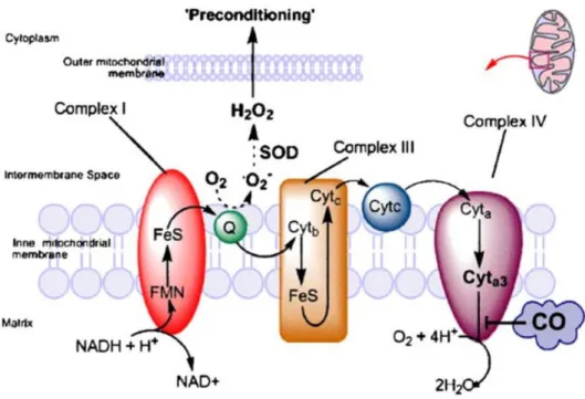
In astrocytes, CO preconditioning acts on mitochondria by a two-step response [16]. Initially, CO slightly inhibits cytochrome c oxidase (complex IV), retarding the rate of transport chain and accumulating the electrons in the complex III. Consequently, the oxygen will be enzimatically reduced to superoxide, been converted into other types of ROS and initiating signaling events that can reprogram gene expression in order to confer protection (figure 1.5) [16][17][23]. After 30 minutes, cytochrome c oxidase activity increases again [16]. This shows that CO acts by partially preventing COX in mitochondria, which generate low amounts of ROS to induce cytoprotection, preventing the excessive production of ROS and oxidative stress capable of causing damage to the cells.
Figure 1.5. Schematic illustration of the inhibitory effect of CO on the electron transport chain in mitochondria (adapted from 17).
1.6. FosB
FosB is an immediate early gene, member of Fos family genes (c-fos, fosB, fra-1 and fra-2)
that responds to a variety of stimuli, like stress and physiological perturbations [24]. Its transcription forms two different mRNAs, FosB and ∆FosB. This last one is a truncated version of the first one,
5 Many studies focus on ∆FosB, since it is mostly known for its involvement in drug addiction mechanisms [26]. However, FosB is a fundamental key player in stress tolerance and neuroprotection [27]
and it was found to be present in some neurons throughout the brain cortex of rodents [24].
In a previous work developed in our laboratory, it was studied the effects of CO-induced preconditioning in gene expression in astrocytes. Among all the differentially expressed genes, FosB showed strong upregulation (unpublished data). However, it is unknown whether CO-induced increased expression of FosB is involved in the cytoprotection role of this gasotransmitter.
As previously seen, CO is also capable of producing low amounts of ROS, being important signaling molecules for CO-induced preconditioning. Thus, one of the hypotheses is that CO promotes FosB expression in a ROS-dependent manner.
1.7. P2X7 receptor
P2X7 is a purinergic receptor, member of the P2X family. There are 7 different receptors (P2X1-7) and they are all expressed in several cell types, including neurons and glial cells [28]. These receptors are ligand-gated cationic channels that are activated in response to the extracellular adenosine 5'-triphosphate (ATP), which is a very important signaling molecule, being responsible for neuron-astroglial communication [29][30]. P2X7, from all the P2X receptors, is the one that needs the greater amount of ATP in order to be stimulated (more than 100 µM) [28][29]. Activation of P2X7 can be prolonged or transient. In the first case, a reversible pore is formed in the membrane, allowing the passage of small molecules. In some circumstances, this pore formation is associated to the induction of apoptosis. The second case reflects better a physiological scenario, only altering the gene transcription [31].
Under normal conditions, P2X7 receptors have a very low density. However, they become dramatically upregulated under pathological conditions [32], due to a great increase of the extracellular ATP following a brain injury [29]. Consequently, these receptors allow calcium influx, activating a variety of signaling processes and changing drastically the intracellular ion homeostasis [28][31-33]. Thus, the stimulation of this receptor has been associated to several nervous system diseases, such as brain ischemia, since it is involved in ATP-mediated cell death, proliferation of immunocompetent cells and also appears to be linked to the syntheses and release of many inflammatory mediators [28][29][31][32]. Astrocytes contain P2X7 receptors in their plasma membrane. Once activated, they may contribute for excitotoxicity, since their stimulation can induce glutamate release [34].
Despite all of this, recent studies have demonstrated that P2X7 receptors are essential for astrocyte-mediated ischemic tolerance [30]. Y. Hirayama et al. showed that P2X7 knockout mice failed
7
2. Objectives
Brain injury due to ischemia is a serious cause of disability and death worldwide. Neuroprotection and brain repair remains a great medical challenge, since pharmacological interventions seem to be ineffective. However, a recent strategy has becoming very popular in science community: preconditioning can induce cerebral tolerance and protection to a subsequent insult such as cerebral ischemia. The majority of studies on preconditioning have focused exclusively on neurons. In this project it will be used primary cultures of astrocytes to disclose CO-induced preconditioning and cytoprotection and the mechanism of action of FosB associated to it. To deeper understand this correlation, three main questions were made:
i) Is FosB protein expression induced by CO?
ii) Is the increased expression of FosB due to ROS produced by CO-induced preconditioning?
iii) Is P2X7 receptor involved in CO-induced FosB expression?
9
3. Materials and Methods
3.1. Cell cultures
Astrocytes were isolated from newborn mice cortex (Bl6/c57 mice) with 1-3 days old. After dissecting the hemispheres, the meninges were carefully removed. The tissue was then washed with ice-cold phosphate-buffered saline (PBS), mechanically disrupted and passed through a 70 µm nylon cell strainer (BD FalconTM) into Dulbecco's minimum essential medium (DMEM; 41966-029, GibcoTM) enriched with 20% of fetal bovine serum (FBS; 10270-106, GibcoTM) and 1% of penicillin/streptomycin solution (Pen Strep; 15140-122, GibcoTM). Cell suspension was then plated in T-flasks (6-8 hemispheres/75 cm2) and stored within controlled conditions (humidified atmosphere of 5% of CO2 at 37°C). After one week in culture, dark phase cells were removed from astrocytic cell layer by vigorous shaking. Subsequently, the medium was changed twice a week for 2 more weeks with gradual reduction in FBS content, 15% in the second week and 10% in the third one. The cultured astrocytes, after confluence, were mild trypsinized with trypsin/EDTA (0.05%; 25300-062, GibcoTM) and subcultured in 6-well plates (approximately 200,000 cells/well) and stored in the same conditions until full confluency.
3.2. Cell Treatment
ROS studies
Astrocytes were treated with β-carotene (500 µM) or N-acetylcysteine (NAC; 1 µM) for 1 hour prior to CO treatment. After, a CO-releasing molecule (CORM-A1; 12.5 µM; Sigma) was administered to the cells and incubated for 40 minutes or 1 hour. Then, cells were washed with ice-cold PBS and submitted to protein extraction. β-carotene and NAC were used as modulators of ROS production induced by CO.
P2X7 studies
Astrocytes were treated with a P2X7 inhibitor (A-438079; 10 µM; Sigma) for 10 minutes, followed by CORM-A1 incubation (12,5 µM) for 40 minutes or 1 hour. Then, cells were washed with ice-cold PBS and submitted to protein extraction.
3.3. Protein extraction and quantification
The proteins were extracted using Radio-Immunoprecipitation Assay (RIPA) lysis buffer (150 mM NaCl; 50 mM Tris-HCl (pH 7.4); 5 mM Ethylene Glycol Tetraacetic Acid (EGTA); 1% Triton; 0.5% sodium deoxycholate (DOC); 0.1% Sodium dodecyl sulfate (SDS); 1X cocktail protease inhibitor (Roche, 11697498001), pH 7.5). Then the cells were scraped and stored in -20°C until used.
10 3.4. Western blot
Protein samples were prepared in order to have 30 µg of protein each and subjected to electrophoresis by a polyacrylamide resolving gel (12%) at 135 V for approximately 1,5 hour. Then, they were electrically transferred into a nitrocellulose blotting membrane (10600018, GE Healthcare), for 1 hour at 500 mA. The membranes were blocked with 5% of bovine serum albumin (BSA) or 5% of light milk diluted in Tris-buffered saline-Tween20® (TBS-T) for 1 hour at RT. Then membranes were incubated with the primary antibody anti-FosB (rabbit monoclonal; 1:333) overnight at 4°C. After that, membranes were washed three times with TBS-T for 15 minutes each and incubated with secondary antibody anti-rabbit (from donkey; 1:5000) for 1 hour at RT. The three washing steps were repeated. Antibody detection was performed with western ECL substrate (170-5061, BioRad; 1:1) and the images were acquired in Chemidoc Touch (BioRad). Image quantification was performed with ImageJ software
3.5. Statistical analysis
11
4. Results
4.1. Primary cultures of astrocytes
Astrocytes are the most abundant cells in the CNS, contributing for the good functioning of neurons in a healthy brain. Herein it will be used primary cultures of astrocytes (figure 4.1). Astrocytes are more resistant than neurons to an insult, being able to give them support during an injury, assuring the minimal conditions for neurons to function and consequently, preventing the worsening of the lesion and promoting neuroprotection. Astrocytic cells are isolated from the cortex of newborn mice with 1-3 days old, staying in culture for 3 weeks to mature before use.
Figure 4.1. Primary culture of astrocytes. Dark regions correspond to confluent astrocytes after three weeks in culture. The lighter spots represent astrocytes under stress due to lack of space and nutrients.
12 4.2. CO-preconditioning induces FosB Protein expression
The base of this work is the potential correlation between CO-induced preconditioning and FosB expression. In a previous work developed in our laboratory, it was observed that exogenous CO was able to increase FosB mRNA expression in astrocytes after 40 and 60 minutes of its administration (data not shown). Herein, this two time points were used to study the same effect of CO but on FosB protein expression, in order to assess if its mRNA and protein expression would have the same behavior.
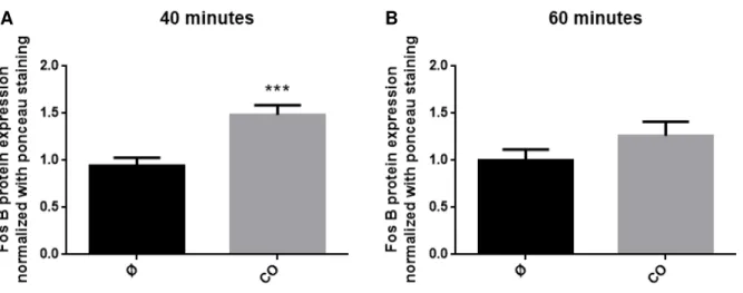
Astrocytes treated with CO during 40 minutes presented a significant increase in FosB expression compared to control (figure 4.2 A). In contrast, it was observed a non-significant increase in FosB protein expression at 60 minutes (figure 4.2 B). Nevertheless, cells treated with CO for 60 minutes showed a decrease in expression compared to astrocytes of 40 minutes. These results are in agreement with the preliminary data generated in our laboratory (data not shown), showing the reproducibility of the experiment.
Figure 4.2. Effect of carbon monoxide in FosB protein expression in a primary culture of astrocytes. Cells were treated with 12.5 µM of CORM-A1 during 40 (A) or 60 minutes (B). Data are mean ± SEM from at least ten independent experiences, *** p < 0.001 versus control.
This confirms that CO is able to induce FosB protein expression in astrocytes during preconditioning and it may have an important contribution for its cytoprotection.
13 4.3. ROS signaling induced by CO-preconditioning is not responsible for the increase of FosB protein expression
One of the aims of this project was to study whether ROS generation and signaling are important for CO-induced increase of FosB expression. Our hypothesis suggests that ROS generation is somehow involved in the increase of FosB expression. To confirm this, astrocytes were treated with β-carotene or N-acetylcysteine (NAC) 1 hour prior CO administration. These are two powerful
antioxidants with distinct mechanism of action. β-carotene, the most abundant carotenoid in human diet, acts as a direct scavenger of free radicals [36][37]. In contrast, the antioxidant properties of NAC are assigned to its ability to increase intracellular glutathione (GSH) levels, an important component of cellular detoxification pathways responsible for protecting cells against oxidative stress [38][39].
14 Figure 4.3. Effect of β-carotene and N-acetylcysteine (NAC) in FosB protein expression in a primary culture
of astrocytes treated with carbon monoxide. Cells were treated with 12.5 µM of CORM-A1 during 40 (A and C) or 60 minutes (B and D) after 1h of incubation with 500 µM of β-carotene (A and B) or 1 µM NAC (C and D). Data are mean ± SEM from at least three independent experiences, * p < 0.05 versus control.
In conclusion, the lack of statistic differences indicates that these two antioxidants have no effect on FosB expression, demonstrating that ROS signaling is not involved in the CO-induced increase of FosB expression.
A B
15 4.4. Role of P2X7 receptor in CO-induced FosB expression
Another hypothesis for CO to promote FosB expression is via activation of P2X7 receptor, since it has been seen that this receptor is essential for astrocyte-mediated ischemic tolerance. Thus, for following this hypothesis, it was used a P2X7 inhibitor, A-438079, in order to asses if its inhibition would affect FosB protein expression.
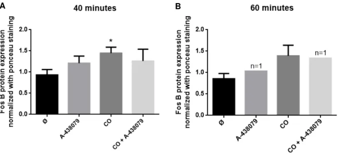
In this experiment, astrocytes pre-treated with A-438079 and then with CO did not show any significant differences in FosB expression compared to control in both 40 and 60 minutes (figure 4.4). However, prevention of P2X7 activation showed a slight tendency to revert CO-induced FosB expression at 40 minutes (figure 4.4 A).
Figure 4.4. Effect of P2X7 inhibitor (A-438079) in FosB protein expression in a primary culture of astrocytes
treated with carbon monoxide. Cells were treated with 12.5 µM of CORM-A1 during 40 (A) or 60 minutes (B) after 10 minutes of incubation with 10 µM of A-438079. Data are mean ± SEM from at least four independent experiences, * p < 0.05 versus control.
These results indicate that A-438079 is not able to revert the expression levels of FosB induced by CO. Nevertheless, it is possible to observe a slight decrease in FosB expression, indicating a possible involvement of P2X7 receptor in CO-induced FosB expression. To confirm this tendency, it would be necessary to repeat the experiment, enhancing the number of experiences, which were only four replicates.
17
5. Discussion
Despite being seen as a toxic and a silent killer, CO has many beneficial effects in the organism when administered below the threshold of injury. In vivo studies have already demonstrated
the ability of CO to protect the brain and reduce the damage in an ischemia model [40]. However, it is still not well known the mechanism(s) by which CO can induce tolerance and protect the brain. The aim of this work was to better understand the way of action of CO-preconditioning.
Herein it was demonstrated that CO increases FosB protein expression after 40 minutes of treatment, while at 60 minutes FosB expression is back to basal levels (figure 4.2). In cell culture, FosB mRNA present a half-life of approximately 90 minutes [41], reason why it was possible to observe an increase in CO-induced FosB transcription (mRNA) after 60 minutes of treatment (previous data generated in the laboratory). However, the same was not observed in FosB protein expression, whose increase was only detectable after 40 minutes of CO administration. FosB proteins have destabilizing elements that lead to rapid degradation by proteossomes [41]. This data could explain why there is an increase in mRNA levels but not in protein level at 60 minutes, returning its levels to basal ones.
It is known that CO directly acts on mitochondria, inducing ROS production, which leads signaling and preconditioning. This could be one of the pathways responsible for the increase of FosB expression. However, neither of the antioxidants used were able to revert the effect of CO on FosB expression, rejecting the previous hypothesis (figure 4.3 A and C). FosB may not be increased via ROS, but its involvement in cytoprotection cannot be discard, since it has been demonstrated its potential importance in neuroprotection [42]. Actually, ROS can be acting downstream FosB, affecting the activator protein-1 (AP-1), for example. Interestingly, NAC, one of the antioxidants used, is able to inhibit the activation of AP-1 [43]. NAC did not affect FosB expression but can act at downstream AP-1 level. This could explain why the antioxidants were not able to decrease the expression levels of FosB.
It has already been demonstrated the importance of P2X7 receptor for astrocyte-mediated tolerance [30], and its interaction with FosB [31], as well as the P2X receptors with CO [20]. Therefore, it led us to hypothesize that this receptor could be involved in CO-induced FosB increased expression. These molecules seem not to be related to each other since inhibition of P2X7 did not significantly alter FosB expression (figure 4.4 A). However, the slight reversion in CO-induced FosB expression left some doubts, having the need to generate more data in order to confirm this ambiguity.
19
6. Conclusion and Future work
Despite being negative, these data elucidated us more about the mechanisms by which CO induces cytoprotection. This enables us to elaborate new questions and to give a step forward into this subject.
It is now known that ROS is not responsible for CO-induced FosB expression. Its role in cytoprotection might follow a different pathway, but it is definitely important in CO-induced brain tolerance [44]. P2X7 receptor seems to be involved in FosB increased expression, although these results are not completely enlightening and more data need to be generated.
In future work, it is essential to demonstrate whether FosB expression is really necessary in CO-induced preconditioning. Through knocking down FosB expression (using for instance siRNA technology), it will be possible to assess if CO still can protect against cell death or not, and to determine FosB importance in this cytoprotective mechanism. It also could be interesting to study the relevance of 1, inhibiting its action in order to prove its relevance for CO-cytoprotection. Since AP-1 can be formed by other proteins belonging to c-Fos family, again, knocking down FosB could reveal if its increased expression induced by CO is important for the AP-1 formation or if it plays a different role.
21
7. References
[1] R. J. Traystman. Animal models of focal and global cerebral ischemia. ILAR Journal. 2003, 44,
85-95
[2] F. Silva. Acidente vascular cerebral isquémico - Prevenção: Aspectos actuais - É preciso agir.
Medicina Interna. 2004, 11, 99-108
[3] Direcção-Geral de Saúde. A Saúde dos Portugueses. Perspetiva 2015. Página 27
[4] U. Dirnagl et al. Pathobiology of ischeamic stroke: an integrated review. Trends in Neuroscience.
1999, 22, 391-397
[5] H. M. Bramlett et al. Pathofysiology of cerebral ischemia and brain trauma: similarities and
differences. Journal of Cerebral Blood Flow & Metabolism. 2003, 24, 133-150
[6] Helena L. A. Vieira et al. Pre-conditioning induced by carbon monoxide provides neuronal
protection against apoptosis. Journal of Neurochemistry. 2008, 107, 375-384
[7] Jin-Moo Lee et al. Brain tissue responses to ischemia. The Journal of Clinical Investigation. 2000,
106, 723-731
[8] A. Graça et al. Hipotermia induzida no tratamento da encefalopatia hipoxico-isquémica neonatal. Consenso Nacional. 2012
[9] K. R. Jessen. Glial cels. The International Journal of Biochemistry & Cell Biology. 2004, 36,
1861-1867
[10] G. Barreto et al. Astrocytes: Targets for neuroprotection in stroke. Central Nervous System Agents in Medicional Chemistry. 2011, 11, 164-173
[11] B. Gabryel et al. Role of astrocytes in pathogenesis of ischemic brain injury. Neurotoxicity Research. 2000, 3, 205-221
[12] M. V. Sofroniew et al. Astrocytes: Biology and pathology. Acta Neuropathologica. 2010, 119, 7-35
[13] Y. Zhao et al. Targeting astrocytes for stroke therapy. The Journal of the American Society for Experimental NeuroTherapeutics. 2010, 7, 439–451
[14] B. Wegiel et al. The social network of carbon monoxide. Trends in Molecular Medicine. 2013, 19,
3-11
[15] L. Rochette et al. Carbon monoxide: Mechanisms of action and potential clinical implications. Pharmacology & Therapeutics. 2013, 137, 133-152
[16] C. S. F. Queiroga et al. Carbon monoxide and the CNS: challenges and achievements. British Journal of Pharmacology. 2014, 172, 1533–1545
[17] M. Bilban et al. Heme oxygenase and carbon monoxide initiate homeostatic signaling. 2008, 86,
267-279
[18] C. S. F. Queiroga et al. Carbon monoxide targeting mitochondria. Biochemistry Research International. 2012, 2012, 749845
[19] C. S. F. Queiroga et al. Glutathionylation of adenine nucleotide translocase induced by carbon
22 [20] A. A. Untereiner et al. The Role of Carbon Monoxide as a Gasotransmitter in Cardiovascular and
Metabolic Regulation. Gasotransmitters: Physiology and Pathophysiology. 2012, 37-70
[21] R. Motterlini et al. CORM-A1: a new pharmacologically active carbon monoxide-releasing
molecule. The FASEB Journal. 2005, 19, 284-286
[22] A. S. Almeida et al. Carbon Monoxide Releasing Molecule-A1 (CORM-A1) improves
neurogenesis: increase of neuronal differentiation Yield by preventing cell death. PLOS ONE. 2016,
11, e0154781
[23] U. Dinargl et al. Preconditioning and tolerance against cerebral ischaemia: from experimental
strategies to clinical use. The Lancet Neurology. 2009, 8, 398–412
[24] K. Tahara et al. DFosB, but not FosB, induces delayed apoptosis independent of cell proliferation
in the Rat1a embryo cell line. Cell Death and Differentiation. 2003, 10, 496–507
[25] H. Kurushima et al. Selective induction of DFosB in the brain after transient forebrain ischemia
accompanied by an increased expression of galectin-1, and the implication of DFosB and galectin-1 in neuroprotection and neurogenesis. Cell Death and Differentiation. 2005, 12, 1078–1096
[26] E. J. Nestler et al.∆FosB: A sustained molecular switch for addiction. Proceedings of the National Academy of Sciences. 2001, 98, 11042–11046
[27] Y. N. Ohnishi et al. FosB Is Essential for the Enhancement of Stress Tolerance and Antagonizes
Locomotor Sensitization by ΔFosB. Biological Psychiatry. 2011, 70, 487–495
[28] A. del Puerto et al. ATP-P2X7 Receptor Modulates Axon Initial Segment Composition and
Function in Physiological Conditions and Brain Injury. Cerebral Cortex. 2015, 25, 2282-2294
[29] J. F. Oliveira et al. Rodent Cortical Astroglia Express In Situ Functional P2X7 Receptors Sensing
Pathologically High ATP Concentrations. Cerebral Cortex. 2011, 21, 806-820
[30] Y. Hirayama et al. Astrocyte-Mediated Ischemic Tolerance. The Journal of Neuroscience. 2015,
35, 3794–3805
[31] L. Y. Lenertz et al. Transcriptional Control Mechanisms Associated with the Nucleotide Receptor
P2X7, a Critical Regulator of Immunologic, Osteogenic and Neurologic Functions. Immunologic Research. 2011, 50, 22–38
[32] C. Ficker et al. Astrocyte–Neuron Interaction in the Substantia Gelatinosa of the Spinal Cord
Dorsal Horn via P2X7 Receptor-Mediated Release of Glutamate and Reactive Oxygen Species. Wiley Periodicals, Inc. 2014, 62, 1671-1686
[33] T. Ozaki et al. The P2X4 receptor is required for neuroprotection via ischemic preconditioning. Scientific Reports - Nature. 2016, 6, 25893
[34] S. S. Kang et al. P2X7 Receptor Inhibition Increases CNTF in the Subventricular Zone, But Not
Neurogenesis or Neuroprotection After Stroke in Adult Mice. Translational Stroke Research. 2013, 4,
533-545
[35] C. S. F. Queiroga et al. Paracrine effect of carbon monoxide: astrocytes promote neuroprotection
via purinergic signaling. Journal of Cell Science. 2016, 129, 3178-3188
[36] L. Mueller, V. Boehm. Antioxidant Activity of β-Carotene Compounds in Different in Vitro Assays.
23 [37] G. Null. Beta Carotene. The Gary Null Blog. 2009
(http://gna.squarespace.com/home/beta-carotene.html)
[38] R. B. Shahripour et al. N-acetylcysteine (NAC) in neurological disorders: mechanisms of action
and therapeutic opportunities. Brain and Behavior. 2014, 4, 108–122
[39] Shi-Yong Sun. N-acetylcysteine, reactive oxygen species and beyond. Cancer Biology & Therapy.
2010, 9, 109–110
[40] C. S. F. Queiroga et al. Preconditioning Triggered by Carbon Monoxide (CO) Provides Neuronal
Protection Following Perinatal Hypoxia-Ischemia. PLOS ONE. 2012, 7, e42632
[41] T. L. Carle et al. Proteasome-dependent and -independent mechanisms for FosB destabilization:
identification of FosB degron domains and implications for ∆FosB stability. European Journal of Neuroscience. 2007, 25, 3009–3019
[42] K. O. Kuroda et al. FosB Null Mutant Mice Show Enhanced Methamphetamine Neurotoxicity:
Potential Involvement of FosB in Intracellular Feedback Signaling and Astroglial Function.
Neuropsychopharmacology. 2010, 35, 641–655
[43] M. Zafarullah et al. Molecular mechanisms of N-acetylcysteine actions. Cellular and Molecular Life Sciences. 2003, 60, 6–20
[44] C. S. F. Queiroga et al. Glutathionylation of adenine nucleotide translocase induced by carbon