UNIVERSIDADE DE LISBOA
FACULDADE DE CIÊNCIAS
DEPARTAMENTO DE BIOLOGIA ANIMAL
Environmental Chemicals and
Fetal Testis Development of Human and Rat
ANA ISABEL ALVES GIL BARRERA CALARRÃO
DISSERTAÇÃO
MESTRADO EM BIOLOGIA HUMANA E AMBIENTE
UNIVERSIDADE DE LISBOA
FACULDADE DE CIÊNCIAS
DEPARTAMENTO DE BIOLOGIA ANIMAL
Environmental Chemicals and
Fetal Testis Development of Human and Rat
ANA ISABEL ALVES GIL BARRERA CALARRÃO
Dissertação orientada por:
PROF.AUGUSTA GAMA PROF.RICHARD M.SHARPE
MESTRADO EM BIOLOGIA HUMANA E AMBIENTE
Para ti, Que do meu lado estiveste E estarás, Sempre.
Se depois de eu morrer, quiserem escrever a minha biografia, Não há nada mais simples Tem só duas datas — a da minha nascença e a da minha morte. Entre uma e outra cousa todos os dias são meus.
Alberto Caeiro “Se Depois de Eu Morrer, Quiserem Escrever a Minha Biografia”
i
Acknowledgements
My appreciation goes toward the Departamento de Biologia Animal and Faculdade de Ciências da Universidade de Lisboa for allowing me to grow academically, intellectually and personally. This has been an invaluable experience for which I can only be grateful. A big thank you to Internal Supervisor Prof. Augusta Gama and to MSc Coordinator Prof. Deodália Dias for all the help and guidance.
Thank you to Dr. Rod Mitchell for running the human tissue xenografting procedures and allowing me to use them.
Thank you to all the members of the Sharpe group, especially Karen Kilcoyne and Afshan Dean, for making me feel welcome and already part of the group even before I met them. Your help at the beginning and all the consequent great fun was crucial to keep me motivated throughout this year. You are the best group I could ask for to work with.
I am deeply thankful to Sheila MacPherson and Chris McKinnell for all the training, guidance, motivation and wise support, regarding working at the lab or not. Your orientation was invaluable as I tried to adapt to a different country and I am so indebted to you for helping me improving my skills.
Saying “thank you” to Professor Richard Sharpe does not even come close to my gratitude. I am truly indebt for the opportunity that was given to me, with the bonus of developing my skills and knowledge. You gave my hopes ground to become reality and that is more than I can ever thank for.
Thanks to all my friends and family, for always supporting and encouraging me. Thanks to Nana, for patience and guidance. To Mum and Dad, I could write a whole thesis with all the things that I thank you – to make a long story short, thank you for everything, you are the best.
Thanks to my love, for being always there for me and never hesitating when rough decisions arise; for fighting and struggling by my side for a better life; for being the best there is.
ii
Table of Contents
Acknowledgements ... i
Table of Contents ... ii
Index of Figures ... iii
Index of Tables ... iii
List of Abbreviations ... iv
Portuguese Abstract ... v
English Abstract ... ix
Introduction ... 1
Testicular Dysgenesis Syndrome ... 2
Testicular Germ Cell Cancer ... 4
Phthalates ... 6
Paracetamol effect through Prostaglandins ... 7
Aims ... 9
Protocols and Procedures ...10
Human studies... 10
Tissue collection and xenografting procedure ... 10
Tissue fixation, microtoming and mounting ... 11
Triple Immunofluorescence for OCT3/4, MAGE-A4 and Ki67 ... 11
Fluorescence capture and germ cell quantification ... 13
Rat Studies ... 14
Tissue collection, fixation, microtoming and mounting ... 14
Immunohistochemistry for VASA ... 14
Staining detection and germ cell quantification ... 15
Statistical analysis... 18
Results ...19
The effect of DBP on germ cell differentiation and proliferation in human fetal testis xenografts ... 19
The effect of paracetamol and indomethacin on germ cell number in e21.5 rat fetal testes ... 25
The effect of fetal exposure to indomethacin on germ cell number in pnd25 rat testes ... 27
Discussion ...30
DBP effects on male human fetal germ cell differentiation and proliferation... 30
Indomethacin effects on male rat fetal germ cell number ... 31
Literature cited ...34
iii
Index of Figures
Figure 1. Fertility rates in 1970 and 2002 ... 1
Figure 2. Sperm counts declined in the last 70 years ... 2
Figure 3. Schematic representation of Testicular Dysgenesis Syndrome ... 3
Figure 4. Current hypothesis of how CIS cells are thought to arise ... 5
Figure 5. Formation of prostaglandins ... 8
Figure 6. Image representative of the xenografting procedure ...10
Figure 7. Series of images of the stereology method ...16
Figure 8. Formulas used to calculate the germ cell number (millions) ...17
Figure 9. Representation of the statistical analysis of human studies ...18
Figure 10. Representation of the steps of data analysis for the human studies 19
Figure 11. Photographs of vehicle control and DBP( MBP) exposed second trimester human fetal testis xenografts ...23
Figure 12. Quantification of germ cell differentiation and proliferation in control and DBP(MBP)-exposed human fetal testis xenografts ...24
Figure 13. Effect of paracetamol or indomethacin on germ cell number in rat e21.5 testes ...26
Figure 14. Images of control and indomethacin rat pnd25 testis sections ...28
Figure 15. Effect of indomethacin on germ cell number of rat pnd25 testis ...29
Index of Tables
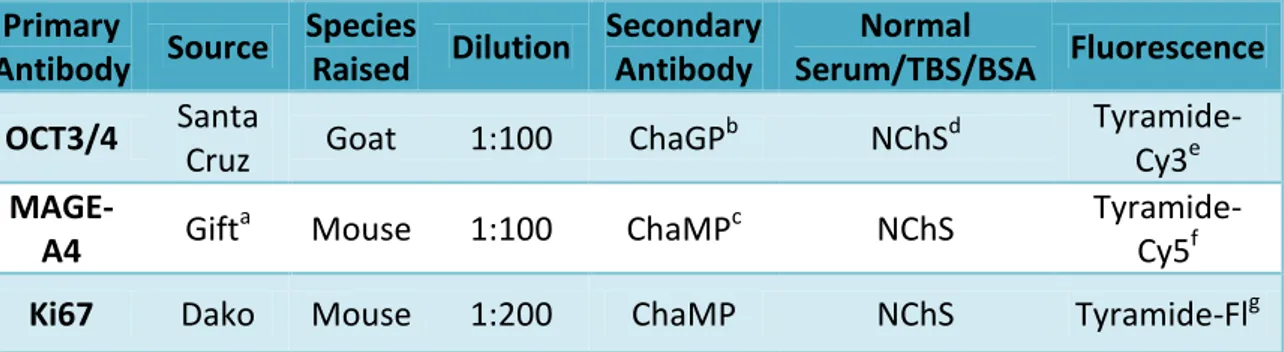
Table I. Details of antibodies used for the triple immunofluorescence ...12Table II. List of the staining combinations counted for the human studies ...13
Table III. Treatment details during rat fetal development regarding the testis collection day of the rat studies ...14
Table IV. Details of antibodies used for the immunohistochemistry ...15
Table V. Classes of germ cells counted for pnd25 rat samples ...17
Table VI. Information gathered from a section of an age-matched control of a 16 week foetus ...20
Table VII. Germ cell values from all treatments for a 16 week foetus ...21
Table VIII. How average means were calculated for the human studies ...21
iv
List of Abbreviations
AA Arachidonic Acid
ANOVA One-way analysis of variance
ChaGP Chicken anti-Goat Peroxidase secondary antibody ChaMP Chicken anti-Mouse Peroxidase secondary antibody
CIS Carcinoma-in-situ
COX Cyclooxygenase
DAB 3,3-diaminobenzidine tetrahydrochloride
DAPI 4',6-diamidino-2-phenylindole
DEHP Diethylhexyl phthalate
DBP Di(n-butyl) phathalate
e Embryonic day
H2O2 Hydrogen peroxidase
Ki67 Ki67 Antigen
MAGE-A4 Homo sapiens melanoma antigen family A, transcript 4
MBP Monobutyl pthalate
MEHP Mono-(2-ethylhexyl) phthalate
MPW Masculinization programming window
NChS/TBS/BSA Normal chicken serum diluted 1:5 in TBS containing 5% (w/v) bovine serum albumin
NGS/TBS/BSA Normal goat serum diluted 1:5 in TBS containing 5% (w/v) bovine serum albumin
NSAID Nonsteroidal anti-inflammatory drugs
OCT4/3 Octamer binding transcription factor 3/4
pnd Postnatal day
SRY Sex-determining region Y
Streptavidin-HRP Streptavidin labelled horseradish peroxidase enzyme conjugate
TBS Tris-buffered saline; 0,05M Tris and 0,85% NaCl; pH 7,6
TBST TBS + 0,05% Tween
TDS Testicular Dysgenesis Syndrome
TGCC Testicular Germ Cell Cancer
Tris Tris(hydroxymethyl)aminomethane; (HOCH2)3CNH2
Tyramide-Cy3 / Cy5 / Fl Tyramide Signal AmplificationTM – Plus Cyanine3 / Cyanine5 / Fluorescein
v
Portuguese Abstract
Ao longo das últimas cinco décadas tem vindo a registar-se um decréscimo da taxa de fecundidade humana. Embora existam vários factores que contribuam para tal (por exemplo, alterações nos comportamentos sociais), são cada vez mais comuns problemas fisiológicos masculinos, como a diminuição da concentração e qualidade espermática, que devem ser também considerados importantes. Adicionalmente, doenças como hipospádias, criptorquidismo e cancro testicular têm também vindo a aumentar de incidência. Devido ao curto período de tempo em que se registam estas grandes diferenças, parece pouco provável que haja apenas influência genética a actuar. Como em tantos outros problemas recentes, a combinação das variações do estilo de vida e da exposição ambiental a moléculas perigosas pode prejudicar o desenvolvimento do sistema reprodutor masculino, diminuindo assim a fertilidade. Foi proposta uma origem comum na vida fetal para as doenças acima referidas, em que uma função deficiente das células somáticas do testículo fetal resultaria numa ruptura da organização e desenvolvimento normal, traduzindo-se em problemas reprodutivos masculinos. Esta teoria dá pelo nome de Síndrome da Disgénese Testicular (Testicular Dysgenesis Syndrome, TDS) e mostra o quão importante é o desenvolvimento fetal das estruturas reprodutivas masculinas para uma saúde e função reprodutiva normal posterior. Uma das doenças incluídas nesta TDS é o cancro testicular com origem na linha germinal (testicular germ cell cancer, TGCC), ao qual é atribuído a maioria das mortes relacionadas com problemas de saúde em homens entre os 15 e os 35 anos e cuja incidência duplicou nos últimos 40 anos. Durante o desenvolvimento fetal no Homem, as células germinais masculinas indiferenciadas mantêm a sua proliferação em níveis baixos após a perda de pluripotência e passagem para um estado diferenciado. Esta perda de pluripotência parece conter a hipótese de como a TGCC surge nos humanos, visto que se o processo for perturbado por factores externos, as células podem entrar num estado intermediário em que mantêm a taxa de proliferação alta, adquirindo características cancerígenas. Um tipo de molécula ambiental ao qual os fetos estão expostos, e que já demonstrou afectar negativamente o sistema reprodutor masculino noutras espécies, são os ftalatos, componentes industriais cuja exposição em roedores levou ao aparecimento de condições semelhantes às da TDS. No entanto, não há informações sobre o seu efeito nos humanos. Outros químicos ambientais cujo efeito no desenvolvimento reprodutivo pode ser negativo são os anti-inflamatórios não esteróides, tais como o paracetamol e a indometacina. Embora o mecanismo exacto ainda não seja conhecido, ambos actuam através da inibição da produção ou acção de prostaglandinas, que são lípidos libertados pelas células quando ocorre uma inflamação, embora a indometacina seja considerada um supressor mais inequívoco do que o paracetamol. A administração de paracetamol e outros analgésicos demostrou ter efeitos negativos na saúde reprodutiva em roedores. Esta tese centrou-se no estudo do efeito da exposição a
vi
químicos ambientais como o ftalato di(n-butyl) (di(n-butyl) phthalate
,
DBP), paracetamol e indometacina no desenvolvimento e diferenciação das células germinais fetais, baseando-se em estudos preliminares e não publicados desenvolvidos pelo grupo de investigação em que me inseri. Para estudar o efeito do DBP, foram recolhidos testículos de fetos humanos (n = 8), com idades compreendidas entre as 14 e 20 semanas de gestação, para serem xeno-enxertados debaixo da pele dorsal de ratos imunodeficientes adultos (machos castrados) em porções pequenas. Sete dias após esta operação, os quais permitem o estabelecimento de irrigação sanguínea nos enxertos, os hospedeiros recebem doses diárias de controlo, DBP ou MBP (ftalato monobutyl, o metabolito activo do DBP) dissolvidos em óleo (500mg/kg/dia), durante 21 dias. Os hospedeiros ainda são sujeitos a três injecções semanais de gonadotropina coriónica humana (20 IU), para replicar as condições normais de gravidez e manter a produção de testosterona. Após o período de tratamento, os enxertos são recolhidos, fixados, cortados em secções e sujeitos a uma imunofluorescência tripla. No total, analisaram-se pelo menos dois enxertos controlos e dois expostos a DBP/MBP para cada feto, resultando, no mínimo, em 32 amostras. O protocolo de imunofluorescência permite visualizar células germinais com antigénios para OCT3/4 (células germinais indiferenciadas), MAGE-A4 (células germinais diferenciadas) e Ki67 (células em proliferação). As imagens foram obtidas por microscopia confocal e as células incluídas num túbulo seminífero definido, que demonstrassem fluorescência para estes antigénios, foram assumidas como pertencendo à linha germinal e contadas manualmente. Os valores registados para os enxertos-controlo e expostos a DBP, do mesmo feto, foram condensados até se obter um valor médio respectivo. A média dos enxertos controlo e dos expostos ao tratamento, de cada um dos fetos, foi utilizada então para análise estatística, usando-se um Teste t para amostras emparelhadas (significância: P<0.05). Os resultados mostraram que os enxertos expostos ao DBP(MBP) registam uma diminuição significativa de mais de 10% na percentagem de células germinais positivas para OCT3/4 (indiferenciadas). Por outro lado, a percentagem de células germinais positivas para MAGE-A4 (diferenciada) em enxertos tratados com DBP(MBP) foi significativamente maior do que os controlos. Porém, a proliferação das células germinais indiferenciadas e diferenciadas não registou alterações significativas em comparação com as amostras controlo. As células germinais reduziram a expressão de OCT3/4 para aumentarem a expressão de MAGE-A4 quando se diferenciaram, o que coincidiu com a redução, e eventualmente a perda, da capacidade proliferativa, demonstrando que o modelo dos xeno-enxertos consegue recapitular o desenvolvimento fetal normal das células germinais masculinas nos humanos. Estes resultados parecem dever-se a uma apoptose selectiva das células indiferenciadas. Se a apoptose fosse geral, a proporção de células germinais indiferenciadas e diferenciadas não mudava após o tratamento com o DBP(MBP), o que não está de acordo com os resultados obtidos, já que há um aumento de células germinais diferenciadas. Devido à falta de dados sobre o efeito do DBP nas gónadasvii
masculinas humanadas, esta hipótese estaria de acordo com dados anteriores de estudos in vitro e in vivo, em humanos e roedores respectivamente, em que a exposição a DBP reduz o número de células germinais fetais positivas para OCT3/4.
O segundo projecto desenvolvido e abordado nesta tese foi analisar os efeitos da exposição ao paracetamol e indometacina no número de células germinais nos testículos fetais de ratos. Fêmeas prenhas de ratos Wistar foram tratadas diariamente com paracetamol (350mg/kg/dia), indometacina (1mg/kg/dia ou 0.8mg/kg/dia) ou tratamento controlo. O tratamento decorreu entre o dia embrionário (e) 15.5 até ao e20.5, sendo os testículos dos descendestes recolhidos ao e21.5. As amostras foram fixadas, seccionadas e processadas através de um protocolo imunohistoquímico para o antigénio VASA (marcador típico de células germinais). O número de células germinais foi obtido por estereologia, que é um método uniforme e sistemático que permite analisar a composição celular de um tecido tridimensional, neste caso o testículo, através da contagem de núcleos de células germinais em secções bidimensionais desse mesmo tecido. Para as amostras fetais e21.5, foram contadas todas as células que expressassem VASA e que estivessem contidas num túbulo seminífero definido. A comparação entre amostras controlo e as expostas a paracetamol/indometacina foi analisada estatisticamente através de uma análise de variância ANOVA (significância: p<0.05). Os resultados mostraram uma redução significativa do número de células germinais em amostras expostas à indometacina, quando comparadas com as de controlo. Embora as gónadas masculinas expostas ao paracetamol não tenham apresentado uma diferença significativa, existe uma tendência para a redução do número de células germinais. O desenvolvimento normal das células germinais no rato passa por perderem totalmente a capacidade proliferativa quando se diferenciam, num processo síncrono e uniforme, ao contrário dos humanos. Não se sabe se o decréscimo do número de células pode ser atribuído a apoptose devido à falta de dados nesta área, mas a indometacina pode induzir um aumento da taxa de diferenciação das células germinais fetais, levando à paragem da divisão celular numa idade anormalmente precoce.
Outro ensaio experimental foi desenvolvido para investigar o efeito da exposição
in utero à indometacina no número de células germinais em testículos de ratos obtidos
no dia 25 após o nascimento (postnatal day, pnd25). O protocolo para investigar este efeito foi em tudo idêntico ao seguido para os testículos e21.5, excepto que não houve amostras expostas ao parecetamol e os testículos foram recolhidos após o nascimento (embora o período de exposição à indometacina tenha sido de e15.5 a e18.5). Visto que a gónada pnd25 já produz espermatozóides, as células germinais foram divididas em categorias que correspondem à progressão temporal da espermatogénese: espermatogónia, espermatócito I e espermatócito II. O número de células germinais de cada categoria, mais o número total de células germinais, foi contado também através de esterologia e a comparação estatística entre amostras controlo e tratadas com
viii
indometacina foi feita com um Teste t de Student (significância: p<0.05). Os resultados mostraram que o número de espermatogónias e o número total de células germinais aumentaram significativamente em amostras tratadas com indometacina, enquanto os valores para espermatócitos I e II não sofreram alterações significativas. Isto sugere que a exposição in utero à indometacina possa induzir apoptose nas células germinais e aquelas que resistem conseguem recuperar, embora faseadamente, os seus números normais após o nascimento e cessação do período de tratamento. Além disso, ambos os ensaios em ratos mostram que, pelo menos em mamíferos, o desenvolvimento fetal normal das células germinais, e posterior fertilidade dos indivíduos, é dependente de prostaglandinas.
Este conjunto de estudos mostrou que a exposição fetal a químicos ambientais comuns, tais como os ftalatos e inibidores de prostaglandinas, pode afectar o desenvolvimento fetal das células germinais nos humanos e no rato, obtendo-se dados sobre este tema pela primeira vez.
ix
English Abstract
Lifestyle and environmental exposure to hazardous molecules have been indicated as a cause for the decrease in human fertility. An increased incidence of hypospadias, cryptorchidism and testicular germ cell cancer has been reported and it has been hypothesized that these may form a Testicular Dysgenesis Syndrome (TDS) with a common origin in fetal life. Although testicular germ cell cancer (TGCC) occurs in young men, it appears that it arises from a failure of fetal germ cells to lose their pluripotency and which then transform into carcinoma-in-situ (CIS) cells, which are germ cells (GC) with pluripotency and proliferative features that probably lead to TGCC in young adulthood. It has been hypothesized that exposure in utero to some environmental factors could play a role in disturbing the testicular endocrine function in the fetus, possibly leading to TGCC. Di(n-butyl) phthalate (DBP), a common environmental chemical, has been shown to affect fetal testis development and endocrine function in the rat. Nonsteroidal anti-inflammatory drugs (paracetamol, indomethacin), which are believed to work through inhibition of prostaglandins, and other inflammation modulators can also affect fetal testis endocrine function in animal models. This study therefore aimed to assess the effect of DBP, paracetamol and indomethacin exposure on fetal GC development and differentiation. To study DBP effects, second-trimester human testis pieces from eight fetuses were xenografted under the backskin of nude castrated male mice, which were then treated with vehicle (control) or DBP for 21 days. Immunofluorescence for OCT3/4 (undifferentiated GC marker), MAGE-A4 (differentiated GC marker) and Ki67 (proliferative cell marker) was used to analyse GC differentiation and proliferation. Exposure to DBP led to a reduction in proportion of undifferentiated GC, although proliferative features were maintained, possibly due to selective apoptosis. To analyse the effect of indomethacin on GC numbers, e21.5 and pnd25 rat testes were evaluated in males exposed in utero to vehicle (control) or indomethacin (1mg/kg or 0.8mg/kg) treatment. Immunohistochemistry for VASA (GC marker) and stereology was used to assess GC number in e21.5 testes and spermatogenic temporal progression in pnd25 testes. Indomethacin exposed e21.5 rat testes showed a significant decrease in GC number, which is in agreement with preliminary results. Pnd25 rat testes from males exposed in utero to indomethacin showed an increase in spermatogonial numbers and total GC number, although the numbers for early and pachytene spermatocytes were not different. This shows that indomethacin affects germ cell development during the exposure period and that there might be a staggered recovery after birth toward normal GC number. Overall, these results show for the first time that exposure to common environmental chemicals affect human and rat male fetal GC development.
1
Introduction
Around 1.2 billion years ago, primitive eukaryotes started to reproduce sexually, which involves the recombination of parental genomes so that a new genotype is created. This allowed the emergence of more dynamic, adaptable life forms in a shorter period of time. So, sex and fertility are key factors to determine the future of our descendants. Being able to maintain fertility rates above levels of sustained population guarantees the existence of a given population as it is and reduces the risk of extinction, if no other external factors interact (such as a flood, for example).
The human species has suffered from variable fertility rates over its history. Nowadays, it is accepted that 2.1 is the minimum fertility rate to guarantee the sustainability of our species. Until two decades ago, some developing countries could reach a fertility rate of 8, and a general decrease has been noticed since. Postponing marriage and first births, associated with more women pursuing careers, are usually the most accepted reasons for the decrease in fertility rates, which in most Western countries do not even reach the replacement level (Skakkebæk et al., 2006; Figure 1).
Figure 1. Fertility rates in 1970 (blue columns) and 2002 (red columns). The minimum fertility rate
2
Although social behaviour can contribute to this change in fertility rates, there are physiological changes that should also be considered, such as falling sperm counts (Figure 2) and other reduced sperm quality features (less motility and/or abnormal shape). Plus, the incidence of hypospadias, cryptorchidism and testicular cancer also seem to be increasing (Giwercman et al., 1993; Skakkebæk et al., 2006). With these great differences in a period of 50-70 years, it seems unlikely that only genetics plays an important role. Instead, changing lifestyle and environmental exposures to hazardous molecules over the years could impair the development of the male reproductive tract and result in a decreased fertility (Skakkebæk et al., 2006).
Figure 2. Sperm counts declined
in the last 70 years. Sperm counts have changed temporally, declining to nearly half in a 70-year period, as shown in a review of 101 studies [adapted from Swan et al., 2000].
Testicular Dysgenesis Syndrome
Although spermatogenesis only starts after puberty and continues for the rest of life, the set-up for it is defined during fetal development (Sharpe, 2010). In humans, the primordial germ cell lineage can be traced back to the 4.5 day-old blastocyst, although it is not fully understood how they develop. It is formed outside of the presumptive gonad, which develops from the coelomic epithelium of the urogenital ridge, and can be found at the yolk sac, by the fourth week of gestation. The germ cells migrate through the gut into the mesoderm and arrive at the genital ridges, where their motile properties are lost. The undifferentiated gonad is then composed of germ cells and supporting coelomic cells (that give rise to Sertoli cells and Leydig cells) as well as other types of interstitial cells and Peritubular Myoid cells (Knobil & Neill, 1994). During the germ cell migration, the Sertoli cells begin to differentiate and aggregate around the germ cells to form spermatogenic cords, a critical process that is independent of testicular hormone production (George & Wilson, 1994; Brennan & Capel, 2004; Sharpe, 2010). After this, the final step is a new wave of cells from the
3
coelomic epithelium which differentiates into Leydig cells. These migrate into the interstitial regions and then begin to produce hormones.
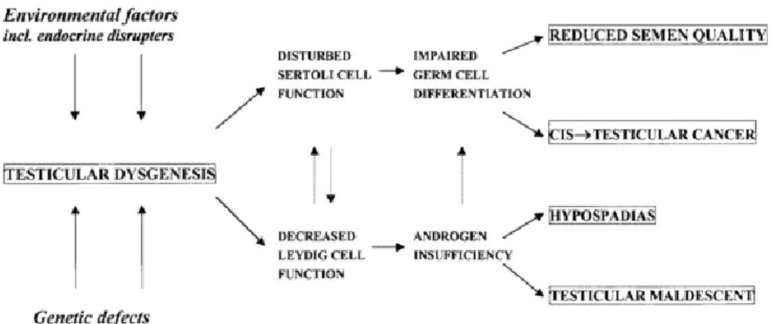
In mammals, the phenotypic development of external and internal reproductive structures in the male is dependent on the hormones the male gonad produces after it has formed (Hughes, 2001; Sharpe & Skakkebæk, 2008). In the rat, it has been shown that there is an early time window, called the masculinization programming window (MPW) in which sufficient androgen action is necessary to set up later masculinization of the reproductive tract and guarantee its proper function later in life (Welsh et al., 2008). Since there is a critical window for hormone action, a risk of disruption if external interferences occur should be considered (Sharpe, 2006). For that reason, reproductive development disorders in males can be caused by deficient action of androgens within the MPW (Sharpe, 2012) and may serve as an explanation for the increasing incidence of reproductive disorders in men. It was proposed that male disorders such as low sperm counts, cryptorchidism, hypospadias and testicular cancer could have a common origin in fetal life, in which testicular dysgenesis results in impairment of fetal testis somatic cell function (Skakkebæk et al., 2001, 2006). This is named the Testicular Dysgenesis Syndrome (TDS) and it is believed that the malformed, or dysgenic, fetal testis will cause faulty Leydig and/or Sertoli cells function, increasing the risk of reproductive disorders (Figure 3; Sharpe & Skakkebæk, 2008). TDS shows how important the normal development of the testis is, along with normal hormone action, for normal reproductive function/health in adulthood (Sharpe, 2006).
Figure 3. Schematic representation of Testicular Dysgenesis Syndrome, where early disruption of
normal testis development due to genetic or environmental factors can result in clinical problems later in life [Image reproduced from Skakkebæk et al., 2001]
4
Testicular Germ Cell Cancer
The normal development of the testis, as previously shown, can be disturbed by environmental factors, leading to impaired germ cell differentiation and resulting in testicular cancer. Testicular germ cell cancer (TGCC) accounts for 1-2% of all cancers in men, but it is the most common type of cancer in early adulthood, being the biggest cause of cancer-related death in 15-35 year-old males (Toppari et al., 1996; Sharpe & Skakkebæk, 2003). TGCC incidence has doubled in the past 40 years and, fortunately, has a high rate of treatment success (Horwich et al., 2006; Bosl & Motzer, 1997).
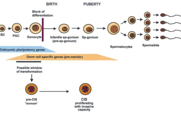
Normal germ cell development during the fetal period is essential to guarantee normal functional sperm during adult life. As said previously, highly proliferative and undifferentiated primordial germ cells migrate to the genital ridge and are enclosed by Sertoli cells, which begin an ongoing differentiation, during fetal life. The primordial germ cells are then called gonocytes and begin to differentiate into spermatogonia, the initial cell type of the spermatogenesis process, during the 2nd and 3rd trimester of pregnancy. The remaining gonocytes disappear throughout the first year of postnatal life (Skakkebæk et al., 1998) and normal spermatogenesis can occur in adulthood (Sharpe, 2010). During fetal rat development, primordial germ cells only proliferate during their pluripotency phase, typified by the expression of pluripotency markers shared with embryonic stem cells, such as octamer-binding transcription factor 3/4 (OCT3/4). The loss of this marker coincides with the quiescence phase of germ cells. In the human, germ cells have a similar behaviour, where they also lose OCT3/4 expression (Cools et al., 2006), though differentiated germ cells (OCT3/4 negative germ cells) still proliferate at a much lower rate (Mitchell et al., 2010). When this happens, they switch on expression of VASA and MAGE-A4 which mark the transition to a differentiated, and still slightly proliferative, state (Rajpert-De Meyts, 2006).
The loss of pluripotency of primordial germ cells holds the hypothesis of how TGCC begins in humans. If the primordial germ cells fail to lose pluripotency (Horwich
et al., 2006; Western et al., 2010) or there is a delay or block in their development
(Cools et al., 2006), they can enter an arrested state where they tend to be more proliferative than normal differentiated germ cells. This is believed to lead to the development of carcinoma-in-situ (CIS) cells (Rajpert-de Meyts & Hoei-Hansen, 2007; Sharpe, 2010; Western et al., 2010), which are germ cells with pluripotency and proliferative features that can spread along a seminiferous tubule throughout childhood, probably leading to TGCC in young adulthood (Sharpe, 2010). Support for this theory has been given by Jørgensen et al. (1995), who showed that CIS cells express a similar profile of markers as do gonocytes and primordial germ cells, having stem cell-like features such as pluripotency that are down-regulated when they differentiate into spermatogonia (Rajpert-de Meyts & Hoei-Hansen, 2007) (Figure 4). It has been hypothesized that impaired function of supporting cells (Sertoli, Leydig and
5
Peritubular Myoid cells) alters normal germ cell differentiation, as conditions of partial androgen insensitivity are associated with substantially increased risk of TGCC (Skakkebæk et al. 1998; Eddy, 2002; Sonne et al., 2009; Wohlfahrt-Veje et al., 2009). Because these events happen during pregnancy, it suggests that there can be environmental or lifestyle effects on the foetus via the mother that are likely to be irreversible (Sharpe, 2010).
Figure 4. Current hypothesis of how CIS cells are thought to arise, becauseof a failure of primordial germ cell/gonocyte differentiation, which would normally result in the loss of pluripotency gene expression. The resulting CIS cells stay dormant until puberty and then develop into a tumour in young adulthood. ESC, embryonic stem cells; PGC, primordial germ cells; CIS, carcinoma in situ cells. [Image reproduced and adapted from Rajpert-De Meyts, 2006].
When it comes to environmental chemicals that might affect testis development, they are considered a growing threat to health due to their wide use in developed countries. Occupational exposure and pollutants have been related to negative effects on sperm counts and spermatogenesis in the human (Sharpe, 2010). Additionally, studies in animal models showed that environmental chemical exposure, either to individual chemicals (although in considerably higher doses than expected to affect humans) or to a combinations of several chemicals, due to lifestyle choices, can induce disorders similar to those of TDS in offspring (Gray et al., 2001; Sharpe, 2012). This study focuses on how environmental chemicals, namely phthalates and indomethacin, have an impact on germ cell development in the human and rat fetal testis.
6
Phthalates
Phthalates are the most abundant man-made environmental chemicals, used extensively in industrial applications, such as solvents, plasticizers, floorings, medical devices, personal-care and cosmetic products and others. The routes of intake include ingestion, inhalation and dermal contact (Diamanti-Kandarakis et al., 2009). Phthalate exposure in fetal life has been shown to be associated with reduced anogenital distance, cryptorchidism, hypospadias and decreased fetal testis testosterone synthesis in rats (Fisher, 2004; Diamanti-Kandarakis et al., 2009; Drake et al., 2009). It is important to note that most experimental studies have used the rat as a model and have used phthalate exposure levels far higher than occur in humans and, consequently, extrapolation to the human situation should be done carefully. There are many different phthalates and not all of them cause effects on reproductive development in rats (Gray et al., 2001). The most common phthalates that humans are exposed to are diethylhexyl phthalate (DEHP) and di(n-butyl) phathalate (DBP), both of which have been shown to have similar effects on the fetal rat testis (Howdeshell et
al., 2008). Both of these phthalates are metabolised to their monoesters,
mono-(2-ethylhexyl) phthalate (MEHP) and monobutyl phthalate (MBP) respectively, which act as active toxicants in the testis (Fisher, 2004).
Phthalates have been used in different animal models to show how they can disrupt the normal development of the reproductive tract. Higuchi et al. (2003) showed that DBP caused reproductive toxicity in rabbits, with a more pronounced effect if the exposure was in utero. Impairment of spermatogenesis and long-term maldevelopment of the male reproductive structures after in utero exposure to DBP in rats was documented by Barlow & Foster (2003). Lee & Veeramachaneni (2005) used low concentrations of DBP during the sexual differentiation period on Xenopus laevis frogs, which revealed a variety of testicular lesions that persisted into adulthood, long after the exposure to DBP had ceased. It was proposed that dermal contact, due to living in an aquatic environment, could have enhanced the DBP effects, which could mimic the exposure of mammals in amniotic fluid. Other studies in rodents concluded that fetal exposure to DBP impaired normal seminiferous cord development, with an increased incidence of multinucleated gonocytes (Ferrara et al., 2006; Gaido et al., 2007; Scott et al., 2007). In utero exposure to DBP also has effects on germ cell differentiation in rats, causing a slight prolongation of pluripotency marker expression and other effects indicative of “delayed” normal development (Ferrara et al., 2006; Jobling et al., 2011). This can be compared with the idea that CIS cells arise from arrested gonocytes, maintaining pluripotency gene expression and behaviour that enhances their potential to become malignant.
Although phthalates act in an anti-androgenic way by suppressing fetal Leydig cell androgen production, their effects on germ cells are independent of androgen action
7
(Lehraiki et al., 2009) and therefore probably involve alteration of Sertoli cell function (Li et al., 2000; Jobling et al., 2011). Therefore, because embryonic development has conserved pathways, altered germ cell development and induction of TDS-like disorders after phthalate exposure in rats points to potential effects also in humans (Fisher, 2004; Carruthers & Foster, 2005; Kleymenova et al., 2005; Gaido et al., 2007). Indeed, Lambrot et al. (2009) have reported an effect in vitro of phthalates on germ cell number in the fetal human testis. In addition, Fisher (2003) concluded, after analysing the effects of DBP exposure in utero on rat testis development, that the results were remarkably similar to human TDS disorders and, since there is no animal model for human TDS currently, studying fetal rat development could provide a model system to understand the underlying mechanisms of human TDS. However, Mitchell et
al. (2012) reported that human fetal testes, which were xenografted subcutaneously
under the dorsal skin of male nude mice, appear to have unaffected steroidogenesis after the host mice are treated with DBP. This is in accordance with previous in vitro data and marmoset studies (Lambrot et al., 2009; McKinnell et al., 2009).
However, we still do not know if the level of phthalate exposure in humans is sufficient to impair testis development and if in utero exposure can affect germ cells during fetal development.
Paracetamol effect through Prostaglandins
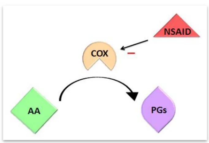
Paracetamol is generally used during pregnancy, which is believed to be safe at therapeutic doses, along with other over-the-counter painkillers (Werler et al., 2005), in response to inflammatory processes such as pain. However, the use of analgesics during pregnancy has been associated with increased risk of asthma (Rebordosa et al., 2008) and cryptorchidism (Kristensen et al., 2011a) in children. Paracetamol is believed to work through the inhibition of prostaglandins, although the exact mechanism of action is still unknown. Prostaglandins are short-lived lipids that are synthesized by many cells (excluding mature erythrocytes) and are released as a response to cell membrane traumas such as inflammation. Nonsteroidal anti-inflammatory drugs (e.g., paracetamol, indomethacin and aspirin) seem to act through the inhibition of prostaglandin release in response to inflammatory signals, thus relieving pain (McDonald-Gibson & Collier, 1979; Vane & Botting, 1987). Prostaglandins have arachidonic acid as a precursor, the production of which is catalyzed by cyclooxygenase (COX), which has two isoforms: COX-1 and COX-2. COX has three different folding units, an epidermal growth factor-like domain, a membrane-binding domain and an enzymatic domain. The COX active site presents itself as a channel, with a hydrophobic profile. COX-2 can bind larger molecules than COX-1, due to its
8
bigger active site (Vane & Botting, 1998). Paracetamol is believed to compete with arachidonic acid for the COX binding site, with some preference for COX-2, hence inhibiting the formation of prostaglandins and reducing the inflammatory response (FitzGerald, 2003; Graham & Scott, 2003) (Figure 5). Indomethacin works by inhibiting directly and non-selectively the catalytic action of COX (although it is more active against COX-1), binding to the upper portion of the active site, preventing its substrate, arachidonic acid, from entering the active site and preventing the formation of prostaglandins (Mitchell et al., 1994). It is considered to be a more unequivocal suppressor of prostaglandins that paracetamol.
Figure 5. Formation of prostaglandins
(PGs) after arachidonic acid (AA) is catalysed by cyclooxygenase (COX). It is believed that nonsteroidal anti-inflammatory drugs (NSAID), like paracetamol and indomethacin, inhibit PGs production by competing with AA for the COX binding site.
Paracetamol exposure in pregnant rats has been shown to cause mild inhibition of
testosterone and prostaglandin D2 production (Kristensen et al., 2011a). Older studies have suggested a link between prenatal exposure to mild analgesics and reduced masculinisation in animals (Gupta & Goldman 1986; Gupta, 1989). This is further supported by the
anti-androgenic effects of paracetamol discovered in rat models leading to a small reduction in anogenital distance (Kristensen et al., 2011a). As prostaglandin receptors are also expressed in Sertoli cells, contributing to cell differentiation and expression of male specific genes in mice (Wilhelm et al., 2005), it is possible that disruption of this system could be caused by exposure to paracetamol or other painkillers during pregnancy, such as indomethacin.
9
Aims
This study is focused on germ cell development during fetal life and on the impact of chemical/medicinal drug exposure, both in the human and in the rat. Therefore, it is my aim to: (1) identify whether DBP exposure affects male human fetal germ cell development and differentiation in a xenograft model; and (2) to analyze the effect of paracetamol and indomethacin on male rat germ cell number. Both studies have been prompted due to preliminary unpublished data developed by Lenka Hrabalkova (2011).
10
Protocols and Procedures
Human studies
Tissue collection and xenografting procedure
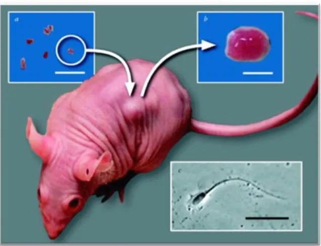
Second-trimester (14 – 20 weeks, n = 8) human fetal testes were obtained following termination of pregnancy, which were unrelated to fetal abnormalities. Women gave consent in accordance with British national guidelines and ethical approval was obtained from the Lothian Research Ethics Committee, as reported by Mitchell et al. (2010). Small portions of each testis were fixed as a pre-graft control. The rest were handled for xenografting, by being placed in ice cold media (containing Liebowitz L-15 with glutamine, 10% fetal bovine serum, 1% penicillin/streptomycin and 1% non-essential amino acids; all Sigma, Poole, UK). Fetal testis specimens were also obtained at gestational ages equivalent to the end of the grafting period and fixed as age-matched controls. Xenografting followed the protocol described in Mitchell et al. (2010; Figure 6), which was shown to recapitulate normal fetal testis growth for 6 weeks or more after grafting.After xenografting 4 – 6 human fetal testis pieces under the back skin of castrate adult male nude mice, and allowing 7 days for grafts to establish a blood supply, the host mice were treated daily by oral gavage with 500mg/kg/day of DBP, MBP or vehicle (control) for a period of 21 days. Additionally, they received injections of 20 IU of hCG three times a week in order to replicate normal pregnancy conditions and to maintain testosterone production, as described in Mitchell et al. (2010).Overall, 32 samples were analyzed (from each of eight foetuses, there were at least 2 vehicle and 2 DBP/MBP-exposed xenografts).
Figure 6. Image representative of the
xenografting procedure.Small pieces of donor human testis tissues are inserted subcutaneously under the dorsal skin of castrated male CD1 nude mice. After the post-surgery, mice are exposed to the treatment explained in the text. Grafts were retrieved and weighted after 6 weeks.
11
Tissue fixation, microtoming and mounting
Pre-grafted, xenografted and age-matched controls and xenografted treated tissues were fixed in Bouin’s fixative for two hours, transferred to 70% ethanol and then processed into paraffin blocks using standard procedures.
For microtoming, all paraffin blocks were chilled by being placed on a metal plate on top of a container full of ice. Blocks were trimmed and cut into 5µm sections on a manual rotative microtome (Leica RM 2135; Leica Microsystems), forming wax ribbons. These were transferred to a 40°C waterbath (Tissue Flotation Bath; ThermoScientific) and then placed on electrostatically positive charged slides (Surgipath X-Tra Adhesive Precleaned Microslides; Leica Microsystems), which were left overnight at 57°C in a fanned convection incubator (Carbolite, UK).
Triple Immunofluorescence for OCT3/4, MAGE-A4 and Ki67
Markers for undifferentiated germ cells (octamer binding transcription factor 3/4; OCT3/4), differentiated germ cells (MAGE-A4) and proliferating cells (Ki67) were used to investigate germ cell expression patterns, as per previous reports (Gaskell et al., 2004; Mitchell et al., 2008; Mitchell et al., 2010). Information regarding antibodies, dilutions, sera and fluorescence used are shown in Table I.
Sections were subjected to a dewax and rehydration process, by immersing in xylene (5 min), then in graded alcohols (100%, 90% and 70%, 20sec each) and finally washed in tap water. Heat-induced antigen retrieval was performed in 0,01M citrate buffer (citrate is diluted in deionized water, 1:10; pH6; Sigma) and, after being left to cool in tap water, the slides were subjected to an endogenous peroxidase block with 3% (v/v) H2O2 (VWR Prolabo) in methanol (Fisher Scientific, UK) for 30min. Two 5min
TBS (Tris-buffered saline; 0,05M Tris and 0,85% NaCl; pH 7,6) washes were performed, and they were repeated between each step of this immunofluorescence protocol. Slides were then incubated for 30min in normal chicken serum diluted 1:5 with TBS containing 5% (w/v) bovine serum albumin (NChS/TBS/BSA). Slides were then incubated overnight (at 4°C) with primary antibody OCT3/4.
12
Table I. Antibodies, dilutions, sera and fluorescence details for the triple immunofluorescence
procedure on human fetal testis. a Gift Dr Guilio Spagnoli; b Chicken anti-Goat Peroxidase; c Chicken anti-Mouse Peroxidase; d Normal Chicken Serum; e Tyramide-Cyanine3; f Tyramide-Cyanine5; g Tyramide-Fluorescein. All secondary antibodies were diluted 1:200 in NChS/TBS/BSA.
Primary Antibody Source Species Raised Dilution Secondary Antibody Normal Serum/TBS/BSA Fluorescence OCT3/4 Santa
Cruz Goat 1:100 ChaGP
b NChSd Tyramide-Cy3e MAGE-A4 Gift a
Mouse 1:100 ChaMPc NChS
Tyramide-Cy5f Ki67 Dako Mouse 1:200 ChaMP NChS Tyramide-Flg
On the next day, slides were incubated with secondary antibody ChaGP for 30min, according to Table I. Then, Tyramide-Cy3 (Tyramide Signal AmplificationTM – Cyanine3; PerkinElmer), which is red detection, was applied for 10min to all slides following the manufacturer’s instructions. Slides, which were kept in the dark from this point onwards, were blocked with 3% (v/v) H2O2 in TBST (TBS plus 0.05% Tween; Tween®20,
Sigma). Normal chicken serum block step was repeated and MAGE-A4 primary antibody was applied to all slides, which were left to incubate overnight (at 4°C).
The next day, slides were submitted to secondary antibody ChaMP for 30 min (see Table I). They were then submitted to Tyramide-Cy5 (blue detection; Tyramide Signal AmplificationTM – Cyanine5; PerkinElmer) for 10 min. After this, slides were microwaved in citrate buffer (with same dilution as for the heat-induced antigen retrieval step) on full power for 2.5min and then left to stand for 20min. Normal chicken serum block was repeated once again and slides were then incubated overnight (at 4°C) with the last primary antibody (Ki67). Afterward, secondary antibody ChaMP was applied the next day for 30 min. Slides were then incubated with Tyramide-Fl (green detection; Tyramide Signal AmplificationTM – Cyanine5; PerkinElmer) for 10min. To stain all cell nuclei, all slides were counterstained for 10 min with DAPI (4',6-diamidino-2-phenylindole; Sigma), which is diluted 1:1000 in TBS. Permafluor Aqueous Mounting Medium (ThermoScientific) was used to mount the slides.
13
Fluorescence capture and germ cell quantification
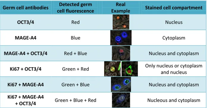
Fluorescence was captured using a Zeiss LSM 710 Axio Observer Z1 confocal laser microscope (Carl Zeiss Ltd.), which scans individual tiles from the full section, without overlap. These tiles were saved and used to count stained germ cells according to Table II. Since Ki67 is not specific for germ cells, it was not considered unless it co-localized with either OCT3/4 and/or MAGE-A4 staining. Overall, there were six different staining classes (Table II):
Table II. List of all the staining combinations counted after scanning fetal human testis
immunofluorescently for three different antibodies (OCT3/4, MAGE-A4, Ki67). Ki67 is a proliferation marker and it is not germ cell specific, so it was only recorded when in combination with either of the other two germ cell specific markers.
Germ cell antibodies Detected germ cell fluorescence
Real
Example Stained cell compartment
OCT3/4 Red Nucleus
MAGE-A4 Blue Cytoplasm
MAGE-A4 + OCT3/4 Red + Blue Nucleus and cytoplasm
Ki67 + OCT3/4 Green + Red Only nucleus or cytoplasm and nucleus
Ki67 + MAGE-A4 Green + Blue Nucleus and cytoplasm Ki67 + MAGE-A4
+ OCT3/4 Green + Blue + Red Nucleous and cytoplasm
All of these combinations were counted manually with a “laboratory counter” (Clay Adams Laboratory Counter). Then, the percentage of OCT3/4 positive (OCT3/4+) and MAGE-A4 positive (MAGE-A4+) cells were calculated, in order to know the fraction of proliferating cells (cells stained for OCT3/4, or MAGE-A4, together with Ki67). Once the information for each age was gathered, average means were calculated to be statistically analysed. For the grafts numbered FT2546/7 (17w/19w), two foetuses were grafted but they were indistinguishable, so they were handled as an 18w foetus. MBP was used in some cases and is considered as being equivalent to DBP.
14
Rat Studies
Tissue collection, fixation, microtoming and mounting
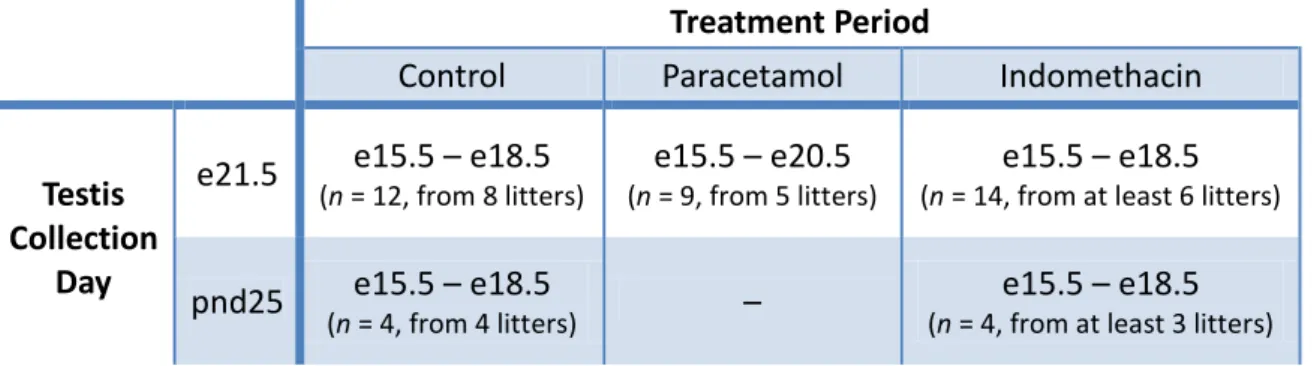
Wistar rats were housed under standard conditions according to UK home office guidelines and had free access to fresh tap water and soy-free food. Time-mated female rats were treated daily with paracetamol (350mg/kg/day) by oral gavage or with indomethacin (1mg/kg/dia or 0.8mg/kg/dia) dissolved in corn oil by subcutaneous injection. Control females were treated with corn oil by oral gavage or subcutaneous injection. Table III shows the gestational ages when the treatments were performed:
Table III. Treatment details during rat fetal development regarding the testis collection day (e,
embryonic day; pnd, postnatal day).
Treatment Period
Control Paracetamol Indomethacin
Testis Collection
Day
e21.5 e15.5 – e18.5
(n = 12, from 8 litters)
e15.5 – e20.5
(n = 9, from 5 litters)
e15.5 – e18.5
(n = 14, from at least 6 litters)
pnd25 e15.5 – e18.5
(n = 4, from 4 litters) –
e15.5 – e18.5
(n = 4, from at least 3 litters)
Tissue collection and fixation was performed in accordance with Dean et al., 2012. To generate slides, the microtoming and mounting technique used previously for human tissue was replicated for rat tissue, with no modifications.
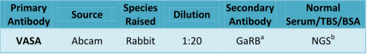
Immunohistochemistry for VASA
Antibody dilutions and serum used are listed in Table IV. Tissue dewax and rehydration, antigen retrieval, methanol/peroxide block and TBS washes were all done as described above for triple immunofluorescence in human testis, with no modifications. For normal serum block, normal goat serum (NGS/TBS/BSA; dilution performed as explained for NChS/TBS/BSA) was used to incubate slides for 30min. Then, primary antibody VASA, a germ cell specific marker (Rajpert-De Meyts, 2006), was added to slides and left overnight (at 4°C).
15
Table IV. Antibody, dilution and serum details for the immunohistochemistry procedure on rat testis. aGoat anti-Rabbit Biotinylated (diluted 1:500 in NGS); b Normal Goat Serum.
Primary Antibody Source Species Raised Dilution Secondary Antibody Normal Serum/TBS/BSA
VASA Abcam Rabbit 1:20 GaRBa NGSb
The next day, after two 5min TBS washes, slides were incubated for 30 min with secondary antibody, diluted 1:500 in NGS. Rat testis sections were then incubated with Streptavidin labelled horseradish peroxidase enzyme conjugate (Streptavidin-HRP; Vector laboratories), diluted 1:1000 in TBS, for 30min. Staining was then developed using 3,3-diaminobenzidine tetrahydrochloride (DAB substrate kit for peroxidise; Vector laboratories) for up to 10min. This reaction was stopped by placing the slides in tap water and there was no need to perform any other TBS wash. The slides were counterstained with haematoxylin and washed in tap water. They were drained as much as possible and submitted to a dehydration process, by being exposed to graded alcohols (70%, 80%, 90%, 100%; 20sec each). Then, the slides were placed in xylene (5min) and mounted using a xylene-based mountant, Pertex (Cellpath plc).
Staining detection and germ cell quantification
All images were captured using a JVC camera attached to an Olympus AX70 microscope and all countings were performed using Axiovision Rel. 4.8. software.
Germ cell quantification followed a stereology method, which is a technique that extracts quantitative information, such as the cellular composition, of a three-dimensional tissue from measurements made on two-three-dimensional planar sections of that tissue. The principle relies on identifying how many cell nuclei are present, as most nuclei are essentially spheroidal and thus the chances of detecting a cell nucleus is directly proportional to the number of such nuclei and their size. Stereology aims to estimate the number of cell nuclei and then, after measuring average nuclear size, to determine the absolute numbers of that cell in a given 2-dimensional area of tissue. The number of such cell per whole organ/tissue specimen can then be determined if
16
the weight or volume of the specimen is known. Stereology does not just count all cell nuclei, it does this in an “unbiased” manner, either by random selection of areas to be counted or by systematic selection. On fields of view, a grid with 25 x 19 points is then superimposed and the percentage of points that fall over the nucleus in question is counted. This enabled the determination of the total germ cell nuclear volume per testis. After this, germ cell mean nuclear volume was measured following the same stereology principles (by measuring the volume of germ cell nucleus from a random selection of two-dimensional fields, which is assumed to be representative of the three-dimensional tissue, and then by calculating the mean volume of all measured germ cell nuclei) (Figure 7).
Figure 7. Series of images of the stereology
method used to estimate germ cell number of the fetal rat testis from a two-dimensional section. (A) There is a random selection of at least 50 fields of view and (B) a grid with 25 x 19 points is superimposed on each field, allowing the number of points that fall over a nucleus to be counted (black stars). After this, (C) the same method is performed to measure the mean germ cell nuclear volume, by creating angled straight lines from the centre of the nucleus and marking its borders on those lines (black arrow). (A) is shown on 2x objective; (B, C) are shown on 63x objective.
B
C
17
The percentage of points that fall over a germ cell nucleus and the germ cell mean nuclear volume are used to calculate the number of germ cells (in millions), through the formulas shown in Figure 8 below.
Figure 8. Formulas used to calculate the germ cell number (millions), which was then used in statistical analysis. GC, germ cell.
For each e21.5 section, between 50 – 100 random fields were selected at 2x objective and all measurements were then performed using a 63x objective. All cells stained with VASA within a clearly defined tubule were assumed to be germ cells and counted as such.
For pnd25 comparisons, each section had between 25 – 65 random fields selected at 2x objective and all measurements were then performed using a 63x objective. Germ cells stained with VASA within a clearly defined tubule were separated into three classes, according to the description in Table V. These criteria follow the temporal appearance of different germ cell types with the progression of spermatogenesis.
Table V. All classes of VASA-stained germ cells counted for pnd25 samples, with morphological
description.
Germ Cell Class Description
Spermatogonia With flat oval nucleus; near or touching the basal
membrane
Early Spermatocyte Round nucleus; close to, but not necessarily
touching, the basal membrane
Pachytene Spermatocyte Big cells with possibly bigger nuclei, darker VASA
staining and always inside the early spermatocyte layer, towards the centre of the tubule
18
Statistical analysis
All statistical analyses were carried out using GraphPad Prism version 5 software and statistical significance was set at P<0.05. To analyse the human samples, a paired
t-test was used after calculating the average means for vehicle-exposed (control) and
DBP-exposed samples from the same fetus. This was performed as shown below (Figure 9).
Fetus A
Vehicle
Nude mouse A Xenograft 1 — GC values
Mean Value Used for statistical analysis Xenograft 2 — GC values
Nude mouse B Xenograft 1 — GC values Xenograft 2 — GC values Nude mouse C Xenograft 1 — GC values Xenograft 2 — GC values
DBP
Nude mouse D Xenograft 1 — GC values
Mean Value Xenograft 2 — GC values
Nude mouse E Xenograft 1 — GC values Xenograft 2 — GC values Nude mouse F Xenograft 1 — GC values Xenograft 2 — GC values
Figure 9. Representation of the statistical analysis of all eight human fetuses. The xenografted pieces were collected from their nude mice hosts and, after counting all types of stained germ cells from that fetus, a mean value was found for: OCT3/4+ germ cells; MAGE-A4+ germ cells; OCT3/4+/Ki67+ germ cells; and MAGE-A4+/Ki67+ germ cells. The control mean value and the DBP-treated mean value for each foetus were then used for statistical analysis. DBP, Di(n-butyl) phathalate; GC, germ cell.
For e21.5 rat testis, comparison between control samples with paracetamol and indomethacin-treated samples was done by one-way analysis of variance (ANOVA). To compare pnd25 controls with indomethacin-treated samples, a Student’s unpaired t-test was performed for each type of germ cell and for the total number of germ cells.
19
Results
The effect of DBP on germ cell differentiation and proliferation in human fetal testis xenografts
The first step of data analysis was to record the values counted for all six types of stained germ cells, for all of the human fetal testis xenografts for each fetus (Figure 10, red box).
Fetus A Vehicle
Nude mouse A Xenograft 1 — GC values
Mean Value Used for statistical analysis Xenograft 2 — GC values
Nude mouse B Xenograft 1 — GC values Xenograft 2 — GC values Nude mouse C Xenograft 1 — GC values Xenograft 2 — GC values
DBP
Nude mouse D Xenograft 1 — GC values
Mean Value Xenograft 2 — GC values
Nude mouse E Xenograft 1 — GC values Xenograft 2 — GC values Nude mouse F Xenograft 1 — GC values Xenograft 2 — GC values
Figure 10. Schematic representation of the first, second and third step of data analysis for the
human studies. Thefirst step (red box) consisted of recording the numbers of all six stained germ cell categories counted for all fetuses. The second step (blue box) simply involved calculation of the mean values/fetus for vehicle (control) and DBP-exposed samples for the six GC staining categories. The mean values were then used for statistical analysis (green box). GC, germ cell.
20
An example of germ cell values recorded for an age-matched control section from a 16 week fetus is shown in Table VI. The same is performed for age-matched controls, pre-grafted controls, vehicle controls and DBP(MBP) exposed xenografts for all foetuses (Table VII). A minimum of 100 germ cells were counted for each human fetal testis xenograft. If that number was not reached with one section, an immunofluorescence protocol was performed on up to 3 sections from the same tissue, until that number was reached, by adding the new values to those previously counted.
Table VI. Germ cell information gathered from a section of an age-matched control of a 16 week
foetus(block 2004-1391, section number 17). Germ cell stain types were counted and absolute values were recorded. The total number of germ cells stained for OCT3/4 (and MAGE-A4), whether they were co-stained with Ki67 or not, was calculated. However, because most sections had very low values for OCT3/4+ / MAGE-A4+ co-stain and triple stain, these values were disregarded when the total OCT3/4+ and MAGE-A4+ values were determined. Then, the percentage OCT3/4+ germ cells and MAGE-A4+ germ cells in the section were calculated. The percentage of proliferative cells found in the OCT3/4+ and MAGE-A4+ pool was also recorded. The highlighted values (red discontinuous line) were the ones used for the next phase of data analysis. MCTRL, age-matched control; %, percentage; GC, germ cells.
Section 2004-1391.17 MCTRL
Germ cell stain types Absolute
values Percentage (%) How (%) is called How (%) was calculated OCT3/4+ 214 % proliferative OCT3/4+ GC i 7 otal 00 OCT3/4+/ Ki67+ 90 29,61
Total OCT3/4+ 304 72,04 % OCT3/4+ GC otal otal 00 MAGE-A4+ 112 % proliferative MAGE-A4+ GC A A i 7 otal A A 00 MAGE-A4+ / Ki67+ 5 4,27
Total MAGE-A4+ 117 27,73 % MAGE-A4+ GC otal A A otal 00
OCT3/4+ / MAGE-A4+ 0
Triple 1
21
Table VII. Germ cell values from all treatments and controls for a 16 weeks foetus. The values highlighted in Table V are shown on the first line of this Table, also highlighted (red discontinuous line). The rest of the table is completed by following the same procedure for all human fetal testis xenograft sections for this 16 week fetus. MCTRL, age-matched control; CTRL, pre-graft control; VEH, vehicle control; DBP (MBP), DBP or MBP exposed samples; GC, germ cell.
16 week
fetus Block % OCT3/4+ GC
% proliferative OCT3/4+ GC % MAGE-A4+ GC % proliferative MAGE-A4+ GC MCTRL 2004-1391 72,04 29,61 27,73 4,27 CTRL 2010-3588 7,99 8,00 92,01 0,00 VEH 2010-3594 A 8,82 55,56 91,18 0,00 2010-3594 C 4,55 83,33 95,45 0,00 2010-3595 A 9,92 58,33 90,08 0,00 2010-3595 C 2,03 100,00 97,97 0,00 DBP (MBP) 2010-3598 A 4,13 80,00 95,87 0,00 2010-3598 C 3,13 85,71 96,88 0,46 2010-3599 A 1,35 100,00 97,97 0,00 2010-3599 C 5,45 35,71 94,16 2,48
The second step was to find the mean values for control and DBP(MBP) exposed human fetal testis xenografts (Figure 10, blue box). For each fetus, a mean percentage was calculated for control and DBP (MBP) exposed human fetal testis xenografts (Table VIII). All means were gathered into Table IX.
Table VIII. How average means were calculated for control and DBP treated groups followingon from the example given for a 16 week fetus in Table VII. The mean percentage of control OCT3/4+ germ cells was found by calculating the average mean of age-matched controls (MCTRL), pre-graft controls (CTRL) and vehicle controls (VEH) (red box). The mean percentage of DBP(MBP) treated OCT3/4+ germ cells was found by calculating the average mean of DBP and MBP exposed xenografts (green box). The same technique was done for % proliferative OCT3/4+ germ cells; % MAGE-A4+ germ cells; % proliferative MAGE-A4+ germ cells. GC, germ cell.
16 week
fetus Block % OCT3/4
+ GC % proliferative OCT3/4+ GC % MAGE-A4+ GC % proliferative MAGE-A4+ GC MCTRL 2004-1391 72,04 29,61 27,73 4,27 CTRL 2010-3588 7,99 8,00 92,01 0,00 VEH 2010-3594 A 8,82 55,56 91,18 0,00 2010-3594 C 4,55 83,33 95,45 0,00 2010-3595 A 9,92 58,33 90,08 0,00 2010-3595 C 2,03 100,00 97,97 0,00 DBP (MBP) 2010-3598 A 4,13 80,00 95,87 0,00 2010-3598 C 3,13 85,71 96,88 0,46 2010-3599 A 1,35 100,00 97,97 0,00 2010-3599 C 5,45 35,71 94,16 2,48
22
Table IX. All mean values for all four control and DBP exposed groups(% OCT3/4+ germ cell; % MAGE-A4+ germ cells; % proliferative OCT3/4+ germ cells; % proliferative MAGE-A4+ germ cells). The control mean average (red box) and the DBP (MBP) treated mean average (green box) for the values highlighted in Table VII are shown for a 16 week fetus. These values were used to perform four paired t-tests, each one corresponding to one of the analysed groups. w, week; GC, germ cell.
Analysed Groups
Ages
% OCT3/4+ GC % MAGE-A4+ GC % proliferative
OCT3/4+ GC % proliferative MAGE-A4+ GC Control DBP (MBP) Control DBP (MBP) Control DBP (MBP) Control DBP (MBP) 14w 24,63 5,92 74,55 94,08 66,51 55,56 3,21 1,59 16w 17,56 3,51 82,40 96,22 18,80 75,36 0,71 0,74 18w 28,75 2,58 70,96 96,97 57,10 87,50 0,63 0,77 18w 16,75 12,17 82,09 85,67 55,32 86,24 3,55 0,78 19w 5,05 3,69 94,95 96,31 27,24 0,00 3,75 3,79 19w 30,89 29,15 69,11 70,72 87,15 27,17 0,97 0,90 20w 43,78 30,27 56,17 69,58 62,07 67,07 0,78 0,61 20w 18,53 15,06 81,47 84,71 59,38 62,76 1,18 0,14
The statistical analysis (Figure 10, green box) was performed by comparing the values from control xenografts with values from the DBP (MBP) exposed xenografts for each analysed group (% OCT3/4+ germ cells; % MAGE-A4+ germ cells; % proliferative OCT3/4+ germ cells; % proliferative MAGE-A4+ germ cells).
Germ cell differentiation in human fetal testis xenografts was assessed by using a germ cell pluripotency marker (OCT3/4) and a differentiated germ cell marker (MAGE-A4) (Figure 11). Human fetal testis xenografts exposed to DBP (or its active metabolite MBP) showed a decline in the percentage of OCT3/4+ germ cells of more than 10% (paired t-test, t=3,25, df=7, p=0,014) (Figure 12, A). In contrast, when comparing the percentage of MAGE-A4+ germ cells, DBP(MBP) treated xenografts show an increase of 10% above control xenografts (paired t-test, t=3,14, df= 7, p=0,016) (Figure 12, B).
The proliferation of undifferentiated (OCT3/4+) and differentiated (MAGE-A4+) germ cells was measured using a proliferation cell marker, Ki67, which is non-specific for germ cells (Figure 11). DBP(MBP) xenografts, when compared to control xenografts, showed no difference in the percentage of proliferating OCT3/4+ germ cells, which was ~60% (paired t-test, t=0,27, df=7, p=0,795) (Figure 12, C). The percentage of MAGE-A4+ germ cells also registered no difference between control and DBP(MBP) xenografts, which was ~2% (paired t-test, t=1,84, df=7, p=0,108) (Figure 12 D).
23
Vehicle
Control
DBP
(MBP)
Figure 11. Photographs of vehicle control and DBP( MBP) exposed second trimester human fetal
testis xenografts after the immunofluorescence protocol for OCT3/4 (red; square labelled arrow - undifferentiated germ cell marker), MAGE-A4 (blue; star - differentiated germ cell marker) and Ki67 (green; circle labelled arrow - proliferating cell marker, and note that it co-stains with OCT3/4). A, C are confocal images of the testis section that result from overlapped individual tiles. B, D are confocal scanned individual tiles used to count germ cells within a clearly defined seminiferous cord and to assess their proliferation. Partial germ cells were counted only if they were on the left or bottom limit of the tile. Germ cells stained positive for the combinations OCT3/4/MAGE-A4, MAGE-A4/KI67 and OCT3/4/MAGE-A4/Ki67 are not shown. Scale bar (A, C) = 500µm; (B, D) = 50µm.
![Figure 1. Fertility rates in 1970 (blue columns) and 2002 (red columns). The minimum fertility rate of 2.1 is also portrayed (green line) [Reproduced from Skakkebæk et al., 2006]](https://thumb-eu.123doks.com/thumbv2/123dok_br/19207748.956846/13.892.170.747.616.923/figure-fertility-columns-columns-fertility-portrayed-reproduced-skakkebæk.webp)
![Figure 2. Sperm counts declined in the last 70 years. Sperm counts have changed temporally, declining to nearly half in a 70-year period, as shown in a review of 101 studies [adapted from Swan et al., 2000].](https://thumb-eu.123doks.com/thumbv2/123dok_br/19207748.956846/14.892.371.761.398.628/figure-sperm-declined-changed-temporally-declining-studies-adapted.webp)