From the Department of Orthopaedics and Traumatology, Hospital das Clínicas, Faculty of Medicine, University of São Paulo.
AN EXPERIMENTAL MODEL FOR THE
TRANSPLANTATION OF FETAL CENTRAL NERVOUS
SYSTEM CELLS TO THE INJURED SPINAL CORD IN
RATS
Tarcísio Eloy Pessoa de Barros Filho, Reginaldo Perilo de Oliveira, Ana Maria Tsanaclis, Erika Meirelles Kalil Pessoa de Barros, Alexandre Fogaça Cristante, Ricardo Moreira Palma, Cristian Vilela dos Santos and Raphael Martus Marcon
RHCFAP/3103 BARROS FILHO TEP de e col. - An experimental model for the transplantation of fetal central nervous system cells to the
injured spinal cord in rats. Rev. Hosp. Clín. Fac. Med. S. Paulo 57(6):257-264, 2002.
INTRODUCTION: Traumatic spinal cord injury is one of the most disabling conditions occurring in man and thus
stimulates a strong interest in its histopathological, biochemical, and functional changes, primarily as we search for preventive and therapeutic methods.
PURPOSE: To develop an experimental model for transplantation of cells from the fetal rat central nervous system to
the site of an injured spinal cord of an adult rat in which the transplanted cells survive and become integrated. This experimental model will facilitate investigations of factors that promote regeneration and functional recovery after spinal cord trauma.
MATERIAL AND METHODS: Fifteen adult Wistar rats underwent laminectomy, and an spinal cord lesion was made
with microdissection. Fetal spinal cord tissue was then transplanted to the site of the injury. The rats were monitored over a 48-hour period, and then their vertebral column was completely removed for histological analysis.
RESULTS: In 60% of transplanted rats, the fetal tissue at the injured site remained viable in the site of the lesion.
DESCRIPTORS: Spinal cord injury. Fetal cells. Rats.
INTRODUCTION
Spinal cord injury has been consid-ered a condition without the possibil-ity of successful treatment15. However,
emerging studies in this field indicate that acute spinal cord injuries can be minimized by the use of pharma-cologic therapy when drugs are admin-istered within a short period of time following the trauma9,23,26,60. This
progress is attributable largely to his-tological observations, which have fa-cilitated better understanding of the sequence of events involved in the spi-nal cord injury10.
The first experiments focusing the
pathophysiologic mechanism of the injured spinal cord were performed at the beginning of this century1, 2.
Nev-ertheless, these works were resumed only in the past decade by investiga-tors who have begun to value the time-dependent changes implicated in the physiopathology of spinal cord trauma3,6,48.
The significant neurological defi-cit related to the spinal cord injury is caused by the sum of two different
conditions: the initial mechanical in-jury and the secondary endogenous injury resulting from the initial in-jury6,7,20,22,32,47,59,63. The primary damage
ef-forts are based to reduce secondary in-jury7,8,24,25,34,42,43,46,53,57,58,61,63. From a
pharmacological standpoint, drugs that modulate the endogenous re-sponses to the primary injury have been progressively introduced into therapeutic protocols to restrict the col-lateral damage and improve the poten-tial for functional recovery of these patients. These drugs are administered to interrupt the pathophysiologic mechanisms of the secondary neuronal injury27,28,51.
Clinical and scientific advance-ments indicate that acutely injured spinal cords can be managed by phar-macological therapeutic procedures used within a short period of time. Methylprednisolone administered within the initial 8 hours after the trauma is the first pharmacological agent to promote significant improve-ment in the recovery of the injured spi-nal cord in human beings 4,11-14,19,35-40,52,56.. Other drugs, such as tirilizad5, 39,41and GM-118, 30, 31, 33, 49, 50, 64,, which
are still under clinical investigation, demonstrate promising preliminary re-sults. These advancements should rep-resent a significant improvement in the quality of life of patients with spinal cord injury once they are employed in current medical practice.
Central nervous system injuries are followed by a deficit condition and a period of variable functional recovery. Such recovery is strongly related to changes that involve unharmed cir-cuits, although the exact mechanism of the recovery has not been completely clarified. Transplanting neural cells is helping investigators to understand the development of the CNS and its re-sponse to injuries. More recently, such transplants have been used to optimize the posttraumatic functional recovery. The specific mechanism through which such transplants act is not completely understood. Present theories posit the influence of trophic activity along with the release of
hor-mones and neurotransmitters, and the re-enervation of host cells by the trans-planted cells16. Current research is
in-tended to determine the degree of re-covery that can be achieved by these transplants.
The possibility of employing fetal nervous system cells for the treatment of a number of CNS pathologies has stimulated several studies on the physiology of the survival and integra-tion of the transplant. Today it is known that transplanting fetal cells potentiates the locomotor recovery both in immature and adult individu-als; However, the mechanism respon-sible for this observation is still un-known44.
Several protocols for transplanta-tion of fetal nervous system cells have been reported, but there is not a con-sensus among physicians on the best method 44.
OBJECTIVE
The purpose of this study was to develop a reproducible method for transplanting cells from the fetal rat central nervous system to the site of an injured spinal cord of an adult rat that results in the survival and integration of the transplanted cells. Using this methodology, researchers will be able to study other factors that support re-generation and functional recovery of the posttraumatic spinal cord.
MATERIALS AND METHODS
Model of an Injured Spinal Cord and Donor Tissue Preparation
One male and two female rats were selected and put into 5 cages. After 12 hours, vaginal swabs from the female rats were taken and analyzed under optical microscopy to verify the pres-ence of spermatozoids. The female rats
whose vaginal swabs presented evi-dence of spermatozoids were consid-ered pregnant and isolated in a differ-ent cage.
The donor tissue was obtained from fetal rats obtained by cesarean section on Day 14 of gestation. Imme-diately after the cesarean, each fetus had its central nervous system re-moved, which was inoculated into the injured spinal cord of the adult rats.
We analyzed the injuries from 15 rats initially, which were produced through hemilaminectomy at the T10 level and aspiration and delicate 05-mm segment microdissection from the rat spinal cord.
The donor tissue was transplanted into the site of the injury using micropipettes, and it was then properly sutured.
The rats were sacrificed 48 hours following transplantation, and their spinal cords were surgically excised and submitted to histopathological analysis to verify the viability of the transplanted cells.
Rats
We used male and female Wistar rats aged 20 weeks and weighing 270 to 315g and 200 to 280g respectively, which were from a single supplier. They were acquired 1 week prior to the sur-gery to become acclimatized and man-ageable.
Laminectomy
The spinal cord was exposed through a laminectomy as follows: • An opening was made in the
me-dian dorsal line of the skin to ex-pose the vertebral column at T10 level (Fig. 1A) .
moved using a small punch. The re-moval began by the caudal edge of T10. Small fragments were delicately removed using the punch guided from the cranial to the caudal half of the sheath at the T9 level. (Fig. 1B). The spinal cord was not harmed (Fig. 2) .
The spinal cord injury procedure
• A 0.05 mm segment of one half of the medula of the rat was removed by aspiration and microdissection using a microscope.
• The rat was removed and placed on
a warm surface. The contusion site was inspected. Any bleeding was stopped, and the contusion site was washed with a saline solution.
Material for the transplantation
The female rats underwent a cesarean section (Fig. 3A) . The fetuses were removed, and embryonic cells of the CNS were harvested with a micro-surgical technique (Fig. 3B). The ma-terial that was obtained was obliquely sectioned into 0.05-mm segments for transplanting into the injured site.
Transplantation of fetal cells into the injured site
• The segment of the fetal CNS that was previously sectioned was im-planted into the site of the injured spinal cord of adult rats.
• After the transplantation, the dural sac was closed using fibrin glue. Muscular, subcutaneous and cuta-neous tissues were sutured.
Posttraumatic procedures
Over a 48-hour period following the injury, the rats were observed and had their deficits registered.
Euthanasia procedure and taking tis-sue samples for acute experiments
Euthanasia was performed 48 hours after surgery. The procedures for the euthanasia and the removal of tissue samples were as follows:
• 48 hours after the injury and trans-plantation, the rat body weight was registered at the time of sacrifice. • The rat was anesthetized using
pentobarbital 40 mg/kg given in-travenously.
• The aorta of the rat was catheter-ized through thoracotomy to allow perfusion with paraformaldehyde solution.
• The vertebral column from C5 to L5 level and most of the muscles were quickly removed (Fig. 4).
• The vertebral column was placed in a centrifuge, covered, and taped
with Parafilm
.
Necropsy
After sacrifice, which occurred 48 hours after the spinal cord was dam-aged and fetal cell transplantation was performed, the animals were weighed and the identity and gender were con-firmed. Then, the vertebral column and the spinal cord were removed.
·
Histological analysis of the trans-planted spinal cord
Histological sections were ob-tained from the area of the spinal cord containing the transplanted cells. The magnitude of the injury and/or the presence, placement, and viability of the implanted cells were analyzed.
RESULTS
The breeding procedure resulted in 4 female rats with vaginal swabs posi-tive for spermatozoids. They all be-came pregnant, and on day 14 of ges-tation, the rats underwent a cesarean section and were sacrificed.
After the production of the spinal cord injury, all the rats presented evi-dence of neurological deficits, which varied from complete monoplegia to complete paraplegia. The clinical find-ing of the magnitude of the spinal cord
injury was later correlated with its his-tological finding.
All the rats were still alive 48 hours after the fetal cells transplanta-tion. Also, there was no evidence of autophagia, pressure ulcers, or infec-tion.
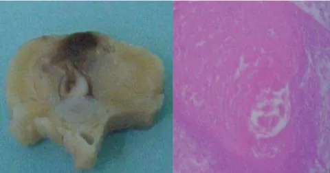
The histological analysis of the spinal cord sections relating to the site of the injury showed that the spinal cord damage resulting from the micro-dissection was not constant, varying from 40% to subtotal injury (Fig. 5). In 3 cases, a hematoma was found at the injured site, which was caused by a broken blood vessel (in this case, the anterior spinal artery).
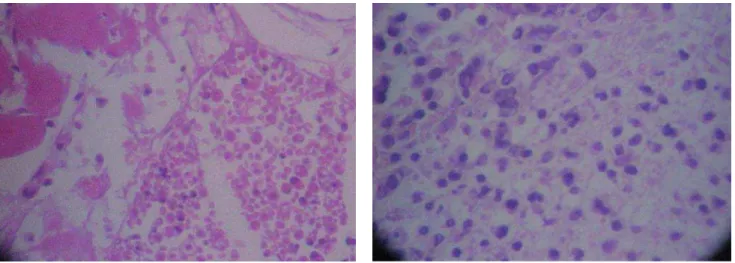
The analysis of the histological sections showed the presence of the implanted fetal cells after a 48-hour period. However, in 40% of the cases, fetal cells were not identified at the site of the injury, but only in its tra-jectory (Table 1 and Fig. 6).
DISCUSSION
Traumatic spinal cord injury is one of the most disabling conditions oc-curring to man, which stimulates a strong interest in its histopathological, biochemical, and functional changes, mainly when we are searching for
pre-Table 1 - Viability and place of the fetal cells transplantation.
RAT TRANSPLANTED FETAL TRANSPLANTED AT THE INJURY SITE
CELLS VIABLE CELLS AT THE INJURY
TRAJECTORY
1 Yes Yes Yes
2 Yes No Yes
3 Yes Yes Yes
4 Yes Yes Yes
5 Yes No Yes
6 Yes No Yes
7 Yes Yes Yes
8 Yes No Yes
9 Yes No Yes
1 0 Yes Yes Yes
1 1 Yes Yes Yes
1 2 Yes No Yes
1 3 Yes Yes Yes
1 4 Yes Yes Yes
1 5 Yes Yes Yes
Figure 4 – Removal of the vertebral column from the C5 to L5 level.
high costs, little availability, handling difficulties, and ethical considera-tions7. Rats are a good alternative in
these experiments because their spinal cord has a cytoarchitectural organiza-tion and vascularizaorganiza-tion similar to that of humans7.
Transplantation of cells from the central nervous tissue has been used for the past 20 years and has helped to increase understanding concerning the development of the nervous system and the neuronal response to injury. More recently, studies involving trans-plantation of cells from the central nervous system have been performed to restore or reduce the loss of func-tion resulting from the injury. It has been proven that transplantation can reduce deficits or even increase the functional recovery following damage of the central nervous system, mainly in cases of degenerative diseases. Transplantation can influence the re-covery of the function after the CNS trauma through several mechanisms including non-specific consequences of transplantation, trophic actions, hormone and neurotransmitter release, and more specific mechanisms involv-ing re-enervation of host cells and es-tablishment of reciprocal connections between the transplanted and the host tissue16,17.
ventive and therapeutic methods for managing the sequelae of the spinal cord trauma.
For treating the spinal cord injury in man, experimental models are nec-essary for testing drugs, surgery tech-niques, and other therapeutic proce-dures, such as cell transplantation to the injured site. In the literature, there is not a universally accepted model of experimental spinal cord injury, which is attributable largely to different pa-rameters that are analyzed and the sig-nificant diversity of therapeutic tech-niques that are tested.
Open experimental spinal cord in-juries produce the best conditions for study. Closed injuries produce frac-tures whose fragments change the natu-ral course that is intended for study. The spinal cord that is exposed can be directly sectioned or contused. Experi-mental methods that use different im-pacts as causative agents for the spi-nal cord injury include falling weights, crushing of the spinal cord using special tweezers for aneurysm57,
extradural balloons that are gradually inflated, and damage caused by radiofrequency and microdissection. The ideal model would include the traumatic mechanism usually found in human beings, reproducibility, and the possibility of a quantitative analysis.
Nevertheless, such model has not yet been described.
We used a laminectomy technique and the 05-mm microdissection of one half of the medula in this preliminary study, and we analyzed the placement and the viability of a 05-mm segment of fetal CNS implanted into the site of the injury.
Although the microdissection was performed using a microscope, the his-tological analysis of the spinal cord injury showed evidence of differences in the magnitude of the injuries that were produced, which were usually larger than 50% of the spinal cord. These differences did not impair the objective of this study, which was to evaluate the histological presence of fetal cells 48 hours after the transplan-tation. However, for better standardiza-tion of the injury, subsequent studies will have to focus on the chronic phase of the injured spinal cord using func-tional evaluations through locomotion scales, electrophysiological, and pathological assessments.
We chose using Wistar rats because of their good availability and the mi-nor technical difficulties in handling these animals. The preferred species for experiments involving spinal cord injury is one of the nonhuman pri-mates, but their use is restricted due to
The requirements for anatomical and functional recovery after spinal cord injury are more complex than from other neurological damage, which usually requires only restoration of the neurotransmitter levels 16.
There are several mechanisms through which the transplantation of fetal cells can affect the response to the injury and mediate the functional re-covery after the injury. The trans-planted fetal cells from the central nervous system are able to connect the spinal cord with the supraspinal struc-tures through the site of the injury. The transplanted cells act as a substratum for restoring cellular communication between upper and lower levels of the
injured tissues. At the cellular level, transplantation can supply trophic support either for mature or immature neurons, inhibit the formation of a glial scar at the site of the injury, and supply a favorable mechanical substra-tum of extracellular matrix for neuro-nal growth16.
Transplantation using cells from the central nervous system can im-prove the locomotor function after a spinal cord injury and provide a more complex microenvironment than that offered by peripheral nerve transplan-tation, cell suspensions, or genetically altered cells44.
In our study, the histological
analy-sis of the injured site showed the pres-ence of viable fetal cells in 9 of 15 rats (60% of cases) that underwent the transplantation of fetal cells from CNS. In 40% of the cases fetal cells were not found at the site of the injury, but only in its trajectory.
This study showed the potential of a rat model of using transplanted fetal spinal cord cells that remain viable for 48 hours after their transplantation. Additional studies on the chronic phase of the spinal cord injury and the short- and long-term viability of fetal cells using functional assessments and histopathological evaluations are planned.
RESUMO RHCFAP/3103
BARROS FILHO TEP de et al. -Modelo experimental de transplante de células do sistema nervoso central fetal para lesão de medula espinal em ratos. Rev. Hosp. Clín. Fac. Med. S. Paulo 57(6):257-264, 2002.
INTRODUÇÃO: A lesão
traumáti-ca da medula espinal consiste numa das mais incapacitantes lesões que o ser humano pode sofrer e tem desper-tado grande interesse no conhecimen-to das alterações hisconhecimen-topaconhecimen-tológicas, bi-oquímicas, funcionais e
principalmen-te na busca de métodos de prevenção e tratamento.
OBJETIVO: Propor um modelo
experimental de transplante de células do sistema nervoso fetal de ratos para o sítio de lesão medular de ratos adul-tos que permitisse sua sobrevivência e integração para possibilitar protocolos de pesquisa para identificar outros fa-tores de regeneração e recuperação funcional pós trauma raquimedular.
MATERIAL E MÉTODOS:
Utili-zaram-se 15 ratos adultos que foram submetidos a laminectomia e lesão de 5mm de hemimelula realizada com
au-xílio de microscópio óptico. Os ratos tiveram seu sítio de lesão medular transplantado com células do sistema nervoso central de fetos de rato. Os ra-tos foram monitorados por 48 horas e tiveram sua coluna vertebral extraída para análise histológica.
RESULTADOS: Demostrou-se que
em 60% dos casos as células transplan-tadas permaneciam viáveis no sítio da lesão.
DESCRITORES: Lesão medular. Células fetais. Ratos.
REFERENCES
1 . ALLEN AR - Remarks on histopathological changes in spinal cord due to impact: An experimental study. J Nerv Ment Dis, 1914; 41:141-147.
2 . ALLEN AR - Surgery of experimental lesions of spinal cord equivalent to crush injury of fracture dislocation. Preliminary report. J Am Med Assoc, 1911; 57:878-880.
3 . ANDERSON DK, MEANS D & SPEARS J - Spinal cord energy metabolism following compression trauma to the feline spinal cord. J Neurosurg, 1980;53:375-380.
4 . ANDERSON DK, MEANS D & WATERS,TR - Microvascular perfusion and metabolism in injured spinal cord after methylprednisolone treatment. J Neurosurg, 1982; 56:106-113.
6 . BALENTINE JD - Hypotheses in spinal cord trauma research. In BECKER PB. Central Nervous System Trauma Status Report, 2nd ed. NIH, 1985. p 455-461.
7 . BALENTINE JD - Pathology of experimental spinal cord trauma. Lab Invest, 1978; 39:236-253.
8 . BEGGS JL & WAGGENER D - Transendothelial vesicular transport of protein following compression injury to the spinal cord. Lab Invest, 1976;34:428-439.
9 . BEHRMANN DL & BEATTIE MS - Modeling of acute spinal cord injury in the rat: neuroprotection and enhanced recovery with methylprednisolone, U-74006F and YM-14673 high 1. Exp Neurol, 1994; 126:61-75.
10.BEHRMANN DL, BRESNAHAN JS, BEATTIE MS et al - Spinal cord injury produced by consistent mechanical displacement of the cord in rats: behavioral and histologic analysis. J Neurotrauma, 1992;9:197-217.
11. BRACKEN MB - Pharmacological treatment of acute spinal cord injury: current status and future projects. J Emerg Med, 1993; 11:42-48.
12.BRAUGHLER JM & HALL ED - Current application of high-dose steroid therapy for CNS injury: A pharmacological perspective. J Neurosurg, 1985; 62:806-810.
13.BRAUGHLER JM & HALL ED - Pharmacokinetics of methylprednisolone in cat plasma and spinal cord following a single intravenous dose of the sodium succinate ester. Drug Met Dispos, 1982; 10:551-552.
14.BRAUGHLER JM & HALL ED - Lactate and pyruvate metabolism in injured cat spinal cord before and after a single large intravenous dose of methylprednisolone. J Neurosurg, 1983; 59:256- 261.
15.BREASTED JH - The Edwin Smith Papyrus, In : BREASTED JH. Spinal Trauma. 1th ed. Chicago, University of Chicago Press,
1930. p. 5.
16.BREGMAN BS & BAGDEN EK - Potential Mechanisms underlying transplant mediated recovery of function after spinal cord injury . In: MARWAH J, TEITELBAUM H, PRASAD KN. Neural transplantation, CNS neuronal injuries and regeneration. 2nd
ed. CRC Press Inc, 1994. p. 81-102.
17.BREGMAN BS & REIER PJ - Neural tissue transplants rescue axotomized rubrospinal cells from retrogade death. J Compar Neurol, 1986; 244:86-95.
18.CUELLO AC, GAROFALO L, KENIGSBERG RL et al. -Ganglioside potentiate in vivo and in vitro nerve growth factor on central cholinergic neurons. Proc. Natl. Acad. Sci. USA, 1989;86:2056-2060.
19.DE LEY G & LEYBAERT,L - Effect of flunarizine and methylprednisolone on functional recovery after experimental spinal injury. J. Neurotrauma, 1993; 10:25-35.
20.DEMOPOULOS HB, FLAMM ES, PIETRONIGRO DD et al. -The free radical pathology and the microcirculation in the major central nervous system disorders. Acta Physiol Scand, 1980; 492:91-119.
21.DENT LJB, MCCASLAND JS, STELZNER DJ et al. - Attempts to facilitate dorsal column axonal regeneration in a neonatal spinal environment.J Compar Neurol, 1996; 372:435-456. 22.DOHRMANN GJ, WAGNER FC, BUCY PC et al. - The
microvasculature in transitory traumatic paraplegia. An electron microscopic study in the monkey. J Neurosurg, 1971; 35:263-271.
23.DUCKER T &, HAMIT HF - Experimental treatments of acute spinal cord injury. J Neurosurg, 1969; 30:603-607. 24.DUCKER T, SALEMAN M, PEROT PL et al. - Experimental
spinal cord trauma: Correlation of blood flow, tissue oxygen and neurological status in the dog. Surg Neurol, 1978; 10:60-63.
25.DUSART I & SCHWAB ME - Secondary cell death and the inflammatory reaction after dorsal hemisection of the rat spinal cord.Eur J Neurosci, 1994; 6:712-724.
26.FADEN AI & SALZMAN S - Pharmacological strategies in CNS trauma.Trends Pharmacol Sci, 1992; 13:29-35.
27.FADEN AI, ELLISON JA, NOBEL LJ et al. - Effects of competitive and non-competitive NMDA receptor antagonist in spinal cord injury. Eur J Pharmacol, 1990; 175:165-174.
28.FADEN AI, JACOBS TP, HOLADAY JW et al. - Endorphins in experimental spinal injury: therapeutic effect of naloxone. Ann Neurol,1982;10:326-332.
29.GARCIA JL, LISTE L, TOBIO JP et al. - Intrathalamic striatal grafts survive and affect circling behaviour in adult rats with excitotoxically lesioned striatum. Neuroscience, 1995; 68:737-749.
30.GEISLER FH, DORSEY FC, COLEMAN WP et al. - Recovery of motor function after spinal-cord injury - a randomized, placebo-controlled trial with G1 ganglioside. N Engl J Med, 1991; 324:1829-1838, 1991.
31.GEISLER FH., DORSEY FC, COLEMAN WP et al. - Past and current clinical studies with G1 ganglioside in acute spinal cord injury. Annals Emergency Medicine, 1993; 22:108-114. 32.GOODKIN R & CAMPBELL JB - Sequential pathological changes
in spinal cord injury. Surgical Forum, 1996; 20:430-432. 33.GORIO A - Gangliosides as a possible treatment affecting neuronal
repair processes. In: GORIO A. Advances in Neurology: Functional Recovery in Neurological Disease. 2nd ed.. New
York, Waxman Raven, 1988, p. 523-530.
34.GREEN BA & WAGNER FC - Evolution of edema in the acutely injured spinal cord: a fluorescence microscopic study, Surg Neurol, 1973; 1: 98-101.
35.HALL E & BRAUGHLER JM - Acute effects of intravenous glucocorticoid pretreatment on the in vitro peroxidation of cat spinal cord tissue. Exp Neurol, 1981; 73: 321-324.
37.HALL ED & BRAUGHLER JM - Effects of intravenous methylprednisolone on spinal cord lipid peroxidation and (Na+, K+)- ATPase activity. Dose-response analysis during the 1st hour after contusion injury in the cat. J Neurourg, 1982; 57:247-253.
38.HALL ED, WOLF DL, BRAUGHLER JM et al. - Effects of a single large dose of methylprednisolone sodium succinate on experimental posttraumatic spinal cord ischemia: dose response and time-action analysis. J Neurosurg, 1984; 61:124-130.
39.HALL ED, McCALL JM, CHASE RL et al. - A nonglucocorticoid steroid analog of methylprednisolone duplicates its high-dose pharmacology in models of central nervous system trauma and neuronal membrane damage. J Pharmacol Exp Ther, 1987; 242:137-142.
40.HALL ED, YONKERS PA, ANDRUS PK et al. - Biochemistry and pharmacology of lipid antioxidants in acute brain and spinal and spinal cord injury. J Neurotrauma, 1992; 9: 425-442. 41.HALL ED - Effects of the 21-aminosteroid U74006 on posttraumatic
spinal cord ischemia in cats. J Neurosurg, 1988; 68:462-465. 42.HERRICK M & MILLS PE - Infarction of spinal cord. Arch Neural,
1971; 24:228-241.
43.HOLTZ AB, NYSTOM N, GERDIN B et al. - Spinal cord blood flow measured by 14C-iodoantipynne autoradiography during
and after graded spinal cord compression in rats. Surg Neurol, 1989; 31: 350.
44.HORNER PJ, POPOVICH PG, REIER PJ et al. - Fetal spinal transplant vascularity: metabolic and immunologic mechanisms. IN: MARWAH J, TEITELBAUM, H, PRASAD KN. Neural transplantation, CNS neuronal injuries and regeneration. 2nd
ed. CRC Press Inc, 1994. p. 81-102.
45.HOULE JD - Regeneration of dorsal root axons is related to specific non-neuronal cells lining NGF-treated intraspinal nitrocellullose implants.Exper Neurol, 1992; 118:133-142.
46.JANSSEN L & HANSEBOUT RR - Pathogenesis of spinal cord injury and newer treatments. Spine, 1987; 14:23-32. 47.KAKULAS BA & BEDBROOCK M - Pathology of injuries of the
vertebral spinal cord-with emphasis on the microscopic aspects. In VINKEN PJ, BRUYN GW. Handbook of Clinical Neurology. Amsterdam, Elsevier, 1976. p. 2-42
48.KAKULAS BA & TAYLOR JR - Pathology of injuries of the vertebral column and spinal cord. In:VINKEN PJ, BRUYN GW. Handbook of Clinical Neurology. Amsterdam, Elsevier, 1992, p. 21-51.
49.KARPIAK SE, WAKADE CG, TAGLIVIA A et al. - Temporal changes in edema, Na+, K+ and Ca++ in focal cortical stroke:
G-1 ganglioside reduces ischemic injury. J Neurosci Res, G-199G-1; 30:512-520.
50.KARPIAKE SE - Ganglioside treatment after global ischemia protects changes in membrane fatty acids properties of Na+/K+
ATPase and Mg++ ATPase. J. Neurosci Res, 1989; 28:402-412.
51.LUER MS, RHONEY DH, HUGHES M et al. - New pharmacological strategies in acute neuronal injury. Pharmacoterapy, 1999; 30: 58-69.
52.MEANS ED, ANDERSON DK, WATERS TR et al. Effect of methylprednisolone in compression trauma to the feline spinal cord.J Neurosurg, 1981; 55:200-208.
53.NOBLE LJ & WRATHALL,JR - Distribution and time course of protein extravasation in the rat spinal cord after contusive injury. Brain Res, 1989; 482:57-56.
54.REIER PJ, PERLOW MJ, GUTH L et al. - Development of embryonic spinal cord transplants in the rat. Develop Brain Res, 1983; 10:201-219.
55.REIER PJ, STOKES BT, THOMPSON FJ et al. - Fetal cell grafts resection and contusion\compression injuries of the rat and cat spinal cord. Exper Neurol, 1992; 115:177-188.
56.RICHARDSON HD & NAKAMURA S - An electron microscopic study of spinal cord edema and the effect of treatment with steroids, mannitol and hypothermia. Proc. Veterans Admin. 18th VA Spinal Cord Injury Conf, 1971; 18:10-16.
57.RIVLIN AS & TATOR CH - Regional spinal cord blood flow in rats after severe cord trauma. J Neurosurg, 1978; 49:844-853. 58.SANDLER NA & TATORCH - Effect of acute spinal cord compression injury on regional spinal cord blood flow in primates.J Neurosurg, 1976; 45:660-676.
59.SPILLER WG - A microscopic study of the spinal cord in two cases of Pott’s disease. Bulletin of the Johns Hopkins Hospital, 1898; 9:125-133.
60.TATOR CH & ROWED C - Current concepts in the immediate management of acute spinal cord injuries. Can Med Assoc J, 1979, 121:1453-1464.
61.TATOR CH, FEHLINGS MG - Review of secondary injury theory of acute spinal cord trauma with emphasis on vascular mechanisms. J Neurosurg, 1991; 75:15-26.
62.TESSLER A, FISCHER I, GIAZTER S et al. - Embryonic spinal cord transplants enhance locomotor performance in spinalized newborn rats. Advances in Neurology, 1997; 72:291-303. 63.WAGNER FC & VANGILDER JC, - Pathological changes from
acute to chronic in experimental spinal cord trauma. J Neurosurg, 1978; 48:92 -98.
64.WALKER JB &, HARRIS M - G1 ganglioside administration combined with physical therapy restores ambulation in humans with chronic spinal cord injury. Neurosci Lett, 1993; 161:174-1 7 8
65.WIRTH D, THEELE DP, MARECI TH et al. - In vivo resonance imaging of fetal cat neural tissue transplants in the adult cat spinal cord. J Neurosurg, 1992; 76:261-274.