Postoperative care and long-term follow-up after a rostral
mandibulectomy to treat an ossifying fibroma in a horse
Cuidados pós-operatórios e acompanhamento a longo prazo após mandibulectomia rostral no tratamento de fibroma ossificante em um equino
Antônio Carlos Lopes Câmara1 Marcel Batista dos Passos1 Anna Beatriz Veltri Peneiras1 Júlio Rafael de Melo Pereira1 Antonio Raphael Teixeira Neto1 Benito Soto-Blanco2*
ISSNe 1678-4596
An ossifying fibroma is a proliferative, fibro-osseous, tumor-like lesion that develops most commonly in the rostral mandible causing distortion
of the lip and adjacent teeth. This neoplasia occurs
more often in horses younger than one year of age
(ROBBINS et al., 1996; SPONSELLER et al.,
2006; CRIJNS et al., 2015), but has been reported
in several other species (ROGERS & GOULD, 1998; McCAULEY et al., 2000). Unilateral rostral
maxillectomy and bilateral rostral mandibulectomy (BRM) with adequate margins have been considered curative surgical procedures and the best approaches
to treat this neoplasia (AUER, 2006; WITTE, 2014).
Radiation alone has also been reported to be successful (ROBBINS et al., 1996) and may be helpful as an
adjuvant therapy in cases of incomplete surgical
resection (WITTE, 2014). Although, successful BRM
has already been performed in horses and is described
in the literature (AUER, 2006; SPONSELLER et al., 2006; DIXON & REARDON, 2015), these reports
fail to mention any complications or the difficulty
of the horse to adapt and feed properly after surgery.
Therefore, this case report describes the postoperative care and long-term follow-up after BRM involved in the treatment of an ossifying fibroma in a horse.
A 3-year-old crossbred horse, weighing 230kg, had a large mandibular mass evaluated. Medical records and information from the owner revealed that dogs had attacked the foal when he was 2 months old causing lacerations in the lips and gum, and complete 1Hospital Escola de Grandes Animais, Faculdade de Agronomia e Medicina Veterinária, Universidade de Brasília (UnB), Granja do Torto,
Brasília, DF, Brasil.
2Departamento de Clínica e Cirurgia Veterinárias, Escola de Veterinária, Universidade Federal de Minas Gerais (UFMG), Av. Presidente
Antônio Carlos, 6627, 31275-013, Belo Horizonte, MG, Brasil. E-mail: benito.blanco@pq.cnpq.br. *Corresponding author.
ABSTRACT: Ossifying fibroma is a disfiguring benign neoplasia of the jaw that affects young animals of several species, including horses. The
present report described the postoperative care and long-term follow-up after a rostral mandibulectomy (RM) that was performed to treat an ossifying fibroma in a horse. A 3-year-old crossbred horse presented a hard, well-defined, 14.5×10.0×9.5cm ulcerated mass attached to the rostral mandible. Radiographic findings were compatible with a nonaggressive mandibular bone deformity (benign neoplasia). Histological features confirmed the diagnosis of the ossifying fibroma. After the RM, the horse slowly adapted to the new feeding conditions and was discharged when it fully recovered and was capable of feeding on the paddock and drinking water on its own on day 60. This slow adaptation was crucial for post-surgical recovery and required hard labor to manage the feeding and hydration by nasogastric tube during the hospital stay.
Key words: benign neoplasia, mandibular growth, postoperative adaptation, rostral mandible.
RESUMO: Fibroma ossificante é uma neoplasia benigna desfigurante da mandíbula, que afeta animais jovens de várias espécies, incluindo
equinos. O presente relato descreve os cuidados pós-operatórios e o acompanhamento a longo prazo após mandibulectomia rostral (MR) no tratamento de um fibroma ossificante em um equino. Um cavalo mestiço de três anos de idade apresentou massa ulcerada, dura, bem definida, medindo 14,5x10x9,5cm, contígua à mandíbula rostral. Os achados radiográficos foram compatíveis com uma deformidade óssea mandibular não agressiva (neoplasia benigna). As características histológicas confirmaram o diagnóstico de fibroma ossificante. Após a MR, o equino adaptou-se lentamente às novas condições de alimentação e recebeu alta clínica totalmente recuperado, sendo capaz de se alimentar em piquetes e beber água por conta própria, no 60o dia. Esta lenta adaptação foi crucial para recuperação pós-cirúrgica e exigiu trabalho árduo
no manejo da alimentação e hidratação por meio de sonda nasogástrica durante o período de internação hospitalar. Palavras-chave: adaptação pós-cirúrgica, crescimento mandibular, mandíbula rostral, neoplasia benigna.
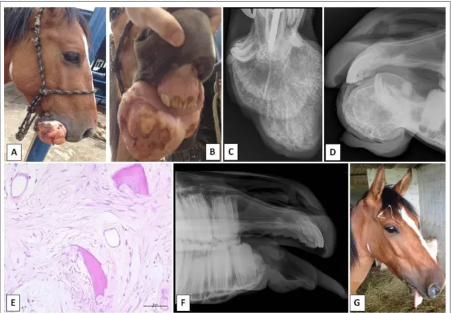
mandibular fracture. At the time, osteosynthesis was performed with transcortical pins and external fixation. Seventy days later, the foal presented with an exuberant gingival tissue growth in the rostral mandible. The foal underwent surgery and the mass was debulked, but no histopathological exam was performed. Two years later, the mandibular growth had increased substantially causing lip distortion and difficulty feeding (Figure 1A and 1B). Physical examination revealed that the horse was alert with a moderate body condition score (scale
5 of 9; HENNEKE et al., 1983). The only change, restricted to the oral cavity, was the presence of a hard,
well-defined, ulcerated mass attached to the rostral mandible. The horse showed no signs of pain upon palpation or manipulation of the mass. Size of the mass hampered adequate mouth opening and oral exploration. Results of hematological analysis (erythrogram and leukogram evaluation) and serum biochemistry
(serum urea, creatinine, total protein, and albumin concentrations, along with alanine aminotransferase, aspartate aminotransferase, and glutamyl transferase activity) showed no significant abnormalities.
A ventrodorsal (intraoral) radiograph
(Figure 1C) of the rostral mandible showed a well-defined osseous mass with intense radiopaque areas and different degrees of ossification, visualized as dispersed radiolucent points inside the mass. Laterolateral radiograph (Figure 1D) showed a single pedunculated well-marginated bony proliferation with a delimited transition zone, a distance of 3cm from premolar teeth 306 and 406. The four mandibular incisors (301, 302, 303 and 401) and the
right canine (304) were not seen in the radiographic
examination. Periodontal ligaments appeared radiographically normal. These radiographic findings were consistent with a nonaggressive mandibular
Figure 1 - A, B. Lateral and frontal aspect of the mandibular mass causing lip distortion and feeding difficulty in a 3-year-old crossbred
bone deformity (benign neoplasia) and/or mineralized mass (dystrophic calcification) of the rostral aspect of the mandible. Based on the horse’s medical history, physical evaluation, and radiographic findings, the tentative diagnosis was a benign mandibular neoplasia as an ossifying fibroma, while osteoma and fibrous dysplasia were considered as the differential diagnoses. Therefore, the mass was surgically removed and sent for pathological examination.
The horse underwent general anesthesia
in dorsal recumbence. After routine pre-operative preparation, mandibular nerves were blocked
with ropivacainea (0.6mg kg-1) and ketamineb
(0.2mg kg-1). The surgical procedure followed the
recommendations of AUER (2006). A transverse incision was made in the oral mucosa on the lingual
side of the incisors. The gingiva was incised on the
labial surface of the incisors ventrally to the lesion, continued laterally and dorsally on both sides, 2cm from premolar teeth 306 and 406. Both incisions were thus connected and continued down to the bone on the mandibular symphysis. Soft tissues were elevated and reflected caudally with a periosteal elevator on the mandibular symphysis to expose the normal bone.
Then, a sterile saw wirec was used to transect the
mandible. The mandibular stumps were rounded and the soft tissue on the lingual surface of the mandible was folded rostrally over the exposed mandible and closed with simple, interrupted, 2-0 polyglycolic acid
sutures. After that, the incisions on the lingual and
labial mucosa were closed with simple, separated, 0 polyglycolic acid sutures. Postoperative antibiotic
therapy consisted of ceftiofurd (4.4mg kg-1, intravenous
[IV], once a day [s.i.d], 7 days). In addition, the
anti-inflammatory and analgesic therapies consisted of
flunixin megluminee (1.1mg kg-1, IV, s.i.d, 5 days) and
dipyroneVI (20mg kg-1, IV, b.i.d, 3 days). Oral lavage
with 0.12% chlorhexidine solutionf was performed
twice daily for 20 days.
After removal, a 14.8×11.5×10.0cm fragment of the mandible was sent for pathological examination. It contained a hard, ulcerated, well-defined mass measuring 14.5×10.0×9.5cm with whitish to brownish coloration. After dissection, the mass had an irregular, heterogeneous features, and
whitish coloration, consistent with osseous tissue.
Samples were taken from the mass, fixed in formalin, decalcified, and sent for further histopathological
analysis. Microscopically, an unencapsulated,
infiltrative, and moderately cellular neoplastic mass, which sharply expanded the lamina propria and adjacent tissues, was observed. The mass was composed of well-differentiated contiguous clusters
of mesenchymal cells (fibroblasts) and joined merged by numerous irregular trabecular bone containing osteocytes surrounded by osteoblasts and osteoclasts (Figure 1E). The histological features confirmed the diagnosis of an ossifying fibroma.
Postoperatively, the horse could not feed
properly or drink water. Therefore, enteral feeds by a nasogastric tube were initiated five times a day (a blended mixture of 5 liters of grass tips, pelletized
ration, and water). Despite the enteral feeds, the horse
lost approximately 40kg during the first 15 days. In this period, the mandibular swelling gradually diminished and the horse was able to capture grass tips and pelleted rations. The feeding management
consisted of 5cm grass tips (Pennisetum purpureum)
and amounts of Tifton grass (Cynodon dactylon)
placed in a 25cm depth feeder to facilitate food capture. Pelleted rations (1kg) were offered four times daily in the same feeder. Concomitantly, the number of nasogastric intubations also diminished gradually, but nasogastric hydration remained. During this period the horse began to gain weight. On day 30, in order to stimulate water ingestion, the
horse was deprived of water for 8h and given 15g of oral electrolyte pasteg. Then a volumetric bucket with
water was offered to measure the amount of ingested water. In the first attempt, the horse began to drink water on its own (approximately 8 liters). Therefore, the horse was accompanied for one more month and
discharged fully recovered 60 days postoperatively. The owner was advised to continue with the feeding
management and offer water ad libitum. During the
hospitalization, the formation of a fibrous-osseous union connecting the mandibular branches was observed (Figure 1F). The only cosmetic sequelae was tongue protrusion (Figure 1G). A new evaluation was performed 12 months after surgery. The horse gained more weight (280kg and body condition score 6 - moderately fleshy) (HENNEKE et al., 1983) and presented no signs of tumor recurrence. A dental examination revealed an overgrowth of the upper incisors and some premolars and molars with dental tips that were properly trimmed. Clinical evaluation of the jaw showed enlargement of the mandibular stumps forming a wider fibrous-osseous union connecting the mandibular branches. The owner stated that the horse is leading a normal life and can also capture short grass in the paddock by itself.
An ossifying fibroma is a disfiguring benign tumor of the jaw that affects young animals of several
species (ROGERS & GOULD, 1998). Although,
food consumption and weight loss. The mass distorts the normal contour of the affected bone resulting in displacement or loss of teeth, disruption of mastication, or obstruction of airflow through the
nares (McCAULEY et al., 2000). The present report is consistent with the characteristics of ossifying
fibromas since the horse was referred after a 2-year progression when it presented with difficulty feeding
and initial weight loss.
Radiography is the most commonly
used ancillary diagnostic technique to investigate
mandibular growths and, in particular, to help differentiate between mandibular swellings caused by apical infections, traumatic lesions, and growths
(DIXON & REARDON, 2015). Lateral, ventrodorsal,
and intraoral radiographs of masses of the skull and mandible are necessary to determine precise location within the bone, extent of bony involvement, margination of the lesion, and the presence or absence of a periosteal reaction or bone lysis (McCAULEY
et al., 2000). In this particular case, radiographic
findings were compatible with a nonaggressive mandibular bone deformity (benign neoplasia) and surgical margins could be precisely planned. The use of computed tomography can greatly help the diagnosis of such lesions, especially to determine the size of the growth and margins, which are important parameters when surgical treatment and/
or radiotherapy are planned (CRIJNS et al., 2015; DIXON & REARDON, 2015). In the future, we
hope that this technology will be more accessible to
veterinary hospitals worldwide.
This report presents an unusual case, in which
the foal was submitted to a mandibular fracture repair, removal of loose incisive teeth, and wound debridement. Subsequently, there was progressive mandibular growth. There are no risk factors associated with ossifying fibromas in horses since the prevalence of mandibular growths in the general equine population is unknown (DIXON & REARDON, 2015). The possibility that previous and repetitive trauma predisposes to the occurrence of ossifying fibromas could not be dismissed and should be investigated.
Successful treatment is especially dependent on early detection and immediate treatment, which helps facilitate complete removal of
the growth without affecting the functional integrity
of the mandible (AUER, 2006; SPONSELLER et
al., 2006). Because of the 2-year progression and the
size of the neoplasia, the BRM reached 2cm from premolar teeth 306 and 406. Stabilization of the remaining mandible was a concern and placement of orthopedic devices was too complex. Nevertheless,
a fibro-osseous union of the transected margins was obtained during hospitalization. In the literature,
several authors stated that horses can survive with few sequelae following BRM (AUER, 2006; SPONSELLER et al., 2006; DIXON & REARDON,
2015), but there are no reports on the difficulties of adapting to new feeding and drinking conditions.
This slow adaptation was the crucial post-surgical
complication and required hard labor to manage the
nasogastric feeding and hydration associated with the
length of the mandible transected.
SOURCES AND MANUFACTURES
a - Cetamin ─ Syntec, Santana da Parnaíba, São Paulo, Brazil. b - Ropi ─ Cristal Pharma, Contagem, Minas Gerais, Brazil. c - Bovivet saw wire ─ Jørgen Kruuse A/S, Langeskov, Denmark. d - Topcef ─ Eurofarma, Itapevi, São Paulo, Brazil.
e - Flunixina Injetável UCB ─ Uzinas Chemicas Brasileiras, Jaboticabal, São Paulo, Brazil.
f - Febrax ─ Lema-InjexBiologic, São Paulo, Brazil. g - Periovet ─ Vetnil, Louveira, São Paulo, Brazil.
h - Eletrolítico Booster JCR ─ Vetnil, Louveira, São Paulo, Brazil.
BIOETHICS AND BIOSECURITY COMMITTEE APPROVAL
We, authors of the article titled “Postoperative care and long-term follow-up afterrostral mandibulectomy to treat an ossifying fibroma in a horse,” declare, for all due purposes, that the project that gave rise to the present data of the same has not been submitted for evaluation to the Ethics Committee of the Universidade de Brasília (UnB), but we are aware of the content of the Brazilian resolutions of the Conselho Nacional de Controle de Experimentação Animal (CONCEA) <http://www.mct.gov.br/ index.php/content/view/310553.html> if it involves animals.
Thus, the authors assume full responsibility for the presented data and are availablefor possible questions, should they be required by the competent authorities.
ACKNOWLEDGEMENTS
The authors thank the Pró-Reitoria de Pesquisa of the Universidade Federal de Minas Gerais (UFMG) for the support for language editing (Edital PRP-UFMG 02/2017).
REFERENCES
AUER, J.A. Craniomaxillofacial disorders. In: AUER, J.A.; STICK, J.A. Equine surgery. St. Louis: Elsevier; 2006. p.1341-1362.
CRIJNS, C.P. et al. Multiple mandibular ossifying fibromas in a yearling Belgian Draught horse filly. Equine Veterinary Education, v.27, n.1, p.11-15, 2015. Available from: <http://onlinelibrary.wiley. com/doi/10.1111/eve.12246/epdf>. Accessed: Sept. 11, 2016.
DIXON, P.M.; REARDON, R.J.M. Equine mandibular growths.
HENNEKE, D.R. et al. Relationship between condition score, physical measurements and body fat percentage in mares. Equine Veterinary Journal, v.15, n.4, p.371-372, 1983. Available from: <http://onlinelibrary.wiley.com/doi/10.1111/j.2042-3306.1983. tb01826.x/epdf>. Accessed: Jan. 02, 2017.
McCAULEY, C.T. et al. Ossifying fibroma in a llama. Journal of Veterinary Diagnostic Investigation, v.12, n.5, p.473-476, 2000. Available from: <http://vdi.sagepub.com/content/12/5/473.full.pdf>. Accessed: Sept. 11, 2016.
ROBBINS, S.C. et al. The use of megavoltage radiation to treat juvenile mandibular ossifying fibroma in a horse. Canadian Veterinary Journal, v.37, n.11, p.683-684, 1996. Available from: <https://www.
ncbi.nlm.nih.gov/pmc/articles/PMC1576516/pdf/canvetj00108-0045. pdf>. Accessed: Sept. 11, 2016.
ROGERS, A.B; GOULD, D.H. Ossifying fibroma in a sheep. Small Ruminant Research, v.28, n.2, p.193-197, 1998. Available from: <http:// www.smallruminantresearch.com/article/S0921-4488(97)00082-5/ pdf>. Accessed: Sept. 11, 2016. doi: 10.1016/S0921-4488(97)00082-5.
SPONSELLER, B.A. et al. Pathology in practice. Ossifying fibroma.
Journal of the American Veterinary Medical Association, v.229, n.11, p.1727-1728, 2006. Available from: <http://avmajournals.avma. org/doi/pdf/10.2460/javma.229.11.1727>. Accessed: Sept. 11, 2016. doi: 10.2460/javma.229.11.1727.