Vol.48, n. 4 : pp. 541-548, July 2005
ISSN 1516-8913 Printed in Brazil
BRAZILIAN ARCHIVES OF
BIOLOGY AND TECHNOLOGY
A N I N T E R N A T I O N A L J O U R N A L
Glucose Metabolism in Rats Submitted to Skeletal Muscle
Denervation
Wilton Marlindo Santana Nunes
*and Maria Alice Rostom de Mello
Departamento de Educação Física; Instituto de Biociências; Universidade Estadual Paulista - UNESP; Av. 24-A, 1515; 13506-900; Rio Claro - SP - Brasil
ABSTRACT
This study analyzed the local and systemic effects of immobilization by denervation of the skeletal muscle on glucose metabolism. The rats were submitted to section of the right paw sciatic nerve. A reduction was observed in glucose uptake by the isolated soleus muscle of the denervated paw after 3 and 7 days, but not after 28 days in relation to the control animals. There was no difference after 3 and 7 days in glucose uptake by the soleus muscle of the opposite intact paw in relation to the control. There was increased glucose uptake in the same paw 28 days after denervation. The rate of glucose removal in response to exogenous insulin after 28 days of denervation was significantly higher than in control animals and those observed after 3 and 7 days of denervation. These results suggest that immobilization by denervation interfered not only in glucose metabolism in the skeletal muscle involved but also in other tissues.
Key words:Denervation, Glucose metabolism, Skeletal muscle
*
Author for correspondence
INTRODUCTION
It is known that glucose is an important energy substrate for the organism and therefore, its utilization has been exhaustively studied in various physiological and pathological situations. Peripheral insulin resistance is characterized mainly by the decreased glucose uptake by the skeletal muscle (Doherty et al., 1997). The decrease in glucose uptake is associated with failures both in the hormone coupling mechanism to its specific receptor and in the post-receptor events (Saad, 1994). Many efforts have been dedicated to understand the mechanisms of insulin action since its discovery in 1921. In the search for understanding the processes that lead to insulin resistance, various models have been proposed over the last few decades, such as, denervation of
the skeletal muscle. The exact intracellular signaling mechanism, which is mediated by the insulin receptor and results in various biological responses, has been partially
glucose transport by the translocation of GLUT-4 molecules coming from intracellular deposits to the plasma membrane (Gumá et al., 1995). According to Turinsky, 1987, the effect of denervation on the muscle metabolism is influenced by the population of muscle fibers. The metabolic disorder caused by the denervation starts between 3 and 6 hours after section of the nerve. As the signs of resistance to insulin occur only in the denervated musculature, the opposite paw muscle is used as control in the skeletal muscle denervation model (Hofmann, 1987). Since the first demonstration that the interruption of the nerve that supplies the muscle results in insulin resistance (Buse and Buse, 1959), denervation has been used as tissue specifc model of insulin resistance (Day et al., 2001, Paassen-Ijkema et al., 2002, Bertelli et al., 2003). Few studies have addressed the effects of the muscular alterations arising from immobilization by denervation on the maintenance of glucose homeostasis by the organism as a whole. Skeletal muscle and adipose tissue are the two main sites of hypoglycemic action of insulin and together compose almost 65% of the body weight (Guyton and Hall, 2000). Thus, any alteration in sensitivity and/or responsiveness of these tissues to the hormone may impair glucose homeostasis. Therefore, this study analyzed the local and systemic effects of immobilization by denervation of the skeletal muscle on the glucose metabolism of rats, to verify if the model was adequate to studies on glucose homeostasis under condition of partial immobilization.
MATERIAL AND METHODS
Male wistar albino rats aged from 3 to 5 months supplied by the UNESP experimental animal facilities were used in the present experiment. The animals had free access to food (Purina for rodents) and water, being submitted to a photoperiodic light/dark cycle of 12 h at a temperature of 25ºC. For denervation, the rats were anesthetized with sodium pentobarbital, (Thiopental, ABBOTT, SP), at a concentration of 50 mg/Kg of body weight, and tricotomized in the posterior portion of the thighs from which a portion of the sciatic nerve from the right paw was
days after denervation; 3)Denervated 7 days: analyzed 7 days after denervation; 4)Denervated 28 days: analyzed 28 days after denervation. In a first series of experiments, glucose metabolism in the isolated soleus muscle was checked after sacrificing the animals by decapitation. The muscle was isolated with the minimum possible lesion and longitudinal strips weighing 25 – 35 mg (wet weight) were first incubated for 30 min at 37ºC in a Dubinoff water bath (FANEN, MODEL 145), inside glass scintillation vials containing 1.5 mL of a Krebs bicarbonate buffer (NaCl 0.6%;
HEPES 6.64 mM; KCl 0.032%; CaCl2 1.14mM;
KH2PO4 0.015%; NaHCO3 0.19%; MgSO4 0.03%)
equilibrated with a mixture of 95%O2-5%CO2, pH
7.4. After this, the muscle strips were transferred to new glass scintillation vials (outer vials) containing 1.5 mL of Krebs-bicarbonate buffer supplemented with glucose 5.5 mM, containing
[U14C]glucose (0.25 mCi/mL) and [3H+]
2-deoxiglucose (2DG, 0.5 µCi/mL) and insulin (100
µU/mL). Inside these scintillation vials, other glass vials (inner vials), which were formed like a scoop with an upwards-directed straight shaft, containing 700mL of hyamine 10-x were installed. The shafts of the inner vials were squeezed about 1 cm through a small hole in a round rubber membrane. The outer vials were sealed with the rubber membrane and lacked with plastic rings. This system, containing the muscle strips, was incubated in the Dubinoff water bath for 60 min. The release of CO2 was stimulated by the injection
of 200 µL of trichloroacetic acid (TCA) 25% into the outer vials and the CO2 was trapped in hyamine
based scintillant (Silva et. al., 2000). Intravenous insulin tolerance test (ittiv) was carried out in a second series of experiments. Animals from all the groups received insulin (mixed regular purified), injected in the penial vein at the concentration of 1UI/mL in the dose of 1UI/Kg of body weight. Blood samples (100 µL) were collected from the femoral vein in 1 mL syringes before (time 0) and after 2:30, 5:00, 10:00, and 20:00 (min) of the administration of insulin, for serum glucose determination. Glucose removal rate (Kitt), during the insulin tolerance test was calculated according to Lundbaek (1962) using the computer program ORIGIN 6.0.After 60 min of insulin infusion for the ittiv, the rats were sacrificed by decapitation for the removal of liver fragments to analyze the effects of the administration of exogenous insulin on the glycogen contents (Lo et al., 1970). Statistical analysis of the data was done by the analysis of variance followed by the Tukey test, wherever appropriate. In all the cases, the critical level of 5% (p<0.05) was fixed.
RESULTS
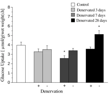
In the first series of experiments, a significant reduction was observed in glucose uptake by the soleus muscle of the denervated paw after 7 days, in relation to the control group (Fig 1). However, there were no significant differences in the denervated paw after 3 and 28 days in relation to the control group. Glucose uptake was also assessed in the soleus muscle of the opposite paw in control and denervated animals groups after 3, 7 and 28 days. The results showed that there was no alteration in the intact opposite paw of the
denervated animals after 3 and 7 days but a significant increase was observed in glucose uptake by the soleus muscle of the opposite paw of the denervated animal after 28 days, in comparison to the control animal (Fig 1). In addition, glucose oxidation and glycogen synthesis by the isolated soleus muscle were also analyzed. There was no difference in glucose oxidation either between the animals denervated paws after 3 and 28 days, as in the opposite soleus of denervated animals after 3, 7 and 28 days in relation to the control animal. However, glucose oxidation diminished in the denervated paw after 7 days (Fig 2).
In regard to glycogen synthesis, there was a decrease in the denervated animals and opposite paws after 3 and 7 days, and increase in the denervated animals and opposite paws after 28 days in relation to the control rat (Fig 3).
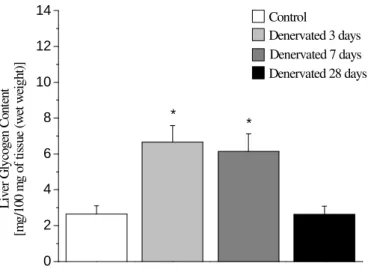
In the second series of experiments, the rate of blood glucose removal (Kitt) was assessed in rats submitted to an intravenous insulin tolerance test (ittiv). The results obtained are described in Figure 5. The animals Kitt values after 28 days of denervation were significantly higher than those of the control animals and those after 3 and 7 days of denervation (Fig 5). Since the liver was exceptionally important in glucose metabolism, and for this reason, liver glycogen contents were checked after the intravenous insulin tolerance test, with the aim of assessing the effects of exogenous insulin.
Figure 1 -Glucose uptake by the soleus muscle (mean ± standard error) for rats in all groups. The (+) sign refers to the denervated paw, while the (-) sign refers to the opposite paw of the same animals. Control refers to intact animals. (*) Significant difference, P< 0.05 in relation to control group. n = 7 to 10 muscle strips per group.
Figure 2 - Glucose oxidation by the soleus muscle (mean ± standard deviation) in rats of
all groups. The (+) sign refers to the denervated paw while the (-) sign refers to the opposite paw of the same animals. Control refers to intact animals. (*) Significant difference, P< 0.05 in relation to the control group. n = 8 to 10 muscle strips.
0 2 4 6 8 10 12
Denervation
Denervated 28 days Denervated 7 days Denervated 3 days Control
-
-- + +
+
*
G
lu
co
se
O
x
id
at
ion
[
µ
m
o
l/
g
(w
et
w
ei
g
h
t)
.h
]
0 1 2 3 4 5 6 7 8
Denervation
Denervated 28 days Denervated 7 days Denervated 3 days Control
*
*
-- +
+ +
-G
lu
co
se
U
p
ta
k
e
[
µ
m
o
l/
g
(w
et
w
ei
gh
t)
.h
Figure 3 - Glycogen synthesis by the soleus muscle (mean ± standard error) in rats of all groups. The (+) sign refers to the denervated paw while the (-) sign refers to the opposite paw of the same animals. Control refers to intact animals. (*) Significant difference, P< 0.05 in relation to the control group. n = 10 to 13 muscle strips.
Figure 4 - Liver glycogen content (mean ± standard error) after 60 minutes of insulin
(1UI/Kg). Administration intravenously to animals of all the groups. Control refers to intact animals. Denervated 28 days rats show contents similar to controls. (*) Significant difference, P< 0.05 in relation to the control rat. n = 5 to 10 rats per group.
0,0 0,5 1,0 1,5 2,0 2,5 3,0 3,5 4,0
Denervation
*
* * *
*
*
-
-- + +
+
Denervated 28 days Denervated 7 days Denervated 3 days Control
G
ly
c
og
e
n
S
yn
th
e
si
s
[
µ
m
o
l/
g
(w
e
t
w
e
ig
h
t)
.h
]
0 2 4 6 8 10 12 14
Denervated 28 days Denervated 7 days Denervated 3 days Control
* *
L
iv
er
G
ly
cog
en
C
on
te
nt
[
m
g
/100
m
g o
f
ti
ss
ue
(w
et
w
ei
g
ht
Figure 5 - Blood glucose removal rate (Kitt), during tolerance to insulin test (mean ± standard error) in rats of all the groups. Control refers to whole animals. (*) Significant difference, P< 0.05 in relation to the control rat. n = 8 to 10 animals per group.
DISCUSSION
In this study, it was found that glucose uptake by the soleus muscle, induced by insulin, underwent variation among the groups. After 7 days of denervation, the animals had diminished glucose uptake by the soleus muscle of the denervated paw. However, the opposite paw continued to show values close to those of the control animal. This reduction in glucose uptake by skeletal muscle after 7 days of denervation has been reported in the literature. Studies have show in decreased glucose uptake in the denervated skeletal muscle of rats after 3 days and 7 days (Turinsky, 1987, Hirose et al., 2001, Turinsky and Damrau-Abney, 1998). The mechanism by which the reduction in glucose uptake occurs in denervated muscle for 3 and 7 days is partly related to the alteration of the internal chemical-enzymatic cascade reaction mechanisms, which culminate in the translocation of the glucose transporter to the plasma membrane (Bertelli et al., 2003 e Hirose et al., 2001). There was no alteration in the glucose uptake by the soleus muscle of the denervated paw after 28 days. On the other hand, glucose uptake by the soleus muscle of the opposite paw of this same group was
the presence of insulin is reduced between the 3rd and 7th day of denervation, increased after 7 days
and increased even more on the 10th day of
denervation. The reduction in glucose transport in response to insulin between the 3rd and the 7th day of denervation suggests resistance to insulin in the denervated skeletal muscle, with a return of the normal response to insulin from the 10th day on
(Davis and Karl, 1988). Denervation inhibits
multiple aspects of glucose metabolism in the skeletal muscle, apart from the uptake, such as: incorporation of the glucose in glycogen, glucose-6-phosphate content and activity of the glucogen synthase enzyme (Burant et al., 1984). These changes have been attributed to the losses of the trophic factor derived from the nerve and/or the electrical stimulation by the nerve (Davis and Karl, 1988).Glucose oxidation was diminished in the animal’s denervated paw after 7 days (Fig 2). This datum suggests that the use of the glycolitic path by glucose was affected. There is a recovery of the use of this path in the soleus muscle of denervated animals after 10 days (Davis and Karl, 1988). This latter information was confirmed in this study, because after 28 days of denervation, the values of glucose oxidation by the soleus muscle returned to normal. Muscle atrophy was generally evident after the 3rd day of denervation
0,0 3,5 4,0 4,5 5,0 5,5 6,0 6,5
Denervated 28 days Denervated 7 days Denervated 3 days Control
*
K
it
decline in glycogen synthetase or phosphorilase activity in the denervated muscle was among the factors responsible for the atrophy. This atrophy occurs to a greater or lesser extent depending on the type of muscle fiber. The data in this study with reference to glycogen synthesis showed its decrease in both the denervated paw which increased after 3 and 7 days after denervation. On the other hand, glycogen synthesis after 28 days increased in relation to the control. In summary, the results of this study, with reference to glucose metabolism in the denervated skeletal muscle, confirmed and broaden information already described in the literature. Once the suitability of the model had been established, one proceeded with analyzing the effect of denervation of the skeletal muscle on the animal’s glucose homeostasis. To do this, blood glucose removal rate (Kitt) as well as liver glycogen contents after “in vivo” administration of exogenous insulin were evaluated. The results showed alteration in the liver glycogen stock of the rats submitted to denervation in relation to the controls after exogenous insulin administration. The significant increase in the liver glycogen content of rats after 3 and 7 days of denervation in relation to the controls and the decrease after 28 days of denervation suggested an alteration in liver sensitivity to the hormone in the rats submitted to denervation. Taken together, the results found in the soleus muscle and in the liver suggest an expected metabolic interrelationship between liver glycogen content after 3, 7 and 28 days and muscle glucose uptake and glycogen synthesis rates. The higher the liver glycogen stores, the lower the muscle glucose uptake and glycogen synthesis rates occurred after 3 and 7 days after denervation, which was reversal 28 days after denervation. Further investigation could be required for this metabolic association. The blood glucose removal rate (Kitt) in response to exogenous insulin administration was significantly higher in the rats after 28 days of denervation in relation to the controls and to the values observed after 3 and 7 days of denervation. Circulating glucose profile observed during the insulin tolerance test seemed to reflect long term adaptations of the organism as a whole to the partial immobilization imposed by denervation of the skeletal muscle. After 3 and 7 days of denervation, the blood glucose removal rate in response to exogenous insulin not alter in comparison with the control animal. Thisd could
be due, at least in part, to the increase in sensitivity to insulin by the hepatic tissue. However, after 28 days of denervation, the increase observed in the blood glucose removal rate in response to insulin, was probably related to the increase in sensitivity to the hormone in the intact skeletal muscle, as suggested by the results of glucose uptake by the isolated soleus muscle in the opposite paw of the
rats submitted to denervation. It could be
concluded that the denervation of the skeletal muscle not only caused local metabolic alterations, but also interfered with the glucose metabolism of the organism as a whole. Thus, rats submitted to partial immobilization by denervation of the skeletal muscle could constitute a useful model for the study of glucose homeostasis under the condition of immobilization.
ACKNOWLEDGMENTS
The authors thank Clarice Y. Sibuya, José Roberto R. da Silva and Eduardo Custódio for technical assistance and Margery J. Galbraith for revising the manuscript. Research supported by the Brazilian Foundations: FAPESP, CNPq and Capes.
RESUMO
REFERENCES
Bertelli, D. F.; Ueno, M.; Amaral, M. E.; Toyama, M. H.; Carneiro, E. M.; Marangoni, S.; Carvalho, C. R.; Saad, M. J.; Velloso, L. A. and Boschero, A. C. (2003), Reversal of denervation-induced insulin resistance by SHIP2 protein synthesis blockade. Am. J. Physiol. Endocrinol. Metab., 284 : (4), E679-87.
Burant, C. F.; Lemmon S. K.; Treutelaar, M. K. and Buse, M. G. (1984), Insulin resistance of denervated rat muscle: a model for impaired receptor-function coupling. Am. J. Physiol., 247 : (5 Pt 1), E657-66. Buse, M. G. and Buse, J. (1959), Glucose uptake and
response to insulin of the isolated rat diaphragm: the effect of denervation. Diabetes., 8 : (3), 218-25. Coderre, L.; Monfar, M. M.; Chen, K. S.; Heydrick,
S. J.; Kurowski, T. G.; Ruderman, N. B. and Pich, P. F.(1992), Alteration in the expression of GLUT 1 and GLUT 4 protein and messenger RNA levels in denervated rat muscle. Endocrinol., 131 : (4), 1821-1825.
Davis, T. A. and Karl, I. E. (1988), Resistance of protein and glucose metabolism to insulin in desnervated muscle. Biochem. J., 254, 667-675. Day, C. S.; Riano, F.; Tomaino, M. N.; Buranatanitkit,
B.; Somogyi, G.; Sotereanos, D. and Huard, J.(2001), Growth factor may decrease muscle atrophy secondary to denervation. J. Reconstru. Microsurg., 1, 51-57.
Doherty, R. O.; Stein, D. and Foley, J.(1997), Insulin resistance. Diabetologia, 40, B10-B15.
Elmerndorf, J. S.; Damrau-Abney, A.; Smith, T. R.; David, T. S. and Turinsky, J. (1997), Phosphatidylinositol 3-kinase and dynamics of insulin resistance in denervated slow and fast muscles in vivo. Am. J. Physiol., 272 : (4 Pt 1), E661-70. Gumá, A.; Zierath, J. R.; Wallberg-Henriksson and
Klip, A. (1995), Insulin induces translocation of GLUT-4 glucose transporters in human skeletal muscle. Am. J. Physiol., 268 : (Endocrinol. Metab. 31), E613-E622.
Guyton, A. C. and Hall, J. E. (2000), Textbook of Medical Physiology. W. B Saunders Company, Philadelphia.
Henriksen, E. J.; Rodnick, K. J.; Mondon, C. E.; James, D. E. and Holloszy, J. O.(1991), Effect of denervation or unweighting on GLUT-4 protein in rat soleus muscle. J. Appl. Physiol., 70 : (5), 2322-2327. Hirose, M.; Kaneki, M.; Sugita, H.; Yasuhara, S.;
Ibebunjo, C.; Martyn, J. A. and Iebunjo, C. (2001), Long-term denervation impairs insulin receptor substrate-1-mediated insulin signaling in skeletal muscle. Metabolism., 50 : (2), 216-22.
Hofmann, W. W. (1987), Musculotrophic effects of insulin receptors before and after denervation. Brain Res., 401 : (2), 312-21.
Sjorgreem, B.; Nordenskjold, D. T.; Holmgren, H. and Wollerstrom, J. (1938), Bertrag zur kentnis des le berrhythmik. Pfluger Arch Gesante Physiol Menschen Tiere, 240, 247.
Lin, Y.; Brady, M. J.; Wolanske, K.; Holbert, R.; Ruderman, N. B. and Yaney, G. C.(2002), Alterations of nPKC distribution, but not Akt/PKB activation in denervation rat soleus muscle. Am. J. Physiol., E318-E325.
Lo, S.; Russell, J. C. and Taylor, A. W. (1970), Determination of glycogen in small tissue samples. J. Appl. Physiol., 28 : (2), 234-6.
Lundbaek, K. (1962), Intravenous glucose tolerance as a tool in definition and diagnosis of diabetes mellitus.
Br. Med. J., 5291, 1507-13.
Paassen-Ijkema, J.; Meek, M. F. and Gramsbergen, A. (2002), Reinnervation of muscle after transection of sciatic nerve in adult rats. Muscle Nerve, 6, 891-897. Saad, M. J. (1994), Molecular mechanisms of insulin
resístanse. Braz. J. Med. Biol. Res., 27 : (4), 941-957. Silva, R. G.; Almeida, C. C. A.; Luciano, E. and Mello,
M. A. R. (2000), Protein malnutrition does not impair adaptations to exercise training. Nutrition Research, 20 : (4), 527-539.
Smith, R. L.; Rach, P. J. and Lawrence, J. C. (1988), Insulin resistance in denervated skeletal muscle. J. Biol. Chem., 263, 658-665.
Sowell, M. O.; Dutton, S. L. and Buse, M. G. (1989), Selective in vitro reversal of the insulin resistance of glucose transport in denervated rat skeletal muscle.
Am. J. Physiol., 257, E418-E425.
Turinsky, J. (1987), Dynamics of insulin resistance in denervated slow and fast muscles in vivo. Am. J. Physiol., 252 : (3 Pt 2), R531-7.
Turinsky, J. and Damrau-Abney, A. (1998), Akt1 kinase and dynamics of insulin resistance in denervated muscles in vivo. Am. J. Physiol., 275 : (5pt2), R1425-R1430.
Wallis, M. G.; Appleby, G. J.; Youd, J. M.; Clark, M. G. and Penschow, J. D. (1999), Reduced glycogen phosphorylase activity in denervated Hindlimb muscles of rat is related to muscle atrophy and fibre type. Live Sciences, 64 : (4), 221-228.