© 2014 Sociedade Brasileira de Hemodinâmica e Cardiologia Intervencionista. Published by Elsevier Editora Ltda. All rights reserved.
Effects of Renal Sympathetic Denervation in Renal Artery
Diameter Evaluated By Quantitative Angiography
Luciana Armaganijan, Rodolfo Staico, Dalmo Moreira, Celso Amodeo, Flávio Borelli, Márcio Sousa,
Amanda Sousa, Alexandre Abizaid
ABSTRACT
Background: Percutaneous renal sympathetic denervation was developed as an adjunct method to treat clinical conditions associated to sympathetic hyperactivity. Percutaneous renal sympathetic denervation increases the renal blood low and reduces vasoconstriction. The effects of percutaneous renal sympathetic denervation in renal artery diameter have not been reported. Our objective was to evaluate such effects by quantitative angiography. Methods: Prospective, observational, study including consecutive patients undergoing percutaneous renal sympathetic denervation. Results: Thirty-one patients were selected, 21 were submitted to percutaneous renal sym-pathetic denervation to control resistant arterial hypertension and 10 to control refractory ventricular arrhythmias. Seventeen patients did not perform renal arteriography in the follow-up due to clinical contraindications or because they did not complete the 6-month period established by the protocol. In addition, one patient performed a unilateral percutaneous renal sympathetic denervation and was also excluded from this analysis. Therefore, 52 renal angiographies (26 pairs) of 13 patients were analyzed. Mean maximal diameter of the right renal artery before the procedure was 4.54 ± 0.21 mm and increased to 5.2 ± 0.44 mm at 6 months (p = 0.01). Likewise, there was a signiicant increase in the diameter of the left renal artery at 6 months of follow-up, increasing from 4.37 ± 0.42 to 5.23 ± 0.77 mm (p = 0.02). Conclusions: The results of this analysis illustrate the signiicant increment in renal artery diameter after percutaneous renal sympathetic denervation. Randomized controlled clinical trials are required to consolidate our observations.
DESCRIPTORS: Hypertension. Drug resistance. Renal artery. Sympathectomy. Catheter ablation. Angiography.
Instituto Dante Pazzanese de Cardiologia, São Paulo, SP, Brazil. Correspondence to: Rodolfo Staico. Avenida Dr. Dante Pazzanese, 500 – Vila Mariana – CEP: 04012-180 – São Paulo, SP, Brazil
E-mail: [email protected]
Received: 03/06/2014 • Accepted: 05/19/2014 RESUMO
Efeitos da Denervação Simpática Renal no Diâmetro da Artéria Renal Avaliados
Pela Angiografia Quantitativa
Introdução: A denervação simpática renal percutânea surgiu como método adjunto no controle de condições clínicas as-sociadas à hiperatividade simpática. Ela resulta em aumento do luxo sanguíneo renal e em redução da vasoconstrição. Os efeitos da denervação simpática renal percutânea no diâmetro da artéria renal ainda não foram descritos. Nosso objetivo foi avaliar tais efeitos por meio da angiograia quantitativa. Métodos: Estudo prospectivo e observacional que incluiu pacientes consecutivos submetidos à denervação simpática renal percutânea. Resultados: Selecionamos 31 pacientes, sendo 21 submetidos à denervação simpática renal per-cutânea para controle da hipertensão arterial resistente e 10 para controle de arritmias ventriculares refratárias. Dezessete pacientes não realizaram arteriograia renal no seguimento por não completarem o período protocolar de 6 meses, ou por contraindicação clínica. Adicionalmente, uma paciente realizou denervação simpática renal percutânea unilateral, sendo também excluída desta análise. Assim, 52 angiograias renais (26 pares) de 13 pacientes foram analisadas. A média do diâmetro máximo da artéria renal direita, antes do procedi-mento, foi de 4,54 ± 0,21 mm e aumentou para 5,2 ± 0,44 mm aos 6 meses (p = 0,01). Da mesma forma, observou-se aumento signiicativo do diâmetro da artéria renal esquerda aos 6 meses de seguimento, ampliando de 4,37 ± 0,42 para 5,23 ± 0,77 mm (p = 0,02). Conclusões: Os resultados desta análise ilustram o incremento signiicativo dos diâmetros das artérias renais após denervação simpática renal percutânea. Ensaios clínicos randomizados e controlados são necessários para consolidar nossas observações.
DESCRITORES: Hipertensão. Resistência a medicamentos. Artéria renal. Simpatectomia. Ablação por cateter. Angiograia.
hyperactivity of the sympathetic autonomic nervous system in the pathophysiology of SAH, percutaneous renal sympathetic denervation (RSD) has emerged as an adjunctive therapeutic strategy in selected groups of refractory hypertensive patients.4 Beneits have also been observed with this method in other clinical contexts associated with chronic hyperactivity of the sympathetic nervous system (SNS), such as cardiac arrhythmias,5 obstructive sleep apnea syndrome,6 heart failure,7 and metabolic syndrome.8
Despite the promising initial results of percutaneous RSD related to SAH, discrepancies have been observed among studies, in large part relecting the variation in the technique used the learning curve, the type of device used, and, especially, the different populations studied, with a wide variation of the importance of the sympathetic system in the genesis of SAH.
Sympathetic activity in the context of percutane-ous RSD is measured by microneurography and by norepinephrine spillover.9 However, although available,
these procedures are not adopted routinely in clinical practice, due to limitations in their execution.
Chronic SNS stimulation progresses with an in-crease in plasma neurohormones, which in turn results in vascular (vasoconstriction, increased thickness, and reduced vascular compliance) and heart (development of myocardial hypertrophy, ischemia, arrhythmias, and heart failure) effects. In kidneys, an increased sympathetic activity leads to the activation of the renin-angiotensin-aldosterone system, increased reabsorption of sodium and water, reduction of renal blood low and glomerular iltration rate, ischemia, and renal failure.10
Percutaneous RSD aims to reduce sympathetic ac-tivity. As a consequence, there is an increase in renal blood low9 and arterial vasodilation.10 In this study; the aim was to evaluate the effects of percutaneous RSD on the diameter of the renal arteries, through the use of quantitative angiography.
METHODS
Selection of patients
Thirty-one consecutive patients undergoing per-cutaneous RSD at the Instituto Dante Pazzanese de
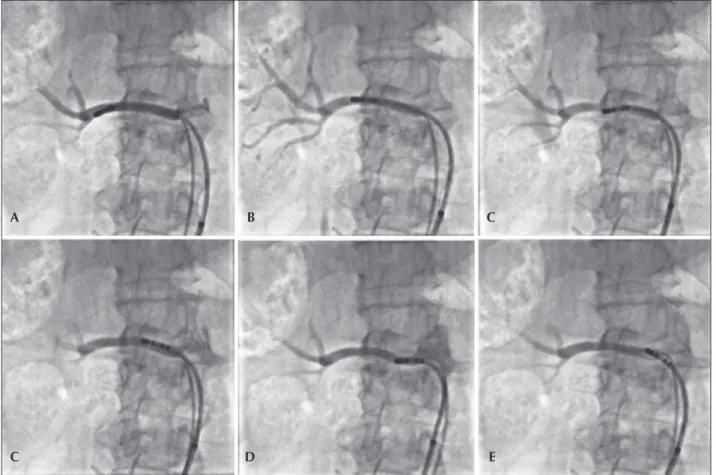
Four to six radiofrequency applications were per-formed in each renal artery with the irrigated catheter, starting at the most distal portion of the vessel, close to the bifurcation, toward the aorta, respecting the mini-mum distance of 5 mm between each application and the helical arrangement of them. The detailed technique for a percutaneous RSD with an irrigated catheter has been described previously elsewhere by our group.11 Figure 1 shows a case in which six radiofrequency applications were conducted in the right renal artery.
EnligHTN
is a dedicated system for percutaneous RSD, designed with the aim of generating better-distributed renal artery lesions, and, therefore, four radiofrequency applications are performed with a minimal manipulation of the catheter. The device consists of a catheter with a basket at its distal end, which has four electrodes that sequentially emit the radiofrequency. Initially, the device was placed distally in the renal artery, wherein a irst section of application was performed. After this step, the device was collapsed and retracted by about 1 cm, for a new application – a total of eight applications per treated renal artery were performed. The technique outlined for percutaneous RSD with the EnligHTN
dedicated system has been described previously elsewhere by the present group.12
Quantitative angiography
Of those 31 patients selected, 17 did not undergo renal arteriography under the protocol during follow-up, or because they did not complete the six-month period, or due to clinical contraindication. Fourteen patients underwent angiography 6 months after percu-taneous RSD, which allowed for the comparison of this examination versus the initial examination performed before the ablation. One case was excluded from this analysis, because the patient was submitted to ablation of only one renal artery. Thus, 52 renal arteriographies (26 pairs) from 13 patients were analyzed by quantita-tive angiography.
Statistical Analysis
Continuous variables were expressed as mean and standard deviation and compared using Student’s t-test. Categorical variables were presented as absolute and relative frequencies. For all parameters compared, p-values < 0.05 were considered statistically signiicant. Data were analyzed using SPSS
, version 16.0 for Windows
(SPSS
Inc., Chicago, Illinois, United States).
RESULTS
Fifty-two renal angiograms were analyzed. The
mean age was 49 ± 10 years and 11 (84.6%) patients
were female. The baseline characteristics of all patients included in the study are shown in Table 1.
Technical characteristics of the procedure
The volume of contrast used was 87.7 ± 29.7 mL
and the luoroscopy time was 21.4 ± 6.6 minutes.
On average, ive applications were performedon each renal artery.
Quantitative angiography
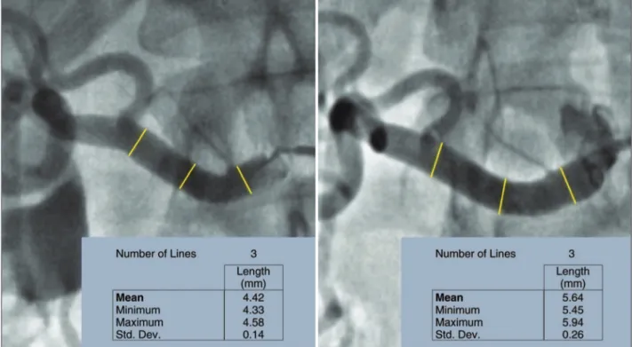
Figure 3 exempliies a quantitative angiography in right renal artery before and 6 months after percutane-ous RSD.
The length of the left and right renal arteries was
26.9 ± 8.9 mm and 36.3 ± 7.4 mm, respectively.
The maximum diameter of the right renal artery before the procedure was 4.54 ± 0.21 mm, showing
an increase to 5.20 ± 0.44 mm at 6 months (p = 0.01).
Similarly, an increase was observed in the diameter of the left renal artery at 6 months of follow-up, which changed from 4.37 ± 0.42 mm to 5.23 ± 0.77 mm
(p = 0.02) (Figure 4).
DISCUSSION
This was the irst study which prospectively as-sessed the effects of percutaneous RSD in the diameter of renal arteries. It was observed, using quantitative angiography, a signiicant increase in diameter, probably
Figure 1 – Sequential radiofrequency applications initiated in the distal segment of the right renal artery. The catheter is pulled and rotated after each application, with the aim of promoting sequential lesions in a helical coniguration (A to E).
A
C D E
through two mechanisms: (1) increased renal blood low and (2) decreased vasoconstriction, both as a result of blocking or reduction of sympathetic activity, caused by percutaneous RSD.
Coronary angiography is the gold standard of imaging for analysis of coronary anatomy; this technique provides deinition of the extent and precise location of coronary artery disease. A well-performed coronary angiography requires a thorough knowledge of anatomy, including its variations, and a protocol for systematic acquisition of sequential images that allows the visualization of all coronary segments, especially in areas with overlapping
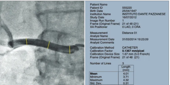
vessels, bifurcations, and tortuous anatomy. Late lumen loss, a procedure applied for years for quantiication of neointimal hyperplasia, has become one of the most sensitive and surgeon-independent angiographic evaluations of the effectiveness of coronary stents, both for drug-eluting devices or other types. Classi-cally deined as the difference between the minimum lumen diameter (MLD) obtained immediately after the procedure (inal MLD) and the MLD obtained during follow-up, late lumen loss is an angiographic measure of the absolute degree of vascular restenosis, different from the binary restenosis calculation.13,14 Renal artery angiography is used in a similar fashion as coronary angiography, and is useful for an accurate analysis of anatomy and also as an adjunctive tool in the evalua-tion of the results of intervenevalua-tions in this territory by means of quantitative angiography. In the present study, accurate renal angiographies were performed, with appropriate angles for better visualization of the renal arteries. The estimates applied in the initial tests were carefully repeated at the six-month follow-up in all patients involved in the study. The analyzes were interpreted with the aid of a contemporary program, QAngio XA, version 7.3. Measurements of maximum diameters in the proximal, middle, and distal seg-ments of each renal artery before and 6 months after percutaneous RSD were performed. These procedures allowed us to assess the changes that took place, by comparing the averages of these measures.
The evaluation of sympathetic activity can be obtained directly by microneurography, usually of the TABLE 1
Baseline characteristics of patients undergoing quantitative renal angiography before and 6 months
after renal sympathetic denervation
Characteristic n = 13
Age, years 49 ± 10
Female, n (%) 11 (84.6)
Diabetes mellitus, n (%) 4 (30.7)
Dyslipidemia, n (%) 10 (76.9)
Smoking, n (%) 0
Prior myocardial infarct, n (%) 1 (7.6)
Previous stroke, n (%) 0
Peripheral arterial disease, n (%) 0
Family history of cardiovascular disease, n (%)
4 (30.7)
ibular nerve, and indirectly by means of norepinephrine spillover.9 Although available, these methods are not
routinely adopted in clinical practice, due to limitations in their implementation.
The development of microneurography, in which the neural activity can be recorded directly by means of microelectrodes inserted percutaneously into a peripheral nerve in humans, has provided valuable information on the control of sympathetic outlow to the muscle. This method allows for measuring the postganglionic efferent sympathetic nerve trafic to skeletal muscles, the so-called muscle sympathetic nerve activity (MSNA). The MSNA in
different pathologies has expanded the knowledge about the functioning of the SNS. Although the quantiication of MSNA is still largely limited to measures of frequency and incidence of bursts (bursts/min and bursts/100 heartbeats, respectively), the development of unit records of MSNA has provided more detailed information on the operation the SNS.15 In healthy subjects, MSNA is activated by decreases in cardiac illing pressure, exercise, hypoxia, hypercapnia, hyperpnoea, and sleep disorders, and is inhibited by lung inlation. Among the main clinical applications of sym-pathetic microneurography, the authors must emphasize the elucidation of the neural mechanisms of control of SAH and of thermoregulation. Despite obtaining direct measures of sympathetic activity (with the result that sympathetic microneurography is regarded as the gold standard), this technique has speciic limitations, such as training and experience of the researcher, critical to the acquisition of high quality signals; reliance of the bursts’ amplitude relative to the position where the electrodes are applied; and the fact that the measure of MSNA in skel-etal muscle may not relect changes in renal and cardiac sites. Furthermore, this is a painful, time-consuming, and impractical method for use in the clinical environment, more suitable for research purposes.16
Historically, the methods used to measure changes of SNS in humans have consisted of determinations of norepinephrine, the primary neurotransmitter released from sympathetic postganglionic nerve endings. Today, the measurement of the excretion of this neurotransmitter
Figure 3 – Quantitative angiography at right renal artery. Left, before percutaneous renal sympathetic denervation with a mean of proximal/middle third/distal diameters of 4.42 mm. Right, 6 months after the procedure, showing increase in mean diameters to 5.64 mm.
Figure 4 – Change in the maximum diameters of the renal arteries, 6 months after percutaneous renal sympathetic denervation (RSD).
5.23 ± 0.77 5.20 ± 0.44
5.4
5 5.2
4.8
3.8
Right renal artery
Pre-RSD 6 months after RSD
Left renal artery 4.6
4.4
4.2
4
Ar
te
ry
Maximum Diameter (mm
)
4.37 ± 0.42 4.54 ± 0.21
0.01
The overlow of norepinephrine (spillover) toward plasma, based on the dilution of radiotracers, is one of the safest ways of assessing regional sympathetic function. Renal norepinephrine spillover consists of the administration of the tritium-marked neurotransmitter at a known concentration, accompanied by the collection of blood samples from renal veins.18 This is a complex method, with little application in clinical practice.
Therefore, the authors believe that, if properly performed by experienced operators and with a care-ful attention to all the recommendations already well established, quantitative angiography of renal arteries can be a practical tool, easy to implement and of low cost, which will allow a better understanding of the results of percutaneous RSD.
Limitations
This study has limitations, for instance, the small sample size, as well as the inherent limitations of observational studies; in particular, the non-use of established methods of measurement of sympathetic activity for comparison with quantitative angiography.
CONCLUSIONS
The results of this analysis illustrate the signiicant increase in diameter of the renal arteries after a renal sympathetic denervation procedure, probably due to the increase of renal blood low and decreased vasocon-striction, both as a result of blockage or reduction of sympathetic activity. Randomized controlled trials are needed to consolidate these observations.
CONFLICTS OF INTEREST
The authors declare no conlicts of interest.
FUNDING SOURCE
None.
REFERENCES
1. Kearney PM, Whelton M, Reynolds K, Muntner P, Whelton PK, He J. Global burden of hypertension: analysis of worldwide
data. Lancet. 2005;365(9455):217-23.
Symplicity HTN-1 study. Lancet. 2014;383(9917):622-9.
5. Staico R, Armaganijan L, Moreira D, Medeiros P, Melo J, Lopes R, et al. Renal sympathetic denervation and ventricular arrhythmias: a case of electrical storm with multiple renal arteries. EuroIntervention.2014;10(1):166.
6. Witkowski A, Prejbisz A, Florczak E, Kądziela J, Śliwiński P, Bieleń P, et al. Effects of renal sympathetic denervation on blood pressure, sleep apnea course, and glycemic control in patients with resistant hypertension and sleep apnea.
Hyper-tension.2011;58(4):559-65.
7. Böhm M, Ewen S, Kindermann I, Linz D, Ukena C, Mahfoud F. Renal denervation and heart failure. Eur J Heart Fail. 2014 Mar 18. [Epub ahead of print].
8. Böhm M, Linz D, Urban D, Mahfoud F, Ukena C. Renal sympathetic denervation: applications in hypertension and beyond. Nat Rev Cardiol. 2013;10(8):465-76.
9. Schlaich MP, Sobotka PA, Krum H, Lambert E, Esler MD. Renal
sympathetic-nerve ablation for uncontrolled hypertension. N
Engl J Med. 2009;361(9):932-4.
10. DiBona GF. Neural control of the kidney: past, present, and future. Hypertension. 2003;41(3 Pt 2):621-4.
11. Armaganijan L, Staico R, Moraes A, Abizaid A, Moreira D, Amodeo C, et al. Renal denervation using an irrigated catheter in patients with resistant hypertension: a promising strategy? Arq Bras Cardiol 2014;102(4):355-63.
12. taico R, Armaganijan L, Moreira D, Medeiros P, Habib R, Melo Neto J, et al. Denervação simpática renal: um novo cateter em
um novo cenário. Rev Bras Cardiol Invasiva.
2013;21(4):396-400.
13. Lansky AJ, Popma JJ, Cutlip D, Ho KK, Abizaid AS, Saucedo J, et al. Comparative analysis of early and late angiographic outcomes using two quantitative algorithms in the Balloon versus Optimal Atherectomy Trial (BOAT). Am J Cardiol.
1999;83(12):1611-6.
14. Mauri L, Orav EJ, Candia SC, Cutlip DE, Kuntz RE. Robustness of late lumen loss in discriminating drug-eluting stents across variable observational and randomized trials. Circulation.
2005;112(18):2833-9.
15. Maceield VG. Sympathetic microneurography. Handb Clin Neurol. 2013;117:353-64.
16. Freeman R, Chapleau MW. Testing the autonomic nervous system. Handb o Clin Neurol. 2013;115:115-36.
17. Esler M, Jennings G, Lambert G, Meredith I, Horne M, Eisenhofer G. Overlow of catecholamine neurotransmitters to the circulation: source, fate, and functions. Physiol Rev.
1990;70(4):963-85.