385 Radiol Bras. 2018 Nov/Dez;51(6):385–390
Pulmonary inhalation-perfusion scintigraphy in the evaluation
of bronchoscopic treatment of bronchopleural fistula
Cintilografia pulmonar inalatória na avaliação do tratamento broncoscópico de fístula broncopleural
Carla Rachel Ono1,a, Miguel Lia Tedde2,b, Paulo Rogerio Scordamaglio3,c, Carlos Alberto Buchpiguel1,d
1. Nuclear Medicine Division, Instituto de Radiologia do Hospital das Clínicas da Faculdade de Medicina da Universidade de São Paulo (InRad/ HC-FMUSP), São Paulo, SP, Brazil. 2. Department of Thoracic Surgery, Instituto do Coração do Hospital das Clínicas da Faculdade de Medicina da Universidade de São Paulo (InCor/HC-FMUSP), São Paulo, SP, Brazil. 3. Respiratory Endoscopy Division, Instituto do Coração do Hospital das Clínicas da Faculdade de Medicina da Universidade de São Paulo (InCor/HC-FMUSP), São Paulo, SP, Brazil.
Correspondence: Dr. Miguel L. Tedde. Rua Itambé, 367, ap. 151-A, Higienópolis. São Paulo, SP, Brazil, 01239-001. E-mail: [email protected]. a. http://orcid.org/0000-0002-0179-7248; b. http://orcid.org/0000-0002-8178-4243; c. http://orcid.org/0000-0001-8971-5333; d. http://orcid.org/0000-0003-0956-2790.
Received 9 August 2017. Accepted after revision 3 November 2017.
How to cite this article:
Ono CR, Tedde ML, Scordamaglio PR, Buchpiguel CA. Pulmonary inhalation-perfusion scintigraphy in the evaluation of bronchoscopic treatment of bronchopleural fistula. Radiol Bras. 2018 Nov/Dez;51(6):385–390.
Abstract
Resumo
Objective: To evaluate the use of pulmonary inhalation-perfusion scintigraphy as an alternative method of investigation and
follow-up in patients with bronchopleural fistula (BPF).
Materials and Methods: Nine patients with BPFs were treated through the off-label use of a transcatheter atrial septal defect oc-cluder, placed endoscopically, and were followed with pulmonary inhalation-perfusion scintigraphy, involving inhalation, via a nebu-lizer, of 900–1300 MBq (25–35 mCi) of technetium-99m-labeled diethylenetriaminepentaacetic acid and single-photon emission computed tomography with a dual-head gamma camera.
Results: In two cases, there was a residual air leak that was not identified by bronchoscopy or the methylene blue test but was de -tected only by pulmonary inhalation-perfusion scintigraphy. Those results correlated with the evolution of the patients, both of whom
showed late signs of air leak, which confirmed the scintigraphy findings. In the patients with complete resolution of symptoms and fistula closure seen on bronchoscopy, the scintigraphy was completely negative. In cases of failure to close the BPF, the scintigraphy confirmed the persistence of the air leak. In two patients, scintigraphy was the only method to show residual BPF, the fistula no
longer being seen on bronchoscopy.
Conclusion: We found pulmonary inhalation-perfusion scintigraphy to be a useful tool for identifying a residual BPF, as well as being an alternative method of investigating BPFs and of monitoring the affected patients.
Keywords: Radionuclide imaging/methods; Radioactive tracers; Bronchial fistula; Septal occluder device; Lung.
Objetivo: Avaliar a cintilografia por inalação-perfusão pulmonar como método alternativo de investigação e acompanhamento em
pacientes com fístula broncopleural (FBP).
Materiais e Métodos: Nove pacientes com FBPs foram tratados de forma endoscópica com o uso off label de um oclusor
transca-teter de defeito do septo interatrial e foram seguidos com cintilografia de inalação-perfusão pulmonar usando tomografia computa -dorizada por emissão de fóton único com câmera de cintilação de duas cabeças e inalação com 900–1300 MBq (25–35 mCi) de ácido dietilenotriaminopentacético marcado com tecnécio-99m, inserido num nebulizador.
Resultados: Broncoscopia e teste de azul de metileno não foram capazes de detectar dois casos de vazamento residual,
detec-tados apenas por cintilografia por inalação-perfusão pulmonar. Esses resuldetec-tados foram correlacionados com a evolução desses pacientes que tardiamente apresentaram sinais de vazamento de ar confirmando os achados da cintilografia. Pacientes com reso
-lução completa dos sintomas e com aspecto broncoscópico do fechamento da fístula apresentaram cintilografia negativa completa. Em casos de falha no fechamento da FBP, a cintilografia por inalação-perfusão confirmou a persistência da fuga de ar. Em dois pacientes, a cintilografia foi o único método a mostrar FBP residual, apesar da ausência da fístula por avaliação broncoscópica. Conclusão: Neste estudo, a cintilografia de inalação-perfusão pulmonar mostrou ser um instrumento útil para identificar FBP resi -dual e como método alternativo de investigação e seguimento de pacientes com FBPs.
Unitermos: Cintilografia/métodos; Traçadores radioativos; Fístula brônquica; Dispositivo para oclusão septal; Pulmão.
parenchyma and the pleura, occurring after infection, rheumatic diseases, necrotizing pneumonia, empyema, ra-diotherapy, bulla rupture, or interventional procedures. A
central BPF is a fistulous communication between the tra -chea or segmental bronchi and the pleura, occurring after
INTRODUCTION
A bronchopleural fistula (BPF) has been defined as a direct communication between a bronchus and the pleural space. A peripheral BPF is a fistulous communication be
-tween the airway distal to the segmental bronchi or lung
lung resection or traumatic disruption of the tracheobron-chial tree. Although rare, a BPF is one of the most serious life-threatening complications of pulmonary resection(1,2).
Dehiscence of the bronchial stump after pulmonary resection continues to be the most common cause of BPF. The reported incidence of BPF ranges from 0.5% to 3.0% after lobectomy and from 2% to 20% after
pneumonec-tomy, its occurrence typically being associated with high morbidity and mortality. In patients with BPF, the air leak through the fistula makes the situation more dramatic be
-cause it impairs ventilation and phonation, as well as in -creasing pleural space secretions(3,4).
Bronchoscopy is one of the most accurate methods to
identify a BPF, which is often challenging, especially when
the BPF is small(5).
A crucial step in the treatment of patients with BPF in
-volves drainage of the pleural space, which, by definition, is
contaminated, the drainage protecting the contralateral lung
from leakage of pleural fluid via the BPF path. When possi -ble, early central BPFs should be treated surgically, through repair of the bronchial stump. Although surgical correction is the treatment of choice, some patients are not suitable candidates for another surgical resection. In this scenario, a variety of minimally invasive transbronchial methods,
in-cluding the use of occlusive agents (e.g., fibrin sealants), coils, stents, or one-way valves, have been employed in order
to close the central BPF directly(6–10). Such bronchoscopic treatments are successful only when the BPF is small. For larger fistulas, such as those caused by complete dehiscence of the stump (the so-called “total” fistulas), none of the en -doscopic treatments have proven effective.
In the present study, we opted to treat patients with
total BPF through bronchoscopic placement of occluders originally developed for percutaneous closure of cardiac septal defects(11). After the treatment, we followed those
patients by performing periodic bronchoscopic evaluations.
However, the presence of the occluder within the bronchial
stump decreases the accuracy of such evaluations.
The purpose of this study was to evaluate the value
and usefulness of nuclear pulmonary inhalation-perfusion scintigraphy as an alternative method of investigation and
follow-up of patients with BPFs.
MATERIALS AND METHODS Study design and patient sample
This was a prospective study evaluating the safety and efficacy of endoscopic treatment of total BPFs through the
off-label use of a transcatheter atrial septal defect occluder
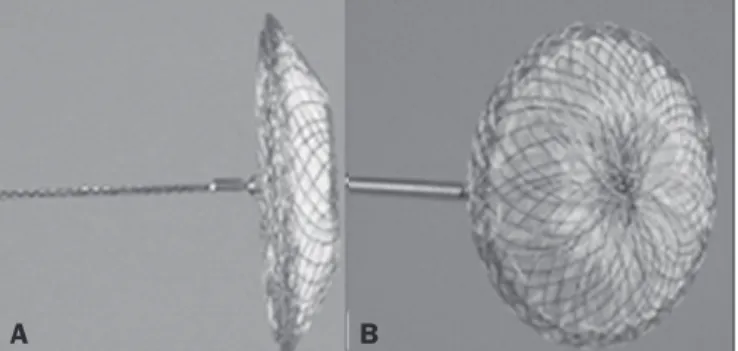
(Figulla ASD N; International Occlutech AB, Helsing
-borg, Sweden), as depicted in Figure 1, and the use of a
pulmonary inhalation-perfusion scintigraphy as a method
of detecting a residual BPF. The study was approved by
the Committee for the Analysis of Research Projects of the Hospital das Clínicas da Faculdade de Medicina da
Universidade de São Paulo (HC-FMUSP), reference no.
1089/09, and was registered with ClinicalTrials.gov (iden
-tifier: NCT01153074; http://www.clinicaltrials.gov/).
The study design included an initial bronchoscopic examination to measure the BPF diameter, assess the
de-gree of mucosal inflammation along the fistula trajectory,
and obtain biopsy samples to exclude the presence of
re-sidual disease. Inhalation-perfusion scintigraphy was per
-formed at baseline (before the occluder was put in place). Patients were followed through periodic bronchoscopy. A final evaluation, conducted at 12 months after the bron
-choscopic occlusion of the fistulas, included a complete clinical interview (to check for residual clinical symptoms such as dyspnea and difficulty in phonation) and a check for air leaks through the thoracostomy or chest tube, as well as bronchoscopy with a methylene blue test and an -other inhalation-perfusion scintigraphy examination.
The study sample comprised nine patients with total BPFs, all of whom had undergone open drainage of the pleural space by pleurotomy or chest tube and were not
suitable candidates for surgical correction. The demo-graphic, anthropometric, and clinical characteristics of the patients, including the Medical Research Council dyspnea score(12), are shown in Table 1.
The underlying disease, the type of surgery performed, the location of the BPF, the treatment of the ipsilateral pleural space, the time since the initial appearance of the
fistula, and the number of previous (surgical or broncho
-scopic) attempts at closure of the fistula are described in
Table 2.
Table 1—Demographic and clinical characteristics of the patients.
Patient 1 2 3 4 5 6 7 8 9 Age (years) 45 43 42 55 58 45 72 38 30 Gender Male Male Male Male Female Male Male Female Male Weight (kg) 62 45 48 74 41 49 48 48 71 Height (m) 1.64 1.74 1.66 1.74 1.74 1.74 1.74 1.74 1.74 BMI (kg/m2)
23.1 14.9 17.4 24.5 17.7 19.9 18.7 20.0 23.2 MRC score 4 5 5 5 5 5 5 4 5 Difficulty in phonation Yes Yes Yes Yes Yes Yes Yes No Yes
BMI, body mass index; MRC, Medical Research Council (dyspnea scale).
Figure 1. The Occlutech Figulla ASD N device. A: Lateral view. B: Frontal view.
The median time from the BPF occurrence to
treat-ment was 5.93 years (range, 0.25–17.0 years), and there had been previous (surgical or bronchoscopic) attempts at closure of the fistula in all of patients except patients 3 and 4. After the first bronchoscopic evaluation, the patients were submitted to a baseline pulmonary inhalation-perfu
-sion scintigraphy. The inclu-sion criterion was having a BPF
detected by scintigraphy. The technique utilized to place the occluders has been previously described in detail(13).
Pulmonary inhalation-perfusion scintigraphy
All scintigraphy procedures were performed in the Nu -clear Medicine Division of the HC-FMUSP Department
of Radiology, using a dual-head gamma camera (E-Cam; Siemens Medical Solutions, Chicago, IL, USA).
Prior to inhalation of the radiopharmaceutical and
the acquisition of images, the thoracostomy was occluded with a bandage or the chest tube was closed. Each patient inhaled, via a nebulizer, 900–1300 MBq (25–35 mCi) of technetium-99m-labeled diethylenetriaminepentaacetic acid (99mTc-DTPA). All patients were kept in the upright
position, and, during tidal breathing with the nose occluded,
the aerosolized 99mTc-DTPA was administered through a mouthpiece over a period of five minutes. The estimated level of activity that was reached and maintained in the lungs was 20–40 MBq (0.5–1.0 mCi).
Image acquisition
After the inhalation of the radiopharmaceutical,
pla-nar images of the chest were obtained in anterior, posterior, lateral, and oblique views. With a 128 × 128 matrix and a low-energy, high-resolution collimator, each planar image accumulated 500 K counts. The system was calibrated for an energy photopeak of 140 keV with a 15% window. One additional image was acquired in the anterior view of the chest with a flood source behind the patient in order to
delineate the contours of the body.
Perfusion scintigraphy
The perfusion scintigraphy scans were obtained af -ter the inhalation scintigraphy scans. The perfusion scans
were acquired after intravenous administration of 185 MBq (5 mCi) of 99mTc-labeled macroaggregated albumin
(99mTc-MAA).
Imaging acquisition
Planar images of the chest were obtained in anterior, posterior, lateral, and oblique views. With a 128 × 128 matrix and a low-energy, high-resolution collimator, each planar image accumulated 1500 K counts. The system was calibrated for an energy photopeak of 140 keV with a 15% window.
Follow-up and statistical analysis
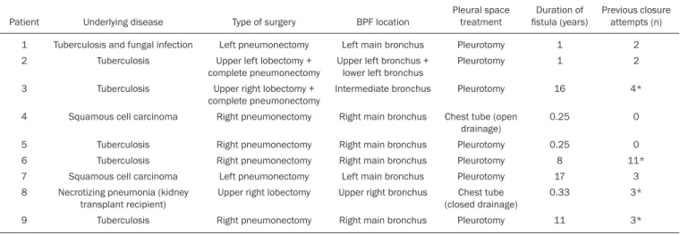
We have provided two examples of scintigraphy exami
-nations. One shows the 99mTc-DTPA activity in the
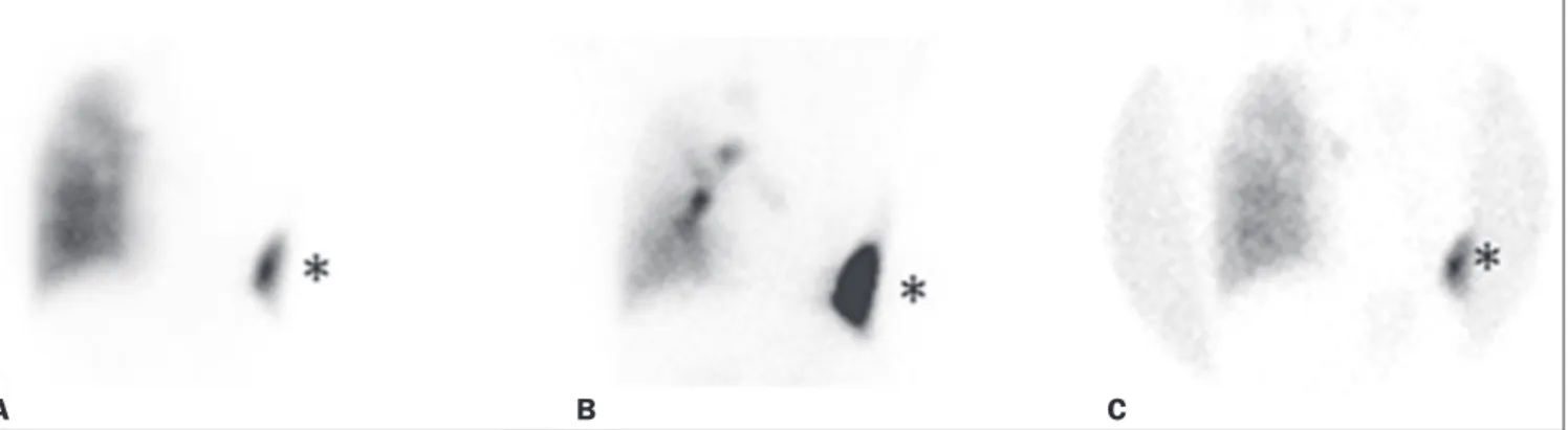
ban-dage occluding the wound on the left side of the chest wall, revealing the BPF (Figure 2). The other example demon -strates no signs of a BPF in a patient submitted to right
pneumonectomy (Figure 3).
As previously mentioned, bronchoscopy was repeated at 12 months after placement of the occluder, in order to eval
-uate the effectiveness of the treatment (Figure 4). The pos
-sibility of residual fistula was explored through instillation of
methylene blue into the treated bronchial stump. The
visu-alization of the dye in the pleural drain or pleurotomy was considered a strong indicator of residual air leak. Another ventilation scan was also acquired at that time.
A favorable outcome was defined as complete closure of the fistula, as confirmed by bronchoscopy, together with a negative methylene blue test or significant improvement of symptoms and the device being well positioned in the stump.
Continuous variables are expressed as means and
standard deviations, whereas categorical variables are ex -pressed as absolute and relative frequencies.
Table 2—The underlying disease, type of surgery performed, characteristics of the BPF, pleural space treatment, time since the occurrence of the fistula, and previous (surgical or bronchoscopic) attempts at closure.
Patient
1 2
3
4
5 6 7 8
9
Underlying disease
Tuberculosis and fungal infection Tuberculosis
Tuberculosis
Squamous cell carcinoma
Tuberculosis Tuberculosis Squamous cell carcinoma Necrotizing pneumonia (kidney
transplant recipient) Tuberculosis
Type of surgery
Left pneumonectomy Upper left lobectomy + complete pneumonectomy
Upper right lobectomy + complete pneumonectomy
Right pneumonectomy
Right pneumonectomy Right pneumonectomy Left pneumonectomy Upper right lobectomy
Right pneumonectomy
BPF location
Left main bronchus Upper left bronchus +
lower left bronchus Intermediate bronchus
Right main bronchus
Right main bronchus Right main bronchus Left main bronchus Upper right bronchus
Right main bronchus
Pleural space treatment
Pleurotomy Pleurotomy
Pleurotomy
Chest tube (open drainage) Pleurotomy Pleurotomy Pleurotomy Chest tube (closed drainage)
Pleurotomy
Duration of fistula (years)
1 1
16
0.25
0.25 8 17 0.33
11
Previous closure attempts (n)
2 2
4*
0
0 11*
3 3*
3*
In two patients (cases 2 and 4) the occluder was with
-drawn: in one, because a wire broke during an attempt to reposition the device; in the other, because the patient was
severely undernourished and had not developed
granula-tion tissue after one year. Two other patients (correspond
-ing to cases 5 and 6) died from unrelated causes, which precluded their inclusion in the final evaluation. Therefore, the final sample comprised five patients. The results of the
baseline pulmonary inhalation-perfusion scintigraphy,
to-gether with aspects observed at 12 months after placement
of the occluder, including the clinical symptoms, the bron-choscopic aspect of the bronchial stump, the methylene
blue test results, and the results of the final scintigraphy
are presented in Table 3.
The placement of the device in the bronchial stump
promoted a significant improvement of clinical symptoms
in the majority of the patients. In most cases, the patients
showed improved respiration and phonation, as well as
a reduction in pleural secretions and an improvement in their nutritional status. The improvement in clinical
symp-toms persisted while the device remained in the stump. In two cases, there was a residual air leak that was not identified by bronchoscopy or the methylene blue test but was detected only by pulmonary inhalation-perfusion scintigraphy. Those results correlated with the evolution of
Figure 2. Patient submitted to left pneumonectomy and inhalation-perfusion scintigraphy. A: Scintigraphy with 99mTc-MAA inhalation, anterior view in the perfusion scan. B: Scintigraphy with 99mTc-DTPA inhalation, anterior view in the ventilation scan. C: Scintigraphy with 99mTc-DTPA inhalation, anterior view and a silhouette contour in the inhalation scan. The images demonstrated no radiopharmaceutical activity in the left lung. Note the intense radiopharmaceutical activity at the bandage occluding the wound (asterisk) on the left side of the chest wall, demonstrating the BPF, in the inhalation scan. In the perfusion scan (A), residual radiopharmaceutical activity from the inhalation scan can be seen.
A B C
Figure 3. Patient submitted to right pneumonectomy and inhalation-perfusion scintigraphy. A: Scintigraphy with 99mTc-MAA inhalation, anterior view in the perfusion scan. B: Scintigraphy with 99mTc-DTPA inhalation, anterior view in the ventilation scan. C: Scintigraphy with 99mTc-DTPA inhalation, anterior view and a silhouette contour in the ventilation scan. D: Coronal computed tomography scan, with a lung window setting. The images demonstrated no radiopharmaceuti-cal activity in the right lung. There were no signs of BPF.
A B C D
Figure 4. Bronchoscopic view of a BPF in the upper right bronchus: A: Pre-treatment. B: At 12 months after placement of the occluder in the fistula
trajectory.
A B
RESULTS
Most (66.7%) of the BPFs evaluated were on the right side, and the mean diameter was 15 ± 3.65 mm (range, 6–17 mm). The initial bronchoscopic evaluation showed that all fistulas presented well-defined edges, suggesting a chronic process, and the biopsy of the mucosa along the fis -tula trajectory excluded active local disease in all cases. The pulmonary inhalation-perfusion scintigraphy obtained at
the patients, both of whom showed late signs of air leak, confirming the scintigraphy findings.
DISCUSSION
A BPF and the subsequent pleural space contamina-tion constitute one of the most serious postoperative com-plications after pulmonary resection. If not drained, the massive secretions from the pleural space can be aspirated
through the fistula, choking the patient or contaminating the contralateral lung. That is why all of the patients in our sample had a chest tube or a pleurotomy. However, drain
-age of the pleural space creates a route for a major air leak
that can hamper respiration and phonation.
The rational for bronchoscopic treatment of BPF in
patients whose clinical condition precludes surgical cor -rection is the fact that the placement of an occluder in the bronchial stump results in rapid improvement of
symp-toms. In a previous study conducted by our group, we re
-ported our experience with the bronchoscopic treatment of fistulas(14–16).
A BPF can be detected by several imaging modali-ties other than bronchoscopy, including chest X-ray and multidetector computed tomography employing advanced
image post-processing techniques. However, the presence of the occluder within the bronchial stump, in the BPF
trajectory, could produce an artifact in the above men-tioned radiographic methods and could preclude better evaluation through bronchoscopy. That is the rationale for choosing lung inhalation scintigraphy as an alterna-tive imaging method to evaluate the BPF before and after endoscopic treatment.
Greyson et al.(17) were the first to demonstrate that scintigraphy with inhalation of a radionuclide (99m
Tc-albu-min colloid fog) is a simple and accurate test for the detec -tion of a BPF.
In addition to 99mTc-albumin colloid fog, a variety of radioactive tracers, including 99mTc-sulfur colloid, 99m
Tc-DTPA, or a gas like 133Xe, could be alternative
radiopharma-ceuticals for use in inhalation-perfusion scintigraphy(18,19). Mark et al.(20) reported their experience using inhala-tion-perfusion scintigraphy with 99mTc-DTPA inhalation in
28 patients who had undergone pneumonectomy, showing
that, for the detection of a BPF, the method had a
sensitiv-ity of 78%, a specificsensitiv-ity of 100%, and an accuracy of 86%. Nevertheless, it lacks accuracy in the detection of very
small BPFs, is time-consuming, and requires patient
co-operation, which can be difficult during the postoperative period, when the patients could be on mechanical ventila -tion, could be critically ill, or could have sepsis.
Pulmonary inhalation-perfusion scintigraphy has other
known limitations. The turbulent flow in the tracheobron
-chial tree, which promotes aerosol deposition, can lead to false-positive results in patients with chronic obstructive
pulmonary disease. Therefore, this modality is currently
used only when conventional bronchoscopy, virtual bron -choscopy, and multidetector computed tomography have all failed to identify a clinically suspected BPF(21).
Although there is no standardization of pulmonary in-halation-perfusion scintigraphy for monitoring these cases, the technique used in the present study provided excellent
results that were strongly correlated with clinical improve
-ment and bronchoscopic findings, even in long-term clini
-cal follow-up.
In the patients with complete resolution of symptoms, closure of the fistula confirmed by bronchoscopy, and no evi
-dence of dye leakage, the inhalation-perfusion scintigraphy was completely negative. In cases of failure to close the BPF, the scintigraphy confirmed the persistence of the air leak.
In two (40%) of the five patients in our sample, scin
-tigraphy was the only method to show residual BPF, which was not detected in the bronchoscopic assessment or in the methylene blue test (no dye extravasation into the pleural space). Therefore, to avoid events related to the severe sep -sis that could occur if the space closed prematurely, the
thoracostomy was maintained, thus minimizing the risk, in those two patients.
In the literature, 99mTc-DTPA aerosol inhalation
scin-tigraphy has been reported to have poor sensitivity. However,
in the present study, the initial inhalation-perfusion scin-tigraphy detected BPF in all of the patients evaluated. That
is probably because open drainage of the hemithorax (with thoracostomy or a chest tube) was employed in all of the pa
-tients in our sample. Such drainage likely promotes the flow directly out of the thoracic cavity, as well as allowing the
detection of radioactive activity in the occlusive bandage or
adjacent to the chest wall (on its outer face). The detection
of radiopharmaceutical activity in the bandage or in the
ad-ditional image obtained with the flood source facilitated the
localization of such activity outside of the body(22).
Our results shows that pulmonary inhalation-perfu -sion scintigraphy is a useful tool to identify residual BPFs,
even when classical methods such as bronchoscopy and
Table 3—Baseline inhalation-perfusion scintigraphy, together with aspects ob-served at 12 months after placement of the occluder, including clinical symp-toms, as well as the results of the bronchoscopic evaluation of the bronchial stump, methylene blue test, and final scintigraphy.
Patient
1 2 3 4 5 6 7 8 9
Baseline scintigraphy
Positive Positive Positive Positive Positive Positive Positive Positive Positive
After occluder placement
Clinical symptoms
Absent Present Improved
Present * * Absent Absent Improved
Bronchoscopy
Closed Device withdrawn
Closed Device withdrawn
Closed Closed Closed
Methylene blue test
Negative Positive Negative Positive
Negative Negative Negative
Final scintigraphy
Negative Positive Positive Positive
Negative Negative Positive
the blue methylene test fail to detect it. Additional studies
with larger patient samples are needed in order to confirm our preliminary findings.
REFERENCES
1. Deschamps C, Bernard A, Nichols FC 3rd, et al. Empyema and bronchopleural fistula after pneumonectomy: factors affecting in-cidence. Ann Thorac Surg. 2001;72:243–7.
2. Sirbu H, Busch T, Aleksic I, et al. Bronchopleural fistula in the surgery of non-small cell lung cancer: incidence, risk factors, and management. Ann Thorac Cardiovasc Surg. 2001;7:330–6. 3. Cerfolio RJ. The incidence, etiology, and prevention of
postresec-tional bronchopleural fistula. Semin Thorac Cardiovasc Surg. 2001; 13:3–7.
4. Turk AE, Karanas YL, Cannon W, et al. Staged closure of compli-cated bronchopleural fistulas. Ann Plast Surg. 2000;45:560–4. 5. Seo H, Kim TJ, Jin KN, et al. Multi-detector row computed
tomo-graphic evaluation of bronchopleural fistula: correlation with clini-cal, bronchoscopic, and surgical findings. J Comput Assist Tomogr. 2010;34:13–8.
6. Gonfiotti A, Santini PF, Jaus M, et al. Safety and effectiveness of a new fibrin pleural air leak sealant: a multicenter, controlled, pro-spective, parallel-group, randomized clinical trial. Ann Thorac Surg. 2011;92:1217–24.
7. Sivrikoz CM, Kaya T, Tulay CM, et al. Effective approach for the treatment of bronchopleural fistula: application of endovascular metallic ring-shaped coil in combination with fibrin glue. Ann Tho-rac Surg. 2007;83:2199–201.
8. Tanaka S, Yajima T, Mogi A, et al. Successful management of a large bronchopleural fistula after lobectomy: report of a case. Surg Today. 2011;41:1661–4.
9. Walsh MD, Bruno AD, Onaitis MW, et al. The role of intrathoracic free flaps for chronic empyema. Ann Thorac Surg. 2011;91:865–8. 10. Wu G, Li ZM, Han XW, et al. Right bronchopleural fistula treated
with a novel, Y-shaped, single-plugged, covered, metallic airway stent. Acta Radiol. 2013;54:656–60.
11. Scordamaglio PR, Tedde ML, Minamoto H, et al. Can total bron-chopleural fistulas from complete stump dehiscence be endoscopi-cally treated? Eur J Cardiothorac Surg. 2017;51:702–8.
12. Fletcher CM, Clifton M, Fairbairn AS, et al. Standardized question-aries on respiratory symptoms. Br Med J. 1960;2:1665.
13. Tedde ML, Scordamaglio PR, Minamoto H, et al. Endobronchial closure of total bronchopleural fistula with Occlutech Figulla ASD N device. Ann Thorac Surg. 2009;88:e25–6.
14. Scordamaglio PR, Tedde ML, Minamoto H, et al. Endoscopic treat-ment of tracheobronchial tree fistulas using atrial septal defect oc-cluders: preliminary results. J Bras Pneumol. 2009;35:1156–60. 15. Tedde ML, Minamoto H, Scordamaglio PR, et al. Broncoscopic
clo-sure of tracheoesophageal fistulas. Ann Thorac Surg. 2011;91:1311. 16. Tedde ML, Scordamaglio PR, Rodrigues A, et al. Minimally invasive
closure of bronchopleural fistulas. Chest. 2011;140:826.
17. Greyson ND, Rosenthal L. Detection of postoperative bronchopleu-ral fistulas by radionuclide fog inhalation. Can Med Assoc J. 1970; 103:1366–8.
18. Nielsen KR, Blake LM, Mark JB, et al. Localization of bronchopleural fistula using ventilation scintigraphy. J Nucl Med. 1994;35:867–9. 19. Pigula FD, Keenan RJ, Naunheim KS, et al. Diagnosis of
postpneu-monectomy bronchopleural fistula using ventilation scintigraphy. Ann Thorac Surg. 1995;60:1812–4.
20. Mark JB, McDougall IR. Diagnosis and localization of broncho-pulmonary air leaks using ventilation scintigraphy. Chest. 1997; 111:286–9.
21. Gaur P, Dunne R, Colson YL, et al. Bronchopleural fistula and the role of contemporary imaging. J Thorac Cardiovasc Surg. 2014;148: 341–7.