Biological Factors and Overestimation of Left Ventricular Ejection
Fraction by Gated SPECT
Marco Antônio Condé de Oliveira
1, Paulo Schiavom Duarte
1, Maria Margarita C. Gonzalez
2, Valdir Ambrósio Moises
2,
Gilberto Alonso
1, Eduardo Vilaça Lima
2, Paola Emanuela Smanio
2, Luiz Roberto Fernandes Martins
1,Carlos A. R.
Oliveira
2, Luiz Eduardo Mastrocolla
2Seção de Medicina Nuclear, Fleury1 - Medicina e Saúde, Seção de Cardiologia, Fleury - Medicina e Saúde2, São Paulo, SP - Brazil
Mailing address: Marco Antônio Condé de Oliveira •
Rua Visconde de Ouro Preto, 138 - Consolação - 01303-060 - São Paulo, SP - Brazil
E-mail: [email protected]
Manuscript received June 11, 2007; revised manuscript received September 4, 2007; accepted November 19, 2007.
Summary
Background: Some patients present an overestimated left ventricular ejection fraction (LVEF) on electrocardiogram-gated myocardial scintigraphy (electrocardiogram-gated SPECT).
Objective: To establish the relationship between biological factors and overestimated LVEF.
Methods: We selected 3838 patients who underwent gated SPECT between May 20, 2000 and September 16, 2005 with
normal perfusion images and LVEF ≥50%. The following variables were analyzed: gender (29.4% females and 70.6%
males), age (from 20 to 94 years – mean: 56 years), weight (from 33.5 to 150 kg – mean: 79.6 kg), height (from 138 to 220 cm – mean: 171 cm) and BMI (from 13.9 to 54 – mean: 27.2). In a subgroup of 1002 patients who underwent echocardiogram, the diastolic diameter (from 36 to 68 mm – mean 47.5 mm) and systolic diameter (from 22 to 41 mm – mean 29.8 mm) variables were included. The patients were divided into two groups: normal LVEF (<80%) and
overestimated LVEF (≥80%). The odds ratio (OR) for presenting an overestimated LVEF was calculated for each variable
using logistic regression.
Results: The following odds ratios were found (p < 0.005): female gender OR = 3.585 (95%CI: 2.745 to 4.683), age in years OR = 1.020 (95%CI: 1.011 to 1.029) and height in cm OR = 0.893 (95%CI: 0.829 to 0.962). Weight and BMI were not significantly associated with LVEF (p>0.2). In the subgroup of 1002 patients, a statistically significant influence was found in overestimated LVEF values for the systolic diameter, gender and height variables.
Conclusion: Although systolic diameter influences the overestimation of LVEF, the gender and height variables have an independent influence on LVEF overestimation by gated SPECT. (Arq Bras Cardiol 2008; 90(5): 305-310)
Key words: Tomography, emission-computed, single-photon; stroke volume.
The minimum value considered normal for LVEF as calculated by gated SPECT is greater than or equal to 50%11, although
some studies in the literature report different values12,13. Normal
individuals may present LVEF values ranging from 55% to 75% as determined by angiocardiography or echocardiogram14.
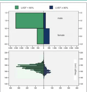
However, in our case series we noticed that some patients – usually short women, have LVEF values above those considerednormal (Figure 1). This association is also observed in other nuclear medicine services and by other authors in the literature15. This study analyzes the influence of several biological
factors such as gender, age, weight, height and body mass index (BMI) in the calculation of LFEV by gated SPECT, determining which factors are associated with overestimated LFEV and the degree of this association. It also analyzes the relationship between ventricular size (diastolic and systolic diameters) and the presence of overestimated LVEF in a subgroup of patients who underwent echocardiogram (Figure 2).
Methods
We retrospectively selected 3838 patients who underwent gated SPECT between May 20, 2000 and September 16, 2005.
Introduction
Myocardial perfusion scintigraphy allows the analysis of regional blood perfusion in myocardial walls, which contributes to the diagnosis, evaluation, and therapeutic and prognostic follow-up of coronary artery disease1. The introduction of
electrocardiographic gating in myocardial perfusion scintigraphy (gated SPECT) added the calculation of left ventricular ejection fraction (LVEF), the determination of systolic and diastolic volumes, and the analysis of myocardial wall motion and thickening to the analysis of perfusion2.In addition to helping
the analysis of perfusion images3-5, these parameters can offer
important prognostic information6,7. Several studies in the
literature have validated the calculation of LVEF by gated SPECT in comparison with other methods (first-pass scintigraphy8,
acquired 30 minutes after intravenous administration of 370 MBq (10 mCi) of 99mTc-MIBI. The stress images were acquired
four hours after the rest images with electrocardiogram gating. Intravenous administration of 1.11 GBq (30 mCi) of 99mTc-MIBI
in the stress phase was made at the peak effortof the exercise test (Bruce protocol, modified Bruce protocol, or Ellestad) or after the administration of dipyridamole (0.56 mg/kg for four minutes). Stress images were acquired 45 to 60 minutes after injection of the radiopharmaceutical. Image acquisition was made in a scintillation camera (Forte™ Phillips), with a low-energy high-resolution collimator. For each patient, 48 25-second projections in a 64 x 64 pixel matrix were obtained. The acquisition angle was 180 degrees (beginning in a right anterior oblique view at 45 degrees and ending in a left posterior oblique view at 45 degrees). Image reconstruction was performed with a filtered back projection technique using a Butterworth filter; LVEF was processed by the Autoquant™ software (Cedars-Sinai’s Quantitative Gated SPECT – QGS) from stress images.
In addition to LVEF, the following variables were included: gender, (29.4% female and 70.6% male), age (from 20 to 94 years – mean: 56 years), weight (from 33.5 to 150 kg – mean: 79.6 kg), height (from 138 to 220 cm – mean: 171 cm) and BMI (from 13.9 to 54 – mean: 27.2) (Table 1). BMI was calculated using the formula BMI = weight / (height x height).
The patients were divided into two arbitrary groups according to LVEF results: normal (< 80%) and overestimated (≥ 80%). Data were analyzed using logistic regression and Pearson’s correlation with the SPSS™ statistical package. The odds ratio for presenting an overestimated LVEF was calculated for each variable.
Of the 3838 patients studied, we selected a subgroup of 1002 patients who had undergone echocardiogram within up to ten days of the gated SPECT (61.4% of the patients underwent both tests on the same day and 95.5% of the patients underwent the tests within up to one week between them – with a mean of 1.57 days). In this subgroup, the values of systolic (from 22 to 41 mm – mean of 29.8 mm) and diastolic (from 36 to 68 mm – mean of 47.5 mm) diameters were listed for each patient (Table 2). These two variables along with the gender (female) and height variables were analyzed using logistic regression and Pearson’s correlation. The odds ratio for presenting an overestimated LVEF was calculated for patients with a < 30 mm systolic diameter; for patients with a < 48 mm diastolic diameter; and for the gender (female) and height variables. These values mentioned for the systolic and diastolic diameters were considered as the cut-off point for the calculation of the odds ratio because they were the
Table 1 - Population Features
All patients n = 3838
Mean ± SD Median
Age (years) 55.82 ± 11.59 56.00
Weight (kg) 79.65 ± 15.06 79.00
Height (cm) 170.71 ± 9.50 172.00
BMI 27.22 ± 4.09 26.70
Figure 1 -Diagram showing the distribution of normal and overestimated LVEF among the male and female genders, and among the different height ranges (all patients n = 3838).
LVEF < 80% LVEF ≥ 80%
male
female
H
e
ig
h
t
(cm)
Figure 2 -Diagram showing the distribution of normal and overestimated LVEF among patients with systolic diameter lower than and higher than 30 mm, and among patients with diastolic diameter lower than and higher than 48 mm (patients undergoing echocardiogram; n = 1002).
LVEF < 90% LVEF ≥ 90%
Systolic diameter < 30mm
Systolic
diameter ≥ 30mm
Diastolic diameter < 48mm
Diastolic
diameter ≥ 48mm
Inclusion criteria were: normal myocardial perfusion images on scintigraphy and LVEF greater than or equal to 50% as calculated by gated SPECT.
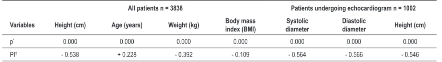
Table 4 - Pearson’s correlation
All patients n = 3838 Patients undergoing echocardiogram n = 1002
Variables Height (cm) Age (years) Weight (kg) Body mass index (BMI)
Systolic diameter
Diastolic
diameter Height (cm)
p* 0.000 0.000 0.000 0.000 0.000 0.000 0.000
PI† - 0.538 + 0.228 - 0.392 - 0.109 - 0.564 - 0.566 - 0.546
* Statistical signiicance. † Pearson’s correlation index.
Table 5 - Logistic regression
Patients undergoing echocardiogram n = 1002
Variables Systolic diameter < 30 mm Diastolic diameter < 48 mm Female gender Height (cm)
p* 0.000 0.113 0.000 0.000
OR† (95,0% C.I.) 4.078 (2.244 – 7.409) 1.600 (0.895 – 2.860) 2.794 (1.598 – 4.884) 0.907 (0.877 – 0.938)
* Statistical signiicance. † Odds ratio for inding an overestimated LVEF.
closest values to the mean. The gender (female) and height variables were included in the logistic regression analysis in the subgroup of 1002patients along with systolic and diastolic diameters, so as to verify whether they had an independent statistical relation to the finding of an overestimated LVEF.
Results
When all patients were considered, the gender, height and age variables showed a statistically significant association with overestimated LVEF (p<0.005), notably the first two (Table 3). Weight and BMI were not significantly associated with the finding of an overestimated LVEF (P>0.2). Among the continuous variables, height had the highest correlation index with overestimated LVEF when all patients were considered (Table 4).
In the subgroup of patients who underwent echocardiogram, the systolic diameter showed a statistically significant relation to overestimated LVEF (Table 5). The diastolic diameter did not independently influence overestimated LVEF. The gender
(female) and height variables still influenced the finding of an overestimated LVEF, although less significantly in relation to the analysis of all patients. The Pearson’s correlation coefficients demonstrated an inverse correlation between overestimated LVEF and height, diastolic diameter and systolic diameter (Table 4).
Discussion
The Autoquant™ software calculates LVEF from the three-dimensional reconstruction of the left ventricular cavity based on gated short-axis images. The end-systolic volume (ESV) and end-diastolic volume (EDV) are determined after epicardial and endocardial edge detection, and LVEF is calculated according to the formula: [(EDV - ESV) / EDV] x 100 (%)8.
In our case series, female patients had an approximately 3.5 times higher chance of presenting an overestimated LVEF in comparison with male patients. Height had an inverse correlation with the finding of an overestimated LVEF. Our data confirm that overestimated LVEF occurs more frequently among short women.
A possible explanation for this finding is the fact that small women have smaller hearts16, which causes the calculated
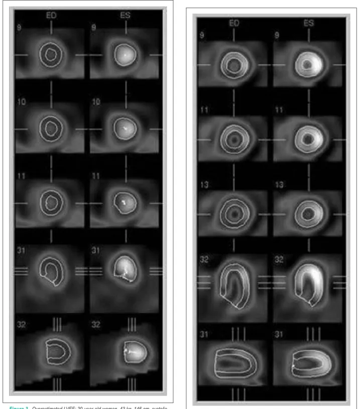
systolic volume to be underestimated, thus consequently overestimating the LVEF. In patients with very small systolic volumes, the automaticdetermination of the endocardial edge by the software is impaired due to low resolution and partial volume effects17 (Figure 3). The endocardial edge, in turn, is
easily determined in patients with greater ventricular volumes
Table 2 - Ventricular diameters
Patients undergoing echocardiogram n = 1002
Mean ± SD Median
Systolic diameter (mm) 29.80 ± 2.93 30.00
Diastolic diameter (mm) 47.47 ± 3.94 48.00
Table 3 - Logistic regression
All patients n = 3838
Variable Female gender Height (cm) Age (years) Weight (kg) BMI
p* 0.000 0.003 0.000 0.473 0.286
OR† (95,0% C.I.) 3.585 (2.745 - 4.683) 0.893 (0.829 - 0.962) 1.020 (1.011 - 1.029) 1.030 (0.950 - 1.118) 0.888 (0.714 - 1.105)
(Figure 4). The analysis of the subgroup of 1002 patients who underwent echocardiogram corroborates the hypothesis of the relationship between ventricular size and overestimated LVEF. The high value of the odds ratio found for the systolic diameter
variable suggests that the main mechanism responsible for LVEF overestimation is possibly the difficulty to determine the endocardial edge in patients with small systolic volumes. However, Yamada et al18 demonstrated that women have
significantly higher LVEF values as calculated both by Segami™
Figure 3 -Overestimated LVEF: 39-year-old woman, 43 kg, 146 cm, systolic diameter (echocardiogram) = 23 mm, diastolic diameter (echocardiogram) = 39 mm, LVEF as calculated by gated SPECT = 97%, systolic volume as calculated by gated SPECT = 1 ml. (First column: diastole views; second column: systole views).
References
1. Clark AN, Beller GA. The present role of nuclear cardiology in clinical practice. Q J Nucl Med Mol Imaging. 2005; 49 (1): 43-58.
2. Maddahi J, Kiat H, Van Train KF, Prigent F, Friedman J, Garcia EV, et al. Myocardial perfusion imaging with technetium-99m sestamibi SPECT in the evaluation of coronary artery disease. Am J Cardiol. 1990; 66 (13): 55E-62E.
3. Sciagra R, Leoncini M. Gated single-photon emission computed tomography: the present-day ‘’one-stop-shop’’ for cardiac imaging. Q J Nucl Med Mol Imaging. 2005; 49 (1): 19-29.
4. Leoncini M, Sciagra R. Role of perfusion myocardial scintigraphy with gated SPECT technique in the diagnostic and prognostic evaluation of patients with chronic coronary disease. Ital Heart J. 2002; 3 (3 Suppl): 309-18.
5. Smanio PE, Watson DD, Segalla DL, Vinson EL, Smith WH, Beller GA. Value of gating of technetium-99m sestamibi single-photon emission computed tomographic imaging. J Am Coll Cardiol. 1997; 30: 1687-92.
6. Sharir T, Kang X, Germano G, Bax JJ, Shaw LJ, Gransar H, et al. Prognostic value of poststress left ventricular volume and ejection fraction by gated myocardial perfusion SPECT in women and men: Gender-related differences in normal limits and outcomes. J Nucl Cardiol. 2006;13: 495-506.
7. Sharir T, Germano G, Kavanagh PB, Lai S, Cohen I, Lewin HC, et al. Incremental prognostic value of post-stress left ventricular ejection fraction and volume by gated myocardial perfusion single photon emission computed tomography. Circulation. 1999; 100 (10): 1035-42.
8. Germano G, Kiat H, Kavanagh PB, Moriel M, Mazzanti M, Su HT, et al.
and Autoquant™ softwares. The Segami™ software defines the ventricular wall position by means of statistical parameters of counting rate distribution and not by the automatic definition of cardiac contours and thus should, at least theoretically, be less influenced by small ventricular volumes.
Our data show that female gender and height independently influenced the finding of an overestimated LVEF in the group of patients who underwent echocardiogram. Despite presenting an inverse correlation with overestimated LVEF (Table 4), the diastolic diameter did not prove to be independently associated with overestimated LVEF in the logistic regression analysis (Table 5).
Other authors have already demonstrated the influence of small hearts in LVEF as estimated by gated SPECT19,20. Germano
et al21 demonstrated an inverse exponential relation between
end-diastolic volume and LVEF, and between end-systolic volume and LVEF in a study with 926 patients in which they used Autoquant™. Some authors suggest techniques for the correction of overestimated LVEF, such as larger acquisition matrices or use of specific filters20. An alternative to prevent
overestimation is the use of techniques that do not use automatic systolic endocardial edge detection17.
In addition to influencing the LVEF calculation, the size of the left ventricular cavity may also influence some other indexes calculated from the automatic detection of the endocardial contours. In a previous study, Duarte et al22 reported the influence of gender in obtaining TID
(transient ischemic dilation) values, an index that measures the difference between ventricular cavities with stress and at rest. In a group of 16 patients with a low pretest probability of ischemia and high TID values (>1.25), 14
were women. According to the authors, a possiblereason
for the predominance of women in this subgroup could be the difficulty to define the endocardial contours in patients with small ventricular cavities, mainly during the resting phase of scintigraphy, in which the radioactive dose administered is approximately three times lower than that administered in the stress phase.
Even in cases of overestimated LVEF, gated SPECT can provide important information on the left ventricular function. In these cases, it is possible to demonstrate the normal
thickening, which is characterized by increased radioactivity by area in myocardial walls during systole in relation to diastole, and the preserved motility as demonstrated by a significant reduction of the ventricular cavity during systole.
A possible study limitation that should be mentioned is the type of study conducted (cross-sectional), which allows the association between independent variables and the dependent variable to be estimated, however without allowing a causal relation between them to be established. Additionally, we were not able to establish a biological mechanism that justifies some associations, such as that between the female gender and LVEF overestimation when this variable is controlled by the systolic diameter. Thus, further studies are necessaryto explain all the causal factors of overestimated LVEF as measured by gated SPECT.
Conclusion
The gender (female), height and systolic diameter variables were associated with the finding of an overestimated LVEF. The age variable contributed less significantly. Weight and BMI did not show a statistically significant relation with the finding of an overestimated LVEF.
This analysis confirms what is observed in clinical practice: short women present overestimated LVEF by gated SPECT. Although a reduced ventricular diameter may be a possible explanation for this phenomenon, the gender and height variables independently influenced the overestimation of LVEF by gated SPECT.
Potential Conflict of Interest
No potential conflict of interest relevant to this article was reported.
Sources of Funding
There were no external funding sources for this study.
Study Association
Automatic quantification of ejection fraction from gated myocardial perfusion SPECT. J Nucl Med. 1995; 36 (11): 2138-47.
9. Cwajg E, Cwajg J, Keng F, He ZX, Nagueh S, Verani MS. Comparison of global and regional left ventricular function assessed by gated-SPECT and 2-D echocardiography. Rev Port Cardiol. 2000;19 (Suppl 1): 139-46.
10. Thorley PJ, Plein S, Bloomer TN, Ridgway JP, Sivananthan UM. Comparison of 99mTc tetrofosmin gated SPECT measurements of left ventricular volumes and ejection fraction with MRI over a wide range of values. Nucl Med Commun. 2003; 24 (7): 763-9.
11. Sociedade Brasileira de Cardiologia. I Diretriz da Sociedade Brasileira de Cardiologia Sobre Cardiologia Nuclear. Arq Bras Cardiol. 2002; 78 (suppl.3): 1-42.
12. Rozanski A, Nichols K, Yao SS, Malholtra S, Cohen R, DePuey EG. Development and application of normal limits for left ventricular ejection fraction and volume measurements from 99mTc-sestamibi myocardial perfusion gates SPECT. J Nucl Med. 2000; 41(9):1445-50.
13. Henzlova MJ, Croft LB. The electrocardiogram as a predictor of left ventricular systolic function: correlation with gated SPECT imaging. Mt Sinai J Med. 2003; 70 (5): 306-9.
14. ZipesDP, Libby P, Bonow RO, Braunwald E. Braunwald’s heart disease: a textbook of cardiovascular medicine. 7th ed. Philadelphia: Saunders; 2005.
15. Ford PV, Chatziioannou SN, Moore WH, Dhekne RD. Overestimation of the LVEF by quantitative gated SPECT in simulated left ventricles. J Nucl Med. 2001; 42 (3): 454-9.
16. Hansen CL, Crabbe D, Rubin S. Lower diagnostic accuracy of thallium-201
SPECT myocardial perfusion imaging in women: an effect of smaller chamber size. J Am Coll Cardiol. 1996; 28 (5): 1214-9.
17. Feng B, Sitek A, Gullberg GT. Calculation of the left ventricular ejection fraction without edge detection: application to small hearts. J Nucl Med. 2002; 43 (6): 786-94.
18. Yamada AT, Campos Neto GC, Soares Júnior J, Giorgi MCP, Araújo F, Meneghetti JC, et al. Diferenças relacionadas ao sexo nos volumes ventriculares e na fração de ejeção do ventrículo esquerdo estimados por cintilografia de perfusão miocárdica: comparação entre os programas quantitative Gated sPeCt (qgS) e Segami. Arq Bras Cardiol. 2007; 88 (3): 285-90.
19. Khalil MM, Elgazzar A, Khalil W, Omar A, Ziada G. Assessment of left ventricular ejection fraction by four different methods using 99mTc tetrofosmin gated SPECT in patients with small hearts: correlation with gated blood pool. Nuclear Medicine Communications. 2005; 26 (10): 885-93.
20. Hambye AS, Vervaet A, Dobbeleir A. Variability of left ventricular ejection fraction and volumes with quantitative gated SPECT: influence of algorithm, pixel size and reconstruction parameters in small and normal-sized hearts. Eur J Nucl Med Mol Imaging. 2004; 31 (12): 1606-13.
21. Germano G, Kavanagh PB, Kavanagh JT, Wishner SH, Berman DS, Kavanagh GJ. Repeatability of automatic left ventricular cavity volume measurements from myocardial perfusion. J Nucl Cardiol. 1998; 5 (5): 477-83.