PATHOLOGICAL FRACTURES DUE TO BONE METASTASES FROM
LUNG CANCER: RISK FACTORS AND SURVIVAL
FRATURA PATOLÓGICA DEVIDO À METÁSTASE ÓSSEA DO CÂNCER
DE PULMÃO: FATORES DE RISCO E SOBREVIDA
M
arceloB
ragança dosr
eiso
liveira1,2, B
runodec
arvalhoM
arques1, r
osaa
urílioM
atos2, c
ésarr
uBensdac
ostaF
ontenelle1,
F
ernandac
arvalhodeq
ueirozM
ello1, M
arcose
duardoM
achadoP
aschoal21. Universidade Federal do Rio de Janeiro, Rio de Janeiro, RJ, Brazil. 2. Escola de Saúde do Exército, Rio de Janeiro, RJ, Brazil.
Citation: Oliveira MBR, Marques BC, Matos RA, F ontenelle CRC, Mello FCQ, Paschoal MEM. Pathological fractures due to bone metastases from lung cancer: risk factors and survival. Acta Ortop Bras. [online]. 2018;26(6):388-93. Available from URL: http://www.scielo.br/aob.
Study performed at the Hospital Universitário Clementino Fraga Filho, Universidade Federal do Rio de Janeiro.
Correspondence: Marcelo Bragança dos Reis Oliveira. Serviço de Traumato-Ortopedia do Hospital Universitário Clementino Fraga Filho da Universidade Federal do Rio de Janeiro. Rua Rodolpho Paulo Rocco, 255, Cidade Universitária, Ilha do Fundão, Rio de Janeiro, RJ, Brazil. 21941-913. marceloreis@hucff.ufrj.br
O
riginala
rticleDOI: http://dx.doi.org/10.1590/1413-785220182606201669
All authors declare no potential conflict of interest related to this article.
Article received in 05/31/2018, approved in 08/20/2018. ABSTRACT
Introduction: Pathological fractures are frequent skeletal-related events among lung cancer patients, which result in high morbidity and decreased overall survival and make operative treatment decisions challenging. Objectives: To identify risk factors associated with the occurrence of pathological fractures in patients with lung cancer and to determine survival. Methods: We conducted a retrospective cohort study with 407 lung carcinoma patients diagnosed between 2006 and 2015. The prevalence of bone metastases and pathological fractures was calculated. Statistical analysis was conducted using a chi-squared test, and the odds ratio and 95% confidence interval were calculated. Overall survival was determined using the Kaplan-Meier method and differences were compared using the log-rank test. Re-sults: The prevalence of bone metastases and pathological fractures was 28.2% (n = 115) and 19.1% (n = 22), respectively. Pathological fractures were more frequent among patients with bone metastases at the time of diagnosis of lung cancer (24.7% [n = 20] vs. 5.9% [n = 2]; p < 0.05). The median overall survival following the diagnosis of lung cancer, bone metastases, and pathological fracture was 6, 4, and 2 months, respectively. Conclusions: Pathological fracture was associated with synchronous bone metastases and overall survival times were considerably reduced. Level of Evidence IV, Case Series.
Keywords: Lung neoplasms, Bone, Fractures.
RESUMO
Introdução: Fratura patológica é um evento esquelético frequente em pacientes com câncer de pulmão, resultando em alta morbidade e sobrevida global reduzida que torna a decisão de tratamento cirúr-gico desafiadora. Objetivos: Identificar fatores de risco associados à ocorrência de fraturas patológicas em pacientes com câncer de pulmão e determinar a sobrevida. Métodos: Conduzimos um estudo retrospectivo de coorte com 407 pacientes diagnosticados com carci-noma pulmonar entre 2006 e 2015. A prevalência de metástase óssea e fratura patológica foi calculada. Análise estatística foi conduzida usando o teste X2, e razão de chances e o intervalo de confiança de 95% foi calculado. A sobrevida global foi determinada usando o método de Kaplan-Meier e as diferenças foram comparadas usando o teste do log-rank. Resultados: A prevalência de metásta-ses ósseas e fraturas patológicas foi de 28,2% (n = 115) e 19,1% (n = 22), respectivamente. Fraturas patológicas foram mais frequentes em pacientes com metástases ósseas ao diagnóstico do câncer de pulmão (24,7 % [n = 20] vs. 5,9% [n = 2]; p < 0.05). A sobrevida global média após o diagnóstico do câncer de pulmão, da metástase óssea e da fratura patológica foram 6, 4 e 2 meses, respectivamente. Conclusão: Fratura patológica foi associada à metástase óssea sincrônica e a sobrevida global consideravelmente reduzida. Nível de Evidência IV. Série de Casos.
Descritores: Neoplasias pulmonares. Osso. Fraturas.
INTRODUCTION
The bone represents one of the most frequent sites of metastases from lung cancer, occurring in approximately 15–40 % of patients.1
Skeletal-related events (SREs) are prevalent among these patients, resulting in high morbidity and a reduced quality of life.2,3 Despite
the negative impact of bone metastases (BMs) on lung cancer prognosis, the unpredictability of survival makes an indication of palliative surgery challenging. Therefore, the elucidation of factors
that can determine which patients will survive for a longer or shorter time is fundamental in guiding optimal treatment decisions. In previous studies conducted by Oliveira et al., adenocarcinoma was associated with the higher risk of developing BMs.3,4 Therefore,
on survival. This analysis can support the decision to surgically treat these patients, or not, and identify in which patient palliative surgery would be better indicated.
MATERIALS AND METHODS
The medical records of 413 patients who had been diagnosed with malignant primary lung tumors at our institution between 2006 and 2015 were retrospectively evaluated. Inclusion criteria included a pathological confirmation of lung carcinoma and complete tumor staging data. Lung carcinomas were stratified based on the World Health Organization’s classification system: adenocarcinoma (ADC), squamous cell carcinoma (SCC), large-cell carcinoma (LCC), NSCLC not otherwise specified (NOS), and SCLC.5
Events of interest included BMs and PF occurrence, and death. BMs were diagnosed by histopathological examination or using Technetium-99m bone scintigraphy with two additional imaging modalities (plain films and computed tomography or magnetic resonance imaging). BMs and PFs were classified, according to the time of diagnosis of the primary tumor, as either synchronous or metachronous. Collected variables included age, sex, smoking status, histological subtype, metastatic sites, a PF, the time of diagnosis of BMs and PFs in relation to the time of diagnosis of the primary tumor, and survival. Patients diagnosed with a second malignant primary tumor who had an unknown date of death were excluded from survival analysis. The minimum follow-up period following a diagnosis of BMs was 24 months, excluding patients with a survival of < 24 months. The proportions of the variables among patients who developed or did not develop a PF were compared using a chi-squared test. The relationship between the variables and the occurrence of a PF was also evaluated using odds ratios (ORs) and 95% confidence intervals (CIs). Overall survival (OS) and associations with the variables were analyzed using the Kaplan-Meier method. Differences between patients
who developed or did not develop a PF, in addition to a synchronous or metachronous PF, were evaluated using the log-rank test. All statistical analyses were performed using Statistical Package for the Social Sciences for Windows, software version 10.0 (SPSS Inc., Chicago, IL, USA). A p < 0.05 was considered statistically significant.
The study protocol was approved by the appropriate Institution-al Review Board committee (protocol number 113/11). All study participants provided written informed consent and research was conducted in accordance with the Declaration of Helsinki.
RESULTS
Among 413 lung cancer patients, 407 (98.5 %) who met the inclu-sion criteria for histological subtype were analyzed. The patients’ clinicopathological characteristics are summarized in Table 1, and a cohort diagram is presented in Figure 1.
ADC was the most prevalent histological subtype (46.7 %; n = 190), followed by SCC (25.1 %; n = 102).
The prevalence of BMs was 28.3 % (n = 115). Synchronous BMs occurred significantly more frequently than metachronous BMs (19.9 % [n = 81] vs. 8.4 % [n = 34]; p < 0.01). In total, 79 pa-tients experienced 122 SREs, with an overall prevalence of 68.7 % among patients with BMs. Radiotherapy was the most frequent SRE (60.7 %; n = 74), followed by PF (19.1 %; n = 22), spinal cord com-pression (9.0 %; n = 11), surgical treatment for BMs (6.6 %; n = 8), and hypercalcemia (5.7 %; n = 7) (Figure 1).
PF fracture was more common among ADC patients (23.9 %; n = 16). The Figure 2 shows the prevalence of PFs according to lung cancer histology. Among 115 patients with BMs, 305 BMs and 22 PFs occurred. The most frequent site of BMs and PFs included the spine (32.1 %; n = 98) and humerus (36.4 %, n = 8), (Figure 3).
To evaluate the relationship between the patients’ clinicopatho-logical characteristics and the occurrence of PFs, we compared the prevalence of histological subtype, smoking, synchronous or metachronous BMs, and the location of BMs in patient groups with or without PFs (Table 3). Among patients who developed a PF, the prevalence of ADC, SCC, LCC, NSCLC, and SCLC was 72.8 % (n = 16), 4.5 % (n = 1), 4.5 % (n = 1), 9.1 % (n = 2), and 9.1 % (n = 2), respectively. The ORs and 95 % CIs are displayed in Table 3. Smoking was significantly associated with the occurrence of a PF. The prevalence of smoking in patients with a PF was substantially higher than the prevalence of smoking in patients without a PF (95.5 % [n = 21] vs. 74.2 % [n = 69]; p < 0.05).
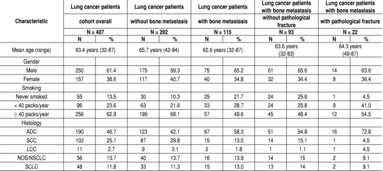
Table 1. Clinicopathological characteristics of different histological subtypes of lung cancer in patients with or without bone metastases and patho-logical fractures.
Characteristic
Lung cancer patients Lung cancer patients Lung cancer patients Lung cancer patients with bone metastasis
Lung cancer patients with bone metastasis
cohort overall without bone metastasis with bone metastasis without pathological
fracture with pathological fracture
N = 407 N = 292 N = 115 N = 93 N = 22
N % N % N % N % N %
Mean age (range) 63.4 years (32-87) 65.7 years (42-84) 62.6 years (32-87) 63.6 years (32-83)
64.3 years (49-87) Gender
Male 250 61.4 175 59.3 75 65.2 61 65.6 14 63.6
Female 157 38.6 117 40.7 40 34.8 32 34.4 8 36.4
Smoking
Never smoked 55 13.5 30 10.3 25 21.7 24 25.8 1 4.5
< 40 packs/year 96 23.6 63 21.6 33 28.7 24 25.8 9 41.0
≥ 40 packs/year 256 62.9 199 68.1 57 49.6 45 48.4 12 54.5
Histology
ADC 190 46.7 123 42.1 67 58.3 51 54.8 16 72.8
SCC 102 25.1 87 29.8 15 13.0 14 15.1 1 4.5
LCC 11 2.7 9 3.1 2 1.8 1 1.1 1 4.5
NOS/NSCLC 56 13.7 40 13.7 16 13.9 14 15 2 9.1
SCLC 48 11.8 33 11.3 15 13.0 13 14 2 9.1
The prevalence of a diaphyseal PF (36.0 %) was significantly higher than that of a metaphyseal PF (9.5 %), demonstrating an increased risk of a PF in the diaphyseal region (OR: 5.34, 95 % CI: 1.65–17.2;
p < 0.01). PFs were more frequently observed in patients with synchronous BMs (24.7 %) compared to patients with metachronous BMs (5.9 %) (OR: 5.24, 95 % CI: 1.15–23.8; p < 0.05).
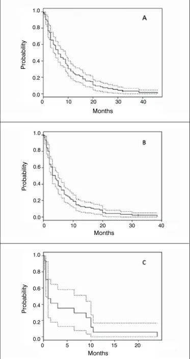
The primary treatment method for PFs was non-operative, based on immobilization and radiation. Surgical treatment was performed in 8 patients (36.3 %) through internal fixation with an intramedullary nail (22.7 %; n = 5) or megaprosthesis replacement (13.6 %; n = 3). The sample size for analysis of OS was 95. Twenty patients (17.4 %) were excluded due to the diagnosis of a second malignant primary tumor (10.4 %; n = 12) or as a result of an unknown date of death (7.0 %; n = 8). The median OS times following a diagnosis of lung cancer, BMs, and PF were 6 months, 4 months, and 2 months, respectively (Table 4). Kaplan-Meier curves with 95 % CIs are presented in Figure 4.
Median OS times following a diagnosis of BMs were compared between patient groups who developed or did not develop a PF. The median OS times were 7.4 months and 6.4 months, respectively, with no statistical significance (p>0.05). The median OS times of patients with synchronous or metachronous PFs were 3.6 months and 10.7 months, respectively, with no statistical significance (p>0.05).
DISCUSSION
The skeleton is one of the most frequent sites of metastatic carci-noma dissemination.6 In the present cohort of lung cancer patients
treated at a single academic general hospital, 28.3 % of lung can-cer patients (n = 115) had secondary bone involvement. Studies comparing the frequencies of metastatic sites in NSCLC patients demonstrated a bone dissemination frequency of 20–40 %.2,3,7-10
Although bone involvement usually means the cancer is incurable, a proportion of patients do survive for prolonged periods. Conse-quently, these patients are more likely to develop SREs.11 Recent
advances in the treatment of cancer have prolonged survival.12 As
survival increases, SREs, including PFs, are gaining increasing importance, since a prolonged survival increases the risk of such complications over a long period. In the present study, the PF rate reached 19.1 % in patients with secondary bone involvement from lung cancer. This finding was comparable to that of Saad et al. (16.6 %; n = 127), who evaluated the significance of PFs in the survival of patients with different primary tumors.11
The patients included in the present study had independent risk factors for the occurrence of PFs: the presence of BMs and a lung cancer diagnosis.13 In the evaluation of variables associated with the
occurrence of PFs, statistical significance was reached for the time of diagnosis or location of BMs and smoking. PFs occurred more frequently in patients with synchronous BMs (24.7 %) compared to patients with metachronous BMs (5.9 %). The frequency of this event was probably higher in the synchronous group, owing to the longer period that these patients are exposed to BMs without specific treatment.
We postulate that the predisposition of cancer patients to developing a PF is multifactorial in origin. In addition to being most obviously and directly related to reduced bone strength, compromised by the presence of metastatic lesions, the risk of developing a PF may also be enhanced by other factors (e.g., immobility, malnutrition, and chemotherapy). Moreover, a major bias is associated with PF risk in lung cancer patients, because of the direct relationship between smoking and lung cancer. However, smoking was independently associated with an increased risk of developing a PF. A previous study reported that smoking increases the PF risk in lung cancer patients, as observed in the present study.14 Other possible factors related to
the occurrence of a PF include the association between lung cancer and chronic obstructive pulmonary disease and, consequently, the use of corticosteroids that result in bone demineralization.15
BMs are associated with a poor prognosis in patients with cancer.16
In a study conducted by Sathiakumar et al., the risk of death was 2.4 times higher in patients who developed BMs compared to those who
407 pts Histologic criteria
237 pts / 58.2% Distant disease*
79 pts / 68.7% SREs 79 pts / 68.7%
SREs
22 pts / 27.8% Pathological fracture
*Stage at cancer diagnosis
115 pts / 28.2% Bone metastases 170 pts / 41.8%
Localized or regional disease*
34 pts / 29.6% Metachronous bone
metastases
81 pts / 70.4% Synchronous bone metastases 413 pts
Lung cancer
6 pts Were excluded
Figure 1. Cohort diagram.
Figure 2. Pathological fracture frequency according to lung cancer histology.
ADC: adenocarcinoma; SCC: squamous cell carcinoma; LCC: large cell carcinoma; NOS not otherwise specified; NSCLC: non-small cell lung cancer, SCLC: small cell lung cancer.
23,9%
6,7% 50,0%
Histologic subtype
Prevalence
100% 90% 80% 70% 60% 50% 40% 30% 20% 10% 0%
12,5% 13,3% 19,1%
Table 2. Analysis of histological subtype, synchronous bone metastases, smoking, and bone location as factors associated with pathological fractures in lung cancer patients.
Pathological Fracture
OR IC (95%) p-value
Yes No
N % N %
Histology
ADC 16 72.8% 51 54.7% 2.19 (0.79-6.11) 0.125
SCC 1 4.5% 14 15.1% 0.26 (0.03-2.16) 0.188
LCC 1 4.5% 1 1.1% 4.38 (0.26-72.9) 0.262
NOS/NSCLC 2 9.1% 14 15.1% 0.56 (0.11-2.68) 0.467
SCLC 2 9.1% 13 14.0% 0.61 (0.12-2.95) 0.540
Bone Metastasis
Synchronous 20 90.1% 61 65.6%
5.08 (1.11-23.1) 0.035 Metachronous 2 9.9% 32 34.4%
Smoking
Yes 21 95.5% 69 74.2%
7.7 (1.01-67.1) 0.029
No 1 4.5% 24 25.8%
Location
Dyaphyseal 9 60.0% 16 22.5%
5.34 (1.65-17.2) 0.005 Metaphyseal 6 40.0% 55 77.5%
ADC: adenocarcinoma; SCC: squamous cell carcinoma; LCC: large cell carcinoma; NOS: not otherwise specified; NSCLC: non-small cell lung cancer, SCLC: small cell lung cancer.
Table 3. Analysis of overall survival following a diagnosis of lung cancer, bone metastases, and pathological fracture.
Survival in months after
Lung cancer Bone metastases Pathological fracture
N 95 95 15
Minimum 0.2 0.2 0.5
Maximum 46 38.5 24
Median 6 4 2
Mean 9.4 6.6 4.7
did not.17 The OS time of patients with BMs from lung cancer was low
in the present study. The median OS times following a diagnosis of lung cancer (6 months) and BMs (4 months) were shorter than the previous study. The median OS time observed following a diagnosis of lung cancer (6 months) in the present study was similar to the OS time predicted (7.2 months) in a study conducted by Wao et al .18 The
present study also demonstrates similar findings to those reported in a French retrospective study in which the median survival time was estimated to be 5.8 months following a diagnosis of BMs.19
The median survival times following a diagnosis of lung cancer (6 months) and BMs (4 months) were very similar. The high proportion of patients who had BMs at time of diagnosis of lung cancer (70.4 %; n = 81) explains this small difference.
The impact of PFs on the outcome of lung cancer patients may be even greater than predicted because the prevalence of PFs in lung cancer patients may be limited by reduced OS times. Therefore, with increasing survival times due to advances in treatment methods, this problem may become even more relevant.
PFs usually occur 5 months after a diagnosis of BMs and the median OS time after the first event in lung cancer is 5 months.20 The median Figure 3. Bone metastases and pathological fracture sites.
Scapular - 4 (1.6%) Skull - 12 (4.8%)
Spine - 80 (32%)
Radius - 1 (0.4%)
Pelvis - 43 (17.2%)
Pathological fracture 1 4.5% Thoracic wall - 38 (15.2%)
Humerus - 25 (10%)
Femur - 43 (17.2%)
Tibia - 4 (1.6%) Pathological fracture 8 36.4%
Proximal 3 13.6% Shaft 5 22.7%
Pathological fracture 6 27.3% Thoracic spine 2 9.1% Lumbar spine 4 18.2%
OS time following a diagnosis of a PF in the present study was even smaller (2 months), as expected, due to the high proportion patients with advanced stage disease. This finding suggests that a PF was a terminal event in our cohort. In the present study, there was no statistically significant difference in the OS times following a diagnosis of BMs in lung cancer patients who developed or did not develop a PF, probably owing to the small number of patients (n = 15) who experienced this SRE. Conversely, we exhibited an opposing trend with a longer OS time in patients who experienced a PF. This result may be absolutely justified by the paradoxical effect of the influence of OS on the patient’s PF risk. The presence of a PF effectively reduces the OS estimated after its occurrence. However, for comparison between the patient groups, the OS time following a diagnosis of BMs was used. Thus, the higher the OS time the longer the patient had to develop a PF and, therefore, the greater the risk of experiencing this SRE. The patient’s OS time before experiencing a PF is correlated with the risk of developing a SRE and is taken into consideration when estimating OS times. According to this method of comparison, when a patient survives with BMs for a longer period before the occurrence of a PF, the effect in reducing the OS time is even more underestimated. In contrast, patients who survive for a short time following a diagnosis of BMs may not have sufficient time to experience a PF. Therefore, the short OS time (2 months) following a diagnosis of a PF should be noted. One interesting observation that eliminates the effect of the OS time prior to the development of BMs and supports the above is the analysis of patients who experienced a synchronous PF. The shortest OS time in the univariate analysis was recorded in this group. The median OS time in this group was 1.5 months, compared to 6.2 months in the patient group who experienced a metachronous PF. Synchronous PFs determine a similar OS time following a diagnosis of lung cancer, BMs, and a PF. The present study did not have sufficient power to detect the statistical significance of differences in OS time in relation to the occurrence of a PF due the small number of patients in each group. However, the observation of this trend is relevant. In addition, the progressively shorter OS times following a diagnosis of lung cancer (6 months), BMs (4 months), and a PF (2 months) reflects the general developmental stages of compatible disease with its natural history. This allows us to infer that the occurrence of a PF is a terminal event in the course of the disease and the OS time after this SRE is considerably shorter. The precise correlation between PFs and decreased OS rates in patients with BMs from lung cancer has yet to be fully elucidated. Several factors may be involved, including increased mortality from PF surgery, loss
of functional independence, increased risk of deep vein thrombosis, and other complications (e.g., clinical changes and disease biology).20
BMs are associated with a poor prognosis in lung cancer patients. Moreover, lung cancer patients with BMs are more likely to experi-ence SREs, including PFs that correlate with increased mortality, the use of financial resources, and a reduced quality of life. With the advancement of other therapies, especially chemotherapy, increased survival and the prevention of bone-related complications through pharmacological and surgical interventions will become even more important in the management of this disease.
In conclusion, the occurrence of PFs in lung cancer patients was as-sociated with synchronous BMs, diaphyseal location, and smoking. OS times following a diagnosis of PF were considerably reduced, suggesting a terminal stage of disease.
Figure 4. Kaplan-Meier survival curves of overall survival following a diagnosis of (A) lung cancer, (B) bone metastases, and (C) patho-logical fracture.
Table 4. A comparison of overall survival following a diagnosis of bone metastases in lung cancer patients who developed or did not develop metachronous and synchronous pathological fractures.
N Survival time* Survival (%) (CI 95%) p-valor Mean Median 6 months 12 months 24 months
Pathological fracture
Yes 15 7.4 3.0 40.0
(15.2-64.8) 13,3 (0-30.5)
13,3 (0-30.5)
0,790
No 80 6.4 4.0 31.2
(21.1-41.4) 13,7 (6.2-21.3)
2,5 (0-5.9)
Sychronous 7 3.6 1.5 28.6
(0-62.0) 0 0
0,093 Methachronous 8 10.7 6.2 50.0
(15.3-84.6) 25,0 (0-55.0)
25,0 (0-55.0)
* months.
0 10 20 30 40
0 10 20 30 40
0 5 10 15 20 1.0
0.8
0.6
0.4
0.2
0.0
1.0
0.8
0.6
0.4
0.2
0.0
1.0
0.8
0.6
0.4
0.2
0.0
Months
Months
Months
Probability
Probability
REFERENCES
1. Kuchuk M, Kuchuk I, Sabri E, Hutton B, Clemons M, Wheatley-Price P. The incidence and clinical impact of bone metastases in non-small cell lung cancer.
Lung Cancer. 2015;89(2):197–202.
2. Langer C, Hirsh V. Skeletal morbidity in lung cancer patients with bone metas-tases: demonstrating the need for early diagnosis and treatment with bisphos-phonates. Lung Cancer. 2010;67(1): 4-11.
3. Oliveira MBR, Mello FCQ, Paschoal MEM. The Relationship between lung cancer histology and the clinicopathological characteristics of bone metastases. Lung Cancer. 2016;96:19-24.
4. Oliveira MB, Souza LC, Sampayo EJG, Carvalho GS, Mello FCQM, Paschoal MEM. O impacto da histologia do carcinoma pulmonar na frequência das metástases ósseas. Rev Bras Ortop. 2018.
5. Travis WD, Brambilla E, Noguchi M, Nicholson AG, Geisinger KR, Yatabe Y, et al. International association for the study of lung cancer/american thoracic society/european respiratory society international multidisciplinary classification of lung adenocarcinoma. J Thorac Oncol. 2011;6(2):244–85.
6. Brodowicz T, O'Byrne K, Manegold, C. Bone matters in lung cancer. Ann Oncol. 2012;23(9):2215-22.
7. Kuchuk M, Addison CL, Clemons M, Kuchuk I, Whatley-Price P. Incidence and consequences of bone metastases in lung cancer patients. J Bone Oncol. 2013;2(1):22-9.
8. Lee DS, Kang JH, Lee CG, Kim SJ, Choi YJ, Lee KY, et al. Predicting Survival in Patients with Advanced Non-squamous Non-small Cell Lung Cancer: Validating the Extent of Metastasis. Cancer Res Treat. 2013;45(2):95-102.
9. Tsuya A, Kurata T, Tamura K, Fukuoka M. Skeletal metastases in non-small cell lung cancer: a retrospective study. Lung Cancer. 2007;57(2):229-32. 10. Kosteva J, Langer C. Incidence and distribution of skeletal metastases in NSCLC
in the era of PET. Lung Cancer. 2004;46(1):S45.
11. Saad F, Lipton A, Cook R, Chen YM, Smith M, Coleman R. Pathologic fractures
correlate with reduced survival in patients with malignant bone disease. Cancer. 2007;110(8):1860-7.
12. Kishimoto Y, Iwase T, Yano T, Mori H. Reossification of osteolytic me-tastases at the acetabulum following gefitinib and multidisciplinary treat-ment for lung cancer: a case report with autopsy findings. J Orthop Sci.
2015;20(5):914–8.
13. Vestergaard P, Rejnmark L, Mosekilde L. Fracture risk in patients with different types of cancer. Acta Oncol. 2009;48(1):105-15.
14. Vestergaard P, Mosekilde L. Fracture risk associated with smoking: a meta--analysis. J Intern Med. 2003;254(6):572-83.
15. Vestergaard P, Rejnmark L, Mosekilde L. Fracture risk associated with systemic and topical corticosteroids. J Intern Med. 2005;257(4):374-84.
16. Sekine I, Nokihara H, Yamamoto N, Kunitoh H, Ohe Y, Tamura T. Risk factors for skeletal-related events in patients with non-small cell lung cancer treated by chemotherapy. Lung Cancer. 2009;65(2):219-22.
17. Sathiakumar N, Delzell E, Morrisey MA, Falkson C, Yong M, Chia V, et al. Mortality following bone metastasis and skeletal-related events among patients 65 years and above with lung cancer: A population-based analysis of U.S. Medicare beneficiaries, 1999-2006. Lung India. 2013;30(1):20-6.
18. Wao H, Mhaskar R, Kumar A, Miladinovic B, Djulbegovic B. Survival of patients with non-small cell lung cancer without treatment: a systematic review and meta-analysis. Syst Rev. 2013;2:10.
19. Decroisette C, Monnet I, Berard H, Quere G, Le Caer H, Bota S, et al. Epide-miology and treatment costs of bone metastases from lung cancer: a French prospective, observational, multicenter study (GFPC 0601). J Thorac Oncol. 2011;6(3):576-82.
20. Cetin K, Christiansen CF, Jacobsen JB, Norgaard M, Sorensen HT. Bone metastasis, skeletal-related events, and mortality in lung cancer patients: a Danish population-based cohort study. Lung Cancer. 2014;86(2):247-54.