Phytophthora parasitica
transcriptome, a new concept
in the understanding of the citrus gummosis
Daniel D. Rosa
1, Magnólia A. Campos
2, Maria Luisa P.N. Targon
3and Alessandra A. Souza
3 1Setor de Defesa Fitossanitária, Departamento de Produção Vegetal, Faculdade de Ciências Agronômicas,
Universidade Estadual Paulista, Botucatu, SP, Brazil.
2
Departamento de Biologia, Universidade Federal de Lavras, Lavras, MG, Brazil.
3
Centro APTA Citros Sylvio Moreira, Instituto Agronômico de Campinas, Cordeirópolis, SP, Brazil.
Abstract
Due to the economic importance of gummosis disease for the citriculture, studies onP. parasitica-Citrus interaction comprise a significant part in the Brazilian Citrus genome data bank (CitEST). Among them, two cDNA libraries con-structed from two different growth conditions of theP. parasitica pathogen are included which has generated the PP/CitEST database (CitEST - Center APTA Citros Sylvio Moreira/IAC- Millennium Institute). Through this genomic approach and clustering analyses the following has been observed: out of a total of 13,285 available in the Phytoph-thora parasitica database, a group of 4,567 clusters was formed, comprising 2,649 singlets and 1,918 contigs. Out of a total of 4,567 possible genes, only 2,651 clusters were categorized; among them, only 4.3% shared sequence simi-larities with pathogenicity factors and defense. Some of these possible genes (103) corresponding to 421 ESTs, were characterized by phylogenetic analysis and discussed. A comparison made with the COGEME database has shown homology which may be part of an evolutionary pathogenicity pathway present inPhytophthora and also in other fungi. Many of the genes which were identified here, which may encode proteins associated to mechanisms of citrus gummosis pathogenicity, represent only one facet of the pathogen-hostPhytophthora - Citrus interaction.
Key words:Citrus disease, elicitins, plant pathogen, gene expression profiles.
Received: August 14, 2006; Accepted: June 13, 2007.
Introduction
In the evolutionary history of eukaryotes, oomycetes are the only organisms with a history of self-sufficiency, due mainly to the genetic distinction and the biochemical
mechanisms of interactions with their hosts (Kamounet al.,
1999). Throughout the world, there are many species of
Phytophthora described as pathogenic to plants, and are
present in over 200 botanical families. ThePhytophthora
complex in citrus crops is a very important disease that
be-longs to this group (Erwin and Ribeiro, 1996).
Phytoph-thora parasiticaDastur (=Phytophthora nicotianaeBreda
de Haan var.parasitica(Dast.) Waterh.) is an oomycete that
belongs to the kingdomStremenopiles, which comprises a
diverse group of organisms which has recently been con-solidated as a result of mitochondrial analysis and
ribo-somal DNA sequences (Gundersonet al., 1987; Försteret
al., 1990; Alexopouloset al., 1996). P. parasiticais the
agent which causes brown rot, foot rot, gummosis and root rot of Citrus species, and the common diseases at high tem-peratures, above 35 °C. It was first reported in 1832 by the Arab botanist Ibn el Awan (Fawcett, 1936). The first to de-scribe the Citrus gummosis in the Brazil was Averna-Saccá
(1917), later identified asP. parasiticaby Müller (1933).
TheP. parasiticaattack on citrus crops led to drastic losses in the field, since the varieties possessing good agrono-mical characteristics have a low resistance to gummosis
(Sivieroet al., 2002).
Despite the investigations related to the biological de-velopment of the citrus gummosis disease, little is known
about the pathogenic determinants ofP. parasitica.
Molec-ular studies on pathogenicity and virulence of oomycetes are relatively rare when compared to those on plant patho-genic fungi, bacteria, and viruses, mainly because they dif-fer in their cell composition, reproduction cycle and also in the genetic composition (Judelson, 1997). In this context, the use of expressed sequence tag (EST) analysis represents an approach that might contribute to the understanding of
the basic biology ofP. parasitica,through the production
of a large volume of sequence information, not available
www.sbg.org.br
previously. The information thus generated may also assist
to establish a database to facilitate further research onP.
parasitica and other related organisms, like P. sojae
(Waughet al., 2000). The understanding of the genetics and
physiology ofP. parasiticamight lead to the development
of control techniques and also provide information for the elucidation of the pathogen during the interaction with cit-rus hosts.
Due to the economic importance of the gummosis
disease in citriculture, studies onP. parasitica-Citrus
inter-action were shown to play a significant role in the Brazilian Citrus genome data bank (CitEST). Among them, two cDNA libraries constructed from two different growth
con-ditions of theP. parasiticapathogen are included, which
generated the PP/CitEST database. This genomic approach is reported in this paper with a number of identified EST
characterizations, possibly involved inP. parasitica-host
interaction, in the PP/CitEST database.
Materials and Methods
Culture, growth conditions, library construction and sequencing
The isolation ofPhytophthora parasitica-IAC 01/95
was cultivated in a medium carrot liquid (50g of triturated cooked carrot, 10 g of the dextrose and distilled water to complete 1liter) for 7 days at 28 °C. Mycelium mass was then cultivated 40 times under the same conditions. The mycelium mass was then filtered through a paper filter and used for RNA extraction. In an attempt to activate the
pathogenicity,P. parasitica-IAC 01/95 was also inoculated
in oranges, recovered from symptoms and cultivated in car-rot medium under the same conditions. In the same way, mycelium mass was filtered through a paper filter and used for RNA extraction. The total RNA was extracted by using Trizol reagent (Life Technologies, Gaithersburg, MD) (10 mL/g of mycelium) and the poly(A+) RNA was iso-lated from 1 mg of the total RNA through the polyATtract mRNA Isolation System (Promega Corporation, Madison, WI). The method is based on a biotinylated oligo(dT) primer to hybridize in solution to the 3’ poly(A) region of the mRNA. The hybrids were retrieved and washed at high stringency using streptavidin coupled with paramagnetic particles and a magnetic separation stand. The mRNA was eluted from the solid phase by adding RNAse-free deionized water.
Two libraries were constructed by using the Super-Script Plasmid System with Gateway Technology for cDNA Synthesis and Cloning (Life Technologies, Gai-thersburg, MD). Complementary DNA (cDNA) was formed from mRNA using a primer consisting of a poly
(dT) sequence with aNotI restriction site.SalI adapters
were connected to the blunt-ended cDNA fragments
fol-lowed by aNotI digestion. The cDNA fragments were
frac-tionated by Sephacryl S cDNA Size Fractionation Columns
(Life Technologies, Gaithersburg, MD) and cloned into the
NotI-Sal I restriction site of the pSPORT 1 vector. The
pSPORT 1 vector (Life Technologies, Gaithersburg, MD) carries an ampicillin-resistance gene necessary for clone selection. The cloned cDNA fragments can be amplified by one of the following pairs of primer vector: SP6 promoter and T7 promoter or M13/pUC forward and M13/pUC re-verse. The connected cDNA fragments were transformed intoE. coliDH5αbacteria through the ice-cold RbCl/CaCl2
solution method (Hanahan, 1983).
The colonies were inoculated into 200 mL of CG me-dium liquid containing 8% of (v/v) glycerol and 100 mg/mL of ampicillin in 96-well-microtiter plates, incu-bated overnight at 37 °C and stored at -80 °C. The sequence reactions were prepared according to the instructions of Applied Biosystems for the DNA sequencing Kit Big Dye Terminator cycle sequence ready reaction. The sequence was accomplished in the ABI 3700-Perkin Elmer.
Trimming and assembly ofPhytophthora parasitica
ESTs into sequence clusters
P. parasiticaexpressed sequence tags (ESTs) were obtained from two cDNA libraries formed by two different growth conditions, and grouped in the PP/CitEST database (Center APTA Citros Sylvio Moreira/IAC- Millennium In-stitute). The clustering of ESTs from PP/CitEST was per-formed in order to estimate the level of redundancy in the libraries. Clustering was the most critical step of the se-quence analyses due to its importance in the reduction in the amount of sequence data. This reduces and organizes the reads into a less redundant set. In an attempt to mini-mize artifacts, the readings were trimmed prior to cluster-ing. Through the cross-match program, the trimming procedure was initiated with vector masking, followed by removal of poly-A signals, vector and adapter regions. A quality trimmer was also applied, removing bases from the sequence ends, one by one, until there were at least 12 bases with quality phred above 15, in a window of 20 bases at the end.
Trimmed readings were assembled using the phrap
program for the PhredPhrap package (Ewinget al., 1998),
with quality and stringent arguments (-penalty -15 -band-width 14 -minscore 100 -shatter greedy). The last assembly was accomplished using phrap program and included all
trimmed readings. After the trimming, clustering of theP.
parasitica13,285 readings was performed using the CAP3 assembler (Huang and Madan, 1999) and its qualities. After clustering, all clusters were analyzed using the BLAST pro-gram and all information was stored in the database.
Database analysis
All the sequences analyzed were obtained from theP.
parasiticaCitEST database. Sequence analyses were
per-formed using the BLAST program (Altschulet al., 1997)
through the BLASTX, version 2.2.10, in the NR database and nucleotide sequences were analyzed through the BLASTN in the EST database, except human ESTs. The re-sults were filtered, restricting the hits to an E-value < 1e-05.
The PP/CitEST database was categorized using a pro-tein database with known functions and defined by 40,000 proteins which had been selected from databases with
ex-amples of each category. The MIPSArabidopsis thaliana
database, Clusters Orthologous Groups-functional annota-tion, and EGAD cellular roles are included. Categorization was achieved through the automatic method followed by the construction of a database containing the proteins se-lected from public databases. Then a BLAST search was
performed in contrast to this database usingP. parasitica
ESTs clusters as input. A cluster was considered to be gorized when matched with a sample protein of that
cate-gory with an E-value = 1e-05and coverage = 60%.
The comparative genomic analysis was performed
between theP. parasitica database and EST collections
COGEME, which comprises 59,765 ESTs obtained from thirteen species of plant pathogenic fungi, two species of phytopathogenic oomycete and three species of sapro-phytic fungi (Soanes and Talbot, 2006). For a comparative
analysis between P. parasitica database and
Saccharomyces cerevisiaegenome, the blastx program was used to compare the databases and also used to categorize pathogenicity-related genes.
For the phylogenetic analysis, multiple alignments between PP sequences and homologies were performed
us-ing ClustalX 1.83 (Thompsonet al., 1997) on a workstation
running Linux (Mandrake 10) with the ToolKit 6.1 (NCBI). Phylogenetic dendrograms were obtained by neighbor-joining analysis using the p-distance method and confi-dence levels assigned at various nodes determined after 10,000 replications or permutation also present in the MEGA (Molecular Evolutionary Genetics Analysis)
soft-ware, version 2 (Kumar et al., 2004) running Windows
2000.
Results and Discussion
The distribution of PP ESTs into clusters and functional annotation
A genomic approach was used to discover novel
genes inP. parasiticathat infect citrus. Out of a total of
15,942 clones sequenced from PP libraries, the 13,285 which expressed sequence tags were grouped into 4,567
clusters,comprising 2,649 singlets and 1,918 contigs, with
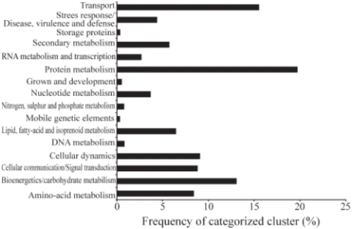
a novelty of 58.0% and a success rate of 83.3%. Then the clusters were submitted to categorization. Among them only 2,651 clusters were categorized. As a result, above 20% were putatively involved with the protein metabolism. These ESTs were linked with the ribosomal proteins and also with other factors which are required for proteins syn-thesis. Also highly expressed were: around 15% related
with carbohydrate metabolism and bioenergetics, 9% with amino acid metabolism, dynamic cell and cellular commu-nication, and 4.5% with the metabolism related to the de-fense system, stress and virulence (Figure 1).
P. parasiticaESTs were distributed between known proteins or hypothetical proteins based on deduced amino acid sequences homologies. It was discovered during the annotation process that 1,915 (41.95%) of all of ESTs did not share sequence similarity with any sequence from the GenBank non-redundant database. This relative portion is consistent with reports of other fungus EST databases. It also depends on other points of the organism such as: the experimental design and the developmental stage (Kamoun
et al., 1999). On the other hand, clusters with E-value < 1e-5 added a total of 2,651 clusters (58.05%), indicating a satis-factory value of known sequences. This percentage
high-lights that less than half of theP. parasiticatranscriptome
is currently unknown. In addition, clusters with full homology with other sequences were spotted, 91 ESTs (0.2%) and 2,641 ESTs (57.83%) with E-values that varied
from 1e-5to 1e-200. These clusters represent probable genes
(Figure 2).
Figure 1- Distribution of categorized clusters according to putative bio-logical function defined by MIPS. Protein matches resulting from BLASTX searches were assigned to one of seven functional categories for comparisons involving life cycle. Percentage of frequency of clusters was shown in each category from a total set of categorized PP clusters.
Comparison of PP/CitEST database with expressed sequence tag collections
The clusters ofP. parasitica/CitEST database were
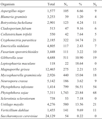
used to search for homologies in the COGEME EST data-base, which consist of 59,765 ESTs from 15 species of phytopathogens and three species of saprophytic fungi. Based on the number of unique sequences found in each species present in the COGEME database, it was possible to individually identify the number and the unique
percent-ages with homology toP. parasiticaclusters, as well as the
number of unique putatively involved in the pathogenicity functions which matched the PP clusters (Table 1).
As a result, it was observed that about 54 unique
se-quences from S. cerevisiae genome have homologies in
PP/CitEST. Comparative analyses with phytopathogen EST database led to the discovery that about 1.2% of
unigenes fromBlumeria graminisESTs have homologies
in PP/CitEST clusters. Similarly, about 56.51% and
23.84% ofP. infestansandP. sojaeunigenes, respectively,
have homologies with PP clusters, among which 54 and 68 homologies are putatively involved with the pathogenicity functions (Table 1).
Through the comparison with saprophytic fungi ESTs, it was discovered that about 2.43% and 6.66% from
Emericella nidulas and Aspergillus niger, respectively,
have homologies with PP ESTs. Among theE. nidulans
homologies, 21 unigenes were discovered putatively
in-volved with the pathogenicity functions, whereas in theA.
nigernine were found (Table 1).
Since the pathogenicity system in a parasite is never single gene-dependent, these data indicate that many genes putatively involved with tpathogenicity functions share se-quence similarities among themselves, and they may have a common ancestor. Unlike some fungi, no pathogenicity
unigenes were found with homologies inP. parasitica.
Ex-amples of this are Sclerotinia sclerotiorum and
Leptosphaeria maculanswith only a few sequences
ana-lyzed andS. cerevisiaewhich is not a plant pathogen.
Genes inP. parasiticainvolved with pathogenicity, host colonization process and defense
As an approach to studying genes possibly involved
with theP. parasiticacolonization process, a number of
clusters coding for wall cell degradation proteins, necro-sis-inducer proteins, elicitins, among others were identified in the analysis. The breakdown of physical barriers during an infection process, penetration process and host tissue colonization involve the secretion of a vast range of degra-dative enzymes. During the process, several ESTs with sig-nificant similarity to degradative enzymes such as amidase, cutinase protein, endo- and exoglucanases, and chitinases have been identified (Table 2).
The degradation of the host cell wall is one of the first steps in disease. The process needs many enzymes, such as phospholipases, ß-glucosidase/ß-xylosidase, exo-1,
3-ß-glucanases, endo-1, 3-ß-glucanase, and
endopolygalactu-ronases (endo-PGs) (Kamoun et al., 1999). Clusters
putatively encoded by all of these enzymes were found in theP. parasitica/CitEST database (Table 2). In addition, two clusters were found sharing sequence similarity with
pectin lyase F isolated fromA. nigerandA. nidulans(Table
2). Pectin lyase F has been described in many plant-pathogenic bacteria and fungi as an enzyme used to
break into the host tissues (Chenet al., 1998). Moreover,
pectolytic enzymes are essential in the decay of dead plant material through nonpathogenic microorganisms and thus
assist carbon compound recycling in the biosphere (Chenet
al., 1998). The low frequency of these genes in the
PP/CitEST database indicates thatP. parasiticamight not
be a pathogenic fungi with great affinity to pectin
degrada-tion. This might be related to the reduced attack of P.
parasiticain citrus fruit.
The important gene that was found in PP/CitEST da-tabases is the CBEL (cellulose binding elicitor lectin) gene (Table 2), with four clusters in the database. This gene
en-codes a protein that binds to cellulosein vitro, suggesting
that CBEL participates in the adhesion ofPhytophthorato
cellulosic substrates (Tucker and Talbot, 2001). Adherence to solid surfaces is a common feature in both saprophytic and parasitic microorganisms. In fungi and oomycetes, ad-herence is mediated by secreted adhesins which are part of the cell wall or it might be physically associated to it
(Gaulinet al., 2002).
Table 1- Comparison ofP. parasiticaclusters with COGEME database.
Organism Total Nu % Np
Aspergillus niger 1,577 105 6.66 9
Blumeria graminis 3,253 39 1.20 4
Botryotinia fuckeliana 2,901 123 4.24 11
Cladosporium fulvum 513 47 9.16 6
Colletotrichum trifolii 550 42 7.64 5
Cryphonectria parasitica 2,185 322 14.74 21
Emericella nidulans 4,805 117 2.43 7
Fusarium sporotrichioides 3,448 111 3.22 10
Gibberella zeae 4,688 511 10.90 19
Leptosphaeria maculans 118 22 18.64 0
Magnaporthe grisea 12,465 275 2.21 15
Mycosphaerella graminicola 2,926 440 15.04 18
Neurospora crassa 5,142 186 3.62 9
Phytophthora infestans 1,414 799 56.51 54
Phytophthora sojae 7,311 1,743 23.84 68
Sclerotinia sclerotiorum 738 53 7.18 0
Ustilago maydis 4,276 580 13.56 21
Verticillium dahliae 1,455 141 9.69 11
Saccharomyces cerevisiae 24,129 54 0.22 0
Total: Total of unigenes; Nu: number of unigenes homology; %: % of
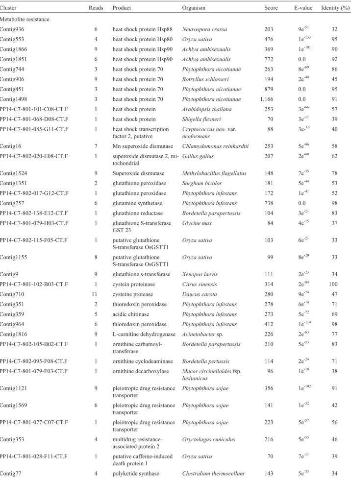
Table 2-Phytophtora parasiticaESTs and their known or predicted function, based on BLASTX results.
Cluster Reads Product Organism Score E-value Identity (%)
Metabolite resistance
Contig936 6 heat shock protein Hsp88 Neurospora crassa 203 9e-51 32
Contig553 4 heat shock protein Hsp80 Oryza sativa 476 1e-133 95
Contig1866 9 heat shock protein Hsp90 Achlya ambisexualis 369 1e-101 90
Contig1851 6 heat shock protein Hsp90 Achlya ambisexualis 772 0.0 92
Contig744 3 heat shock protein 70 Phytophthora nicotianae 263 8e-69 86
Contig906 9 heat shock protein 70 Botryllus schlosseri 194 2e-48 45
Contig451 3 heat shock protein 70 Phytophthora nicotianae 879 0.0 95
Contig1498 3 heat shock protein 70 Phytophthora nicotianae 1,166 0.0 91
PP14-C7-801-101-C08-CT.F 1 heat shock protein Arabidopsis thaliana 253 3e-66 57
PP14-C7-801-068-D08-CT.F 1 heat shock protein Shigella flexneri 70 3e-11 39
PP14-C7-801-085-G11-CT.F 1 heat shock transcription
factor 2, putative
Cryptococcus neo.var. neoformans
88 3e-16 40
Contig16 7 Mn superoxide dismutase Chlamydomonas reinhardtii 253 5e-66 58
PP14-C7-802-020-E08-CT.F 1 superoxide dismutase 2, mi-tochondrial
Gallus gallus 207 2e-60 62
Contig1524 9 Superoxide dismutase Methylobacillus flagellatus 148 7e-35 78
Contig1351 2 glutathione peroxidase Sorghum bicolor 181 5e-44 53
PP14-C7-802-017-G12-CT.F 1 glutathione peroxidase Phytophthora infestans 172 1e-41 52
Contig757 6 glutamine synthetase Phytophthora infestans 738 0.0 98
PP14-C7-802-138-E12-CT.F 1 glutathione reductase Bordetella parapertussis 104 3e-21 83
PP14-C7-801-079-H03-CT.F 1 glutathione S-transferase GST 23
Glycine max 84 4e-15 37
PP14-C7-802-115-F05-CT.F 1 putative glutathione
S-transferase OsGSTT1
Oryza sativa 103 6e-21 33
Contig1155 8 putative glutathione
S-transferase OsGSTT1
Oryza sativa 99 8e-20 33
Contig9 9 glutathione s-transferase Xenopus laevis 111 2e-23 34
PP14-C7-801-102-B03-CT.F 1 cystein proteinase Citrus sinensis 314 2e-84 100
Contig710 11 cysteine protease Daucus carota 280 9e-74 47
Contig351 2 thioredoxin peroxidase Phytophthora infestans 278 6e-74 71
Contig359 5 acidic chitinase Phytophthora infestans 273 5e-72 69
Contig964 6 thioredoxin peroxidase Phytophthora infestans 412 1e-114 98
Contig1816 9 L-carnitine dehydrogenase Acinetobactersp. 226 2e-63 77
PP14-C7-802-105-B02-CT.F 1 ornithine
carbamoyl-transferase
Bordetella parapertussis 210 5e-53 83
PP14-C7-802-095-F08-CT.F 1 ornithine cyclodeaminase Bordetella pertussis 114 2e-24 71
PP14-C7-801-079-F03-CT.F 1 ornithine decarboxylase Mucor circinelloidesfsp. lusitanicus
96 1e-18 38
Contig1121 9 pleiotropic drug resistance
transporter
Phytophthora sojae 356 1e-102 91
Contig1569 6 pleiotropic drug resistance
transporter
Phytophthora sojae 141 1e-32 42
PP14-C7-801-077-C07-CT.F 1 pleiotropic drug resistance transporter
Phytophthora sojae 223 5e-57 56
Contig353 4 multidrug
resistance-associated protein 2
Oryctolagus cuniculus 216 5e-55 46
PP14-C7-801-028-F11-CT.F 1 putative caffeine-induced death protein 1
Oryza sativa 70 7e-11 39
Table 2 (cont.)
Cluster Reads Product Organism Score E-value Identity (%)
Contig735 7 pepsinogen C Monodelphis domestica 57 6e-07 36
PP14-C7-801-042-F12-CT.F 6 1 elicitin protein Phytophthora parasitica 180 7e-45 100
Contig1181 2 elicitin protein Phytophthora sojae 134 2e-30 57
Contig739 11 elicitin protein Phytophthora sojae 55 1e-06 35
Contig133 13 elicitin
gamma-megaspermin pro-tein
Phytophthora megasperma 210 3e-53 86
Contig888 9 elicitin INF2A protein Phytophthora infestans 201 1e-50 85
Contig987 6 elicitin INF7 protein Phytophthora infestans 113 4e-24 56
PP14-C7-801-101-H07-CT.F 1 wound-inducible basic
pro-tein
Phaseolus vulgaris 88 1e-16 85
Contig1413 6 necrosis-inducing-like
pro-tein
Phytophthora sojae 107 2e-22 32
PP14-C7-802-125-A07-CT.F 1 crinkling and
necro-sis-inducing protein CRN2
Phytophthora infestans 66 1e-09 34
PP14-C7-802-082-A05-CT.F 1 crinkling and
necro-sis-inducing protein CRN2
Phytophthora infestans 100 9e-20 38
Contig1550 4 crinkling and
necro-sis-inducing protein CRN1
Phytophthora infestans 90 4e-17 47
Contig1422 3 crinkling and
necro-sis-inducing protein CRN1
Phytophthora infestans 72 4e-11 30
Hydrolytic enzymes
Contig1603 3 acidic chitinase Phytophthora infestans 387 1e-106 81
PP14-C7-801-048-C04-CT.F 1 acidic chitinase Phytophthora infestans 238 1e-61 66
Contig1114 3 amidases related to
nicotinamidase
Burkholderia cepacia 177 2e-43 59
PP14-C7-801-047-A01-CT.F 1 amidases related to
nicotinamidase
Burkholderia cepacia R1808 109 1e-22 49
PP14-C7-801-032-F02-CT.F 1 beta(1-3)endoglucanase Aspergillus fumigatus 80 5e-14 30
PP14-C7-802-111-C10-CT.F 1 beta(1-3)endoglucanase Aspergillus fumigatus 61 1e-08 30
Contig201 6 beta-glucosidase Phytophthora sojae 104 1e-21 44
PP14-C7-802-030-F10-CT.F 1 beta-glucosidase Bacillus clausii 62 2e-08 34
PP14-C7-801-042-E05-CT.F 1 beta-glucosidase precursor Tenebrio molitor 77 5e-13 46
Contig659 6 beta-glucosidase/xylosidase Phytophthora infestans 213 9e-60 60
Contig618 9 CBEL protein Phytophthora parasitica 505 1e-142 89
Contig1583 11 CBEL protein Phytophthora parasitica 448 1e-125 87
PP14-C7-801-040-C07-CT.F 1 CBEL protein, formerly
GP34
Phytophthora parasitica 224 3e-57 45
Contig381 2 CBEL protein, formerly
GP34
Phytophthora parasitica 76 7e-13 27
Contig543 6 cutinase (CutB) Phytophthora brassicae 402 8e-12 83
Contig373 3 endo alpha-1,4
poly-galactosaminidase
Idiomarina loihiensis 89 3e-34 43
Contig1609 4 endo-1,3-beta-glucanase Phytophthora infestans 388 1e-127 79
PP14-C7-801-016-D05-CT.F 1 endo-1,3-beta-glucanase Phytophthora infestans 140 3e-32 41
PP14-C7-801-016-D05-CT.F 1 endo-1,3-beta-glucanase Phytophthora infestans 140 3e-32 41
Contig1624 8 endo-1,4-beta-glucanase Pyrococcus horikoshii 127 7e-28 26
Contig1622 9 endo-1,4-beta-glucanase Pyrococcus horikoshii 82 3e-14 25
PP14-C7-802-135-B12-CT.F 1 endoglucanase Clostridium thermocellum 62 1e-08 39
Table 2 (cont.)
Cluster Reads Product Organism Score E-value Identity (%)
Contig1330 3 exo-beta-1,3-glucanase Magnetospirillum
magnetotacticum
79 3e-13 25
Contig764 2 exopolyphosphatase Bordetella parapertussis 230 6e-60 87
PP14-C7-801-079-C06-CT.F 1 exopolysaccharide
biosynthesis protein
Mesorhizobium loti 116 9e-25 38
PP14-C7-802-098-H03-CT.F 1 lipase, putative Paramecium tetraurelia 89 1e-16 32
PP14-C7-801-105-A03-CT.F 1 pectine lyase F Aspergillus niger 492 4e-48 44
Contig420 6 pectine lyase F Aspergillus nidulans 135 8e-31 42
Contig1852 6 putative 1,3-beta-glucan
synthase
Oryza sativa 325 2e-87 56
PP14-C7-802-076-B03-CT.F 1 putative beta-1,3-glucan
synthase
Nicotiana alata 171 2e-41 38
Contig585 7 putative
endo-1,3;1,4-beta-glucanas e
Phytophthora infestans 58 5e-08 96
PP14-C7-801-064-A02-CT.F 1 putative esterase Oryza sativa 117 3e-25 38
PP14-C7-801-007-E03-CT.F 1 putative
exo-1,3-beta-glucanase
Phytophthora infestans 345 5e-95 74
PP14-C7-802-074-B12-CT.F 1 putative
exo-1,3-beta-glucanase
Phytophthora infestans 325 3e-88 98
Contig1478 16 putative
exo-1,3-beta-glucanase
Phytophthora infestans 270 3e-71 65
Contig258 23 putative
exo-1,3-beta-glucanase
Phytophthora infestans 919 0.0 88
Contig258 3 putative
exo-1,3-beta-glucanase
Phytophthora infestans 919 0.0 88
PP14-C7-801-023-D06-CT.F 1 putative
exo-1,3-beta-glucanase
Phytophthora infestans 100 3e-20 40
PP14-C7-801-013-E08-CT.F 1 related to amidase Neurospora crassa 81 4e-14 39
Contig1326 3 urea amidolyase Xanthomonas axonopodispv.
citri
50 1e-05 44
Others
Contig1574 8 CTR1-like kinase kinase
kinase
Oryza sativa 196 1e-48 43
Contig946 5 CTR1-like protein kinase Oryza sativa 135 2e-30 29
Contig336 3 CTR1-like kinase kinase
kinase
Brassica juncea 79 3e-13 35
Contig810 5 CTR1-like kinase kinase
kinase
Brassica juncea 97 6e-19 30
Contig1781 8 CTR1-like kinase kinase
kinase
Oryza sativa 87 3e-16 38
PP14-C7-801-036-D03-CT.F 1 MAPK-related kinase Tetrahymena thermophila 129 1e-28 39
PP14-C7-802-140-G08-CT.F 1 MAP kinase 4 Zea mays 84 3e-15 59
PP14-C7-802-010-E02-CT.F 1 MAP kinase kinase Yarrowia lipolytica 77 1e-13 43
PP14-C7-801-066-F02-CT.F 1 MAP3K beta 1 protein
kinase
Brassica napus 82 2e-14 37
Contig1084 5 cyst germination specific
acidic protein
Phytophthora infestans 92 3e-17 42
PP14-C7-801-090-C09-CT.F 1 ascus development protein
1
Neurospora crassa 100 1e-21 33
PP14-C7-801-099-F07-CT.F 1 ethylene-inducible
CTR1-like protein kinase
Six putative genes which belonged to the complex family of elicitin-like proteins were also found in the PP/CitEST database (Table 2). Elicitin-like genes encode putative extra cellular proteins which share the 98 amino-acid elicitin domains, which correspond to the mature INF1 elicitin. Fiveinfgenes (inf2A,inf2B,inf5,inf6, andinf7) encode predicted proteins with a C-terminal domain in ad-dition to the N-terminal elicitin domain. The elicitins genes are classified into four classes, class IA, class IB, class II
and class III, based on peptide signal sequence (Baillieulet
al., 2003). These proteins may form a `lollipop on a stick’
structure, formed by disulfide bonds in cysteine residues (Figure 3), on which an O-glycosylated domain forms an extended rod that holds the protein to the cell wall causing the extra cellular N-terminal domain to be left exposed on the cell surface. Therefore, these atypical INF proteins may be associated with the surface or cell wall glycoprotein that
interacts with plant cells during infection (Kamounet al.,
1997).
Elicitins are extracellular proteins with still unknown functions, but it has already been proven that they induce a hypersensitive reply in the host, as already proven in
to-bacco by Qutobet al.(2003). It is believed that elicitins are
lipid binding-related proteins and that they present func-tions of phospholipid; thus they are able to cross cell mem-branes, by an interaction with ergosterol in residues present
in amino acid sequences (Kamounet al., 1997). Other
stud-ies suggest that multiple layers of INF elicitin recognition
and late blight resistance occur inNicotiana tabacum
(Ba-keret al., 1997).
Experiments with elicitins in tobacco have shown that elicitins are either proteins which cause hypersensitive re-sponses in the plant or they are virulence factors. Such mol-ecules are typically secreted into the intercellular interface between the pathogen and the plant, or they are taken up into the host cell to reach their cellular target. Interactions between plants and microbial pathogens involve complex signal exchange on the plant surface and in the intercellular space interface. The elicitins are considered only one signal in this complex communication (Parniske, 2000; Hahn and Mendgen, 2001).
Phylogenetic analysis of the six PP/CitEST elicitins and homologies has grouped the sequences into four clades,
except for the outgroup (Figure 4). One PP elicitin cluster (PP14-C7-801-042-F12-CT.F) was grouped in the clade of
the class IA which has 75.4% of homology with P.
cinnamomi.A second clade consisting of two PP clusters (Contig 987, Contig 739) similar to elicitins class IB was
close toP. megasperma, with 82% and 79% sequence
iden-tity, respectively (Figure 4). In a third clade, one PP cluster
(contig 133) grouped with class IIP. cinnamomielicitins,
with 75% sequence similarity. In the last clade, two PP clusters, consisting of contig 888 and contig 1181, were
grouped together with the class III P. infestans and P.
brassicaeelicitins, which share 78% and 84% similarity,
Figure 4- Phylogenetic dendrogram of elicitin amino acid sequences. Multiple alignment of selected elicitin amino acid sequences from PP/CitEST and homology was performed in Clustal X. Dendrogram was constructed and visualized by Mega programs using neighbor-joining method. The following sequences were obtained from EMBL GeneBank: Phytophthora megasperma1 (AJ493606), 2 (AJ493607), 3 (gi|544239), P. cryptogea1 (gi|599947), 2 (gi|21466142),P cinnamomi1 (gi|4469292), 2 (gi|4469290), P. dreschslei (gi|544238),P. sojae (gi|27922903),P. infestans1 (gi|51832281), 2 (gi|2707621), 3 (gi|16225870),P. brassicae1 (gi|29838396), 2 (gi|29838400),Pythium vexans(gi|945184). There were six clusters fromPhytophthora parasitica/CitEST: one sharing sequence similarity with elicitin protein class IA (PP14-C7-801-042-F12-CT.F), two clusters with elicitins class IB (Contig 987, Contig 739), one cluster with elicitin class II (Contig 133) and two clusters with elicitins class III (Contig 888, Contig 1181). The numbers indicate percentages supporting the branches by 10,000 bootstrap replicates (bar corresponds to 0.1 substi-tutions per site).
respectively (Figure 4). Huitemaet al.(2005) demonstrated that elicitins class III induced hypersensitive response ac-tivity that led to cell death and showed a resistance charac-ter. The class I elicitins are known by their interaction with
non-host and they are probably used byP. parasiticato
sur-vive in the saprophytic form.
Necrosis-inducer proteins are related to the necrotic responses in plant. Many genes related to this induction are
characterized asavrgenes (MacGregoret al., 2002). There
are countless Avr loci, but there are only fewavrgene
se-quences known.P. sojaehas more than 13avrgenes, but
they have not been isolated yet, although a recent study has shown that Avr loci may be successfully identified by
posi-tional cloning methods (Tyleret al., 1995). This has led to
both the isolation of the Avr1a and Avr1b/Avr1k loci, and
also the identification of the Avr1b protein (Tyleret al.,
1995; MacGregoret al., 2002). No work has reported that
these molecules have been described to elicit host and non-host responses, although they seem to be specific Avr
gene for definitive races (Cheonget al., 1991; Nürnberger
et al., 1994).
The analyses of the responses induced by the crin-kling and necrosis-inducers (CRN) cDNAs in many plants, suggest that they are general elicitors that trigger necrotic responses nonspecifically, both in resistant species and also
in the susceptible host (Kamounet al., 1998; Kamounet al.,
1999; Qotubet al., 2002). It is believed that CRNs differ
from specific elicitors, such as INF1, which induce defense
responses only in specific plant genotypes (Kamounet al.,
1998; Kamoun et al., 1999), but it also resembles NIP,
which functions in several dicotyledonous plants (Qutobet
al., 2002). The general elicitors of plant pathogens were
re-cently compared to pathogen-associated molecular patterns (PAMPs), which are surface-derived molecules that induce the expression of defense-response genes as well as the pro-duction of antimicrobial compounds in both animal and plant cells (Gomez-Gomez and Boller, 2002; Nürnberger and Brunner, 2002). Whether the CRN proteins function as PAMPs still remains unclear. It is supported by observation
that CRN genes are found in severalPhytophthoraspecies.
In addition, CRNs could aid in a colonization process of plant tissue during the late necrotrophic phase of the
infec-tion, as proposed for the NIP protein (Qutobet al., 2002).
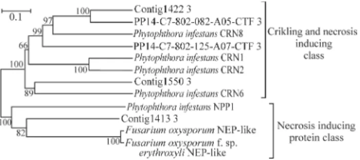
Four EST clusters similar to CRN proteins (Figure 5,
Table 2) were found in theP. parasitica/CitEST database.
Three of these clusters (Contig 1422, PP14-C7-802-082-A05-CT.F and PP14-C7-802-125-A07-CT.F) were
phylogenetically related to CRN8 ofP. infestans;, whereas
the fourth cluster (Contig 1550) showed similarity to CRN6 ofP. infestans(Figure 5). Strange as it may seem, the
ob-servation that CRN genes were found in theP. parasitica
virulent strain during colonization of the media culture in-dicates that it is only expressed during the infection process is incorrect. In fact, what is correct is the importance of the factor for the expression and secretion during the infection
process. In the future, additional functional analyses of the
CRN genes inP. parasitica and theP. parasitica-Citrus
system will aid in determining the nature of the contribu-tion of these genes in the infeccontribu-tion process
Another necrosis-inducing protein in the P.
parasitica/CitEST database was NIP (necrosis-inducing protein), which is a secreted protein of 60 amino acids. This protein was detected in other pathogens, and besides that, there is the hypothesis that this gene product has a dual function in both fungal avirulence and virulence (Tyler,
2002). In barley cultivars expressing theRgeneRrs1,the
protein elicits defense reactions ofin the plant (avirulence function). However, in a concentration dependent manner, and without considering the plant resistance genotype, the formation of necrotic lesions is induced similar to the scald symptoms. This occurred in barley cultivars. as well as in other cereal species; however, it did not occur in the
dico-tyledonous species Arabidopsis thaliana
(virulence-associated function). This toxic activity seems to be medi-ated by the stimulation of the plant plasma
membrane-localized H+-ATPase (Tyler, 2002).
In Fusarium oxysporum f. sp. erythroxyli, a NEP1 protein (necrosis-inducing protein) was found which causes cell death in many different plant species when ap-plied as a foliar spray. In other studies, orthologues of
NEP1 gene were cloned and characterized inPhytophthora
megakarya; indicating that it is a fungal agent for black pod
disease inTheobroma cacao(cacao). After observing the
necrotic lesions in cacao leaves sprayed with NEP1 (Baeet
al., 2005) for 10 days, the constitutive expression of this
protein was noted. This is directly involved with the transi-tion between the hemibiotrophic and the necrotrophic phases.
In theP. parasiticadatabase, one cluster with
homo-logy toP. sojaeprotein (Table 2) was detected, but in the
phylogenetic analyses this cluster appeared in the clade
with NEP ofF.oxysporumwith 64% similarity (Figure 5).
The production of polypeptides and polyamines is also an important factor in pathogenicity. In the PP/CitEST database, one singleton read was found sharing a sequence
similar to the ornithine decarboxilase (ODC) of Mucor
circinelloidesf.lusitanicus, (Table 2), and other enzymes of this pathway. ODC is an important enzyme in polyamine production. The inhibition of this enzyme is an effective therapy in the treatment of Trypanosomiasis and also other
diseases caused byPlasmodia, Giardia, andLeishmania
and in Stagonospora (Septoria) nodorum, a phytopathogenic fungi. This is probably a target for chemi-cal control because of the need for this enzyme in virulence
and growth (Baileyet al., 2000).
In yeast, a pleiotropic drug resistance transporter sys-tem is responsible for the protection of microorganism cell
against antibiotic and heavy metals, such as cadmium. InP.
parasitica,three clusters that have homology with these
genes were noted. These were also found inP. sojae(Table
2). This system probably aids in its survival in soil with high levels of heavy metal or exposure to fungicides.
Another group of expressed genes found in P.
parasiticatranscripts are heat shock proteins (HSP), also called stress proteins. This is a group of proteins that is present in all cells in all kinds of organisms. They are in-duced when a cell undergoes different kinds of environ-mental stresses such as heat, cold and oxygen deprivation. Heat shock proteins are molecular chaperones. They are usually cytoplasmic proteins and they perform func-tions in many of the intra-cellular processes. They play an important role in protein-protein interactions, such as fold-ing and assistfold-ing in the establishment of proper protein con-formation (shape) and also in the prevention of undesirable protein aggregation. Through the partial stabilization of the unfolded proteins, HSPs aid to transport proteins across intreacellular membranes. Some members of the HSP fam-ily are expressed from low to moderate levels in all organ-isms due to their essential role in protein maintenance (Lund, 2001).
Here, eleven clusters related to HSPs were found in theP. parasiticadatabase, with evidence pointed out by
Ja-cobsonet al.(1994) thatP. parasiticaprobably uses these
proteins in melanin metabolism and in the infection process (Table 2). Moreover, several clusters were also found in the
P. parasiticadatabase (Table 2), showing significant se-quence similarity to other genes related to infection and host colonization processes, such as cystein proteinase, pepsinogen, proteases and acidic chitinase.
There is evidence linking melanin biosynthesis to
vir-ulence in Aspergillus fumigatusconidia. Superoxide
dis-mutases, glutathione S-transferase GST, glutathione peroxidase and glutamine synthetase are important
clean-ing antioxidants and they have an additional hypothetical role in virulence. However, although these enzymes have
been biochemically characterized in Aspergillus and
Cryptococcus, there is no concrete evidence that these en-zymes are involved in pathogenicity. Catalase production
may play some role in the virulence ofCandida albicans,
but this enzyme has not yet been proven to have some kind
of influence in the virulence ofA. fumigatus. There is data
supporting an antioxidant function of the acyclic hexitol
mannitol inC. neoformans, however, further investigation
is required. Research on the putative antioxidant activities in a range of other fungal enzymes, like acid phosphatases, currently still limited (Hamilton and Holdom, 1999).
Eleven genes of this group were detected in theP.
parasiticadatabase (Table 2). These genes are important in the pathogen’s defense system because their products pro-tect the organism against reactive oxygen species or induce
cell death in the host. Jacobsonet al.(1994) report the
pro-duction of superoxide dismutase (SOD) and melanin in pathogenic fungi as important factors for basidiomycetes, considering that melanin production is an established viru-lence factor and that pathogenic fungi produce melanin
(Ja-cobsonet al., 1994).
Three clusters spotted in theP. parasitica database
share sequence similarity with a superoxide dismutase
fam-ily inP. infestans(67%) and inC. reinhardtii(58%). In
ad-dition, we found: four clusters showing similarity with glutathione S-transferase, two clusters similar to gluta-thione peroxidase, one cluster similar to glutamine syn-thetase, one glutathione reductase and two similar to thioredoxin peroxidase (Table 2). The expression of
oxida-tive stress-related genes in vitro could be related to P.
parasiticamelanin production, and it could also be related to an increase in expression during the infection process.
This is the first report on global gene expression inP.
parasitica,a causal agent of gummosis in citrus. Here,
sev-eral genes were identified which may contribute to the
un-derstanding of pathogenicity mechanisms ofP. parasitica
and which also may represent new possible tags for chemi-cal control. The understanding of this pathogenicity could aid in the development of new methods or new chemical control tags for citrus gummosis. For instance, the develop-ment of molecules that deactivate the pathogenicity factors presented here.
Acknowledgements
We would like to thank CNPq/ Millennium Institute /Citrus and FAPESP for financial support.
References
Altschul S, Madden T, Schaffer A, Zhang J, Zhang Z, Mille W and Lipman DJ (1997) Gapped BLAST and PSI-BLAST: A new generation of protein database search programs. Nucleic Acids Res 25:3389-3402.
Averna-Saccá R (1917) Moléstias das laranjeiras. Boletim Agrí-cola 18:33-36.
Bae H, Bowers J, Tooley P and Bailey B (2005) NEP1 orthologs encoding necrosis and ethylene inducing proteins exist as a multigene family inPhytophthora megakarya, causal agent of black pod disease on cacao. Mycol Res 109:1373-1385. Bailey BA, Apel-Birkhold PC, Akingbe OO, Ryan JR, O’Neill
NR and Anderson JD (2000) Nep1 protein fromFusarium oxysporumenhances biological control of opium poppy by
Pleospora papaveracea. Phytopathol 90:812-818.
Baillieul F, de Ruffray P and Kauffmann S (2003) Molecular cloning and biological activity of Alpha, Beta, and Gama-megaspermin, three elicitins secreted by Phytophthora megaspermaH20. Plant Physiol 131:155-166.
Baker B, Zambryski P, Staskawicz B and Dinesh-Kumar SP (1997) Signalling in plant - Microbe interactions. Science 276:726-733.
Chen W, Hsieh H and Tseng T (1998) Purification and character-ization of a pectin lyase fromPythium splendensinfected cucumber fruits. Bot Bull Acad Sin 39:181-186.
Cheong J, Bierberg W, Fügedi P, Pilotti A, Garegg PJ, Hong N, Ogawa T and Hahn MG (1991) Structure-activity relation-ships of oligo-b-glucoside elicitors in phytoalexin accumu-lation in soybean. Plant Cell 3:127-136.
Erwin DC and Ribeiro OK (1996)Phytophthora parasitca. In: Erwin DC and Ribeiro OK (eds) Phytophthora Diseases Worldwide. APS Press, St. Paul, pp 347-478.
Ewing B, Hillier L, Wendl M and Green P (1998) Basecalling of automated sequencer traces using phred. I. Accuracy assess-ment. Genome Res 8:175-185.
Fawcett HS (1936) Citrus Disease and Their Control. 2nd edition. McGraw-Hill Book Company, New York, 312 pp. Förster H, Coffey MD, Elwood H and Sogin ML (1990) Sequence
analysis of the small subunit ribosomal RNAs of three zoos-poric fungi and implications for fungal evolution. Myco-logia 82:306-312.
Gaulin E, Jauneau A, Villalba F, Rickauer M, Esquerré-Tugayé MT and Bottin A (2002) The CBEL glycoprotein of Phy-tophthora parasiticavar-nicotianaeis involved in cell wall deposition and adhesion to cellulosic substrates. J Cell Sci 115:4565-4575.
Gomez-Gomez L and Boller T (2002) Flagellin perception: A par-adigm for innate immunity. Trends Plant Sci 6:251-256. Gunderson JH, Elwood H, Ingold A, Kindle K and Sogin ML
(1987) Phylogenic relationships between chlorophytes, chrysophytes, and oomycetes. Proc Natl Acad Sci USA 84:5823-5827.
Hahn M and Mendgen K (2001) Signal and nutrient exchange at biotrophic plant-fungus interfaces. Curr Opin Plant Biol 4:322-327.
Hamilton A and Holdom MD (1999) Antioxidant systems in the pathogenic fungi of man and their role in virulence. Med Mycol 37:375-89.
Hanahan D (1983) Studies on transformation ofEscherichia coli
with plasmids. J Mol Biol 166:557-580.
Huang X and Madan A (1999) CAP3: A DNA Sequence Assem-bly Program. Genome Res 9:868-877.
Huitema E, Vleeshouwers VGANA, Cakir C, Kamoun S and Govers F (2005) Differences in intensity and specificity of hypersensitive response induction in Nicotiana spp. by INF1, INF2A, and INF2B ofPhytophthora infestans. Mol Plant Microbe Interact 18:183-193.
Jacobson ES, Jenkins ND and Todd JM (1994) Relationship be-tween superoxide dismutase and melanin in a pathogenic fungus. Infect Immun 62:4085-4086.
Judelson HS (1997) The genetics and biology ofPhytophthora infestans: Modern approaches to a historical challenge. Fun-gal Genet Biol 22:65-76.
Kamoun S, Lindqvist H and Govers F (1997) A novel class of elicitin-like genes fromPhytophthora infestans. Mol Plant Microbe Interact 10:1028-1030.
Kamoun S, van West P, Vleeshouwers VGAA, de Groot K and Govers F (1998) Resistance ofNicotiana benthamianato
Phytophthora infestansis mediated by the recognition of the elicitor protein INF1. Plant Cell 10:1413-1425.
Kamoun S, Huitema E and Vleeshouwers VGAA (1999) Resis-tance to oomycetes: A general role for the hypersensitive re-sponse? Trends Plant Sci 4:196-200.
Kumar S, Tamura K and Nei M (2004) MEGA3: Integrated Soft-ware for Molecular Evolutionary Genetics Analysis and Se-quence Alignment. Brief Bioinformatics 5:150-163. Lund PA (2001) Microbial molecular chaperones. Adv Microb
Physiol 44:93-140.
MacGregor T, Bhattacharyya M, Tyler B, Bhat R, Schmitthenner AF and Gijzen M (2002) Genetic and physical mapping of Avr1a inPhytophthora sojae. Genetics 160:949-959. Müller AS (1933) Observations and notes on citrus disease in
Minas Gerais, Brasil. Phytopatholgy 23:734-737.
Nürnberger T and Brunner F (2002) Innate immunity in plants and animals: Emerging parallels between the recognition of gen-eral elicitors and pathogen-associated molecular patterns. Curr Opin Plant Biol 4:318-324.
Nürnberger T, Nennstiel D, Jabs T, Sacks WR, Hahlbrock K and Scheel D (1994) High affinity binding of a fungal oligo-peptide elicitor to parsley plasma membranes triggers multi-ple defense responses. Cell 78:449-460.
Parniske M (2000) Intracellular accommodation of microbes by plants: A common developmental program for symbiosis and disease? Curr Opin Plant Biol 3:320-328.
Qutob D, Kamoun S and Gijzen M (2002) Expression of a Phy-tophthora sojae necrosis-inducing protein occurs during transition from biotrophy to necrotrophy. Plant J 32:361-373.
Qutob D, Huitema E, Gijzen M and Kamoun S (2003) Variation in structure and activity among elicitins from Phytophthora sojae. Mol Plant Pathol 4:119-124.
Siviero A, Furtado EL, Boava LP, Barbasso DV and Machado MA (2002) Avaliação de métodos de inoculação de Phy-tophthora parasiticaem plântulas e plantas jovens de citros. Fitopatologia Brasileira 27:574-580.
Soanes DM and Talbot NJ (2006) Comparative genomics analysis of phytopathogenic fungi using expressed sequence tag (EST) collections. Mol Plant Path 7:61-70.
Tucker SL and Talbot NJ (2001) Surface attachment and pre-penetration stage development by plant pathogenic fungi. Annu Rev Phytopathol 39:385-417.
Tyler BM (2002) Molecular basis of recognition between
Phytophtora pathogens and their hosts. Annu Rev Phyto-pathol 40:137-167.
Tyler BM, Förster H and Coffey MD (1995) Inheritance of avi-rulence factors and restriction fragment length polymor-phism markers in outcrosses of the ÖomycetePhytophthora sojae. Mol Plant Microbe Interact 8:515-523.
Waugh M, Hraber P, Weller J, Wu Y, Chen G, Inman J, Kiphart D and Sobral B (2000) The Phytophthora genome initiative da-tabase: Informatics and analysis for distributed patho-genomic research. Nucleic Acids Res 28:87-90.
Internet Resources
http://citest.centrodecitricultura.br - Center APTA Citros Sylvio Moreira/IAC - Millennium Institute, Database webpage (ve-rified January 28, 2005).
http://www.phrap.org - PhredPhrap package (verified March 25, 2004).
http://www.ncbi.nlm.nih.gov - GenBank database and BLAST tools (verified January 28, 2005).
http://www.ncbi.nlm.nih.gov/COG/ - Clusters of Orthologous Groups of proteins (verified February 28, 2005).
http://www.tigr.org/docs/tigr-script/edga_scripts/roles_report.spl - Script tool (verified March 2, 2005).
http://cogeme.ex.ac.uk/ - EST collections COGEME (verified March 2, 2005).