University of Lisbon
Faculty of Sciences
Department of Plant Biology
Characterization and functional analysis of biomarkers in
endocrine resistance of breast cancer treatment
Carla Maria Lourenço Alves
M. Sc. Thesis
M. Sc. in Molecular Biology and Genetics
1
University of Lisbon
Faculty of Sciences
Department of Plant Biology
Characterization and functional analysis of biomarkers in
endocrine resistance of breast cancer treatment
Carla Maria Lourenço Alves
M. Sc. Thesis supervised by Prof. Margarida Telhada, Prof. Henrik Ditzel and
Post doc Daniel Elias
M. Sc. in Molecular Biology and Genetics
2
ACKNOWLEDGEMENTS
The work presented in this thesis was carried out in the Ditzel Laboratory, Department of Cancer and Inflammation Research, Institute of Molecular Medicine, University of Southern Denmark through the Erasmus Exchange Programme.
I want to express my sincere gratitude to my supervisor Prof. Henrik Ditzel for providing me the opportunity to work in his laboratory and for the interesting project I was entrusted. I am very grateful for his help, valuable guidance and support. I would also like to extend my appreciation to my internal supervisor of the University of Lisbon, Prof. Margarida Telhada, for her guidance throughout this year and especially for her teaching skills and useful paper discussions in her lessons that increased my interest in research.
A special thanks to my co-supervisor Post doc Daniel Elias, who has always been there to listen and give advice. I am deeply grateful to him for the rewarding discussions during this study and for carefully reviewing this thesis.
To the professors, students and technicians in Ditzel group, I am grateful for the chance of being a part of the lab. Thank you for welcoming me as a friend and helping me every time I needed. Further, I wish to thank all the WP25 3rd floor staff for assistance in the lab and pleasant work environment.
Finally, I sincerely thank my family and friends whose support and understanding were essential throughout my entire degree. To my parents and brother, thank you for helping me with the submission of this work, among many other things in my life. And a special acknowledge to João Fonseca whose patience and love are invaluable to me.
I am grateful for the financial support through a scholarship from the Erasmus Programme and afterwards from the Faculty of Health Sciences of SDU.
Carla Alves September 2013
3
TABLE OF CONTENTS
ABSTRACT ... 5
RESUMO ... 6
RESUMO DA TESE EM PORTUGUÊS ... 7
ABBREVIATIONS ... 11
1.INTRODUCTION ... 12
1.1 Breast cancer biology and treatment ... 12
1.2 Estrogen receptor function ... 13
1.3 Molecular mechanism of endocrine resistance ... 13
1.4 Fulvestrant resistance ... 14
1.5 Cell line model of fulvestrant resistance ... 14
1.6 Genes upregulated in fulvestrant-resistant cell lines ... 15
1.7 The present study ... 16
2.MATERIALS AND METHODS ... 17
2.1 Cell lines and culture conditions ... 17
2.2 RNA expression analysis ... 17
2.2.1 RNA extraction ... 17
2.2.2 cDNA synthesis ... 18
2.2.3 RT-qPCR ... 18
2.3 Protein expression analysis ... 19
2.3.1 Cell extracts ... 19
2.3.2 Protein concentration ... 19
2.3.3 SDS Page and Western Blotting ... 19
2.4 Immunocytochemistry (ICC) ... 21
2.5 Target gene knock down using siRNA ... 21
2.6 Functional assays ... 22
2.6.1 Proliferation assay ... 22
2.6.2 Cell death assay ... 22
2.6.3 Effect of chemical inhibitor on cell proliferation ... 23
2.7 Statistical Analysis ... 23
4
3.1 Evaluation of proliferation of resistant cell lines and parental
fulvestrant-sensitive cell line in different medium conditions ... 24
3.2 Validation of altered expression of selected genes and proteins in fulvestrant-resistant breast cancer cell lines ... 25
3.2.1 Validation of the altered expression of selected genes in fulvestrant-resistant breast cancer cell lines by RT-qPCR ... 25
3.2.2 Validation of the altered expression of selected proteins in fulvestrant-resistant breast cancer cell lines by Western blotting ... 27
3.2.3 Validation of the altered expression of selected proteins in fulvestrant-resistant breast cancer cell lines by ICC ... 28
3.3 Knock down experiments ... 29
3.3.1 Optimization of transfection conditions ... 29
3.3.2 CDK6 knock down ... 29
3.3.2.1 Validation of CDK6 mRNA and protein expression after CDK6 knock down in fulvestrant-resistant and MCF-7/S0.5 breast cancer cell lines ... 30
3.3.2.2 Evaluation of the effect of CDK6 knock down in cell proliferation and death in fulvestrant-resistant and MCF-7/S0.5 breast cancer cell lines ... 31
3.3.3 SNAI2 knock down ... 33
3.3.3.1 Validation of SNAI2 mRNA and protein expression after SNAI2 knock down in fulvestrant-resistant and MCF-7/S0.5 breast cancer cell lines ... 33
3.3.3.2 Evaluation of the effect of SNAI2 knock down in cell proliferation and death in fulvestrant-resistant and MCF-7/S0.5 breast cancer cell lines ... 34
3.4 Effect of chemical inhibitor on cell proliferation ... 37
3.4.1 CDK4/6 inhibitor ... 37
3.4.2 Evaluation of the effect of CDK4/6 inhibitor in proliferation of fulvestrant-resistant and MCF-7/S0.5 breast cancer cell lines ... 38
4.CONCLUSION AND FUTURE PERSPECTIVES ... 39
REFERENCES ... 41
APPENDICES ... 43
Appendix I: Preliminary evaluation of proliferation of fulvestrant-resistant cell lines and parental fulvestrant-sensitive cell line in different medium conditions... 43
Appendix II: Optimization of transfection conditions ... 43
Appendix III: Evaluation of the effect of CDK6 and SNAI2 knock down in cell death of fulvestrant-resistant and MCF-7/S0.5 breast cancer cell lines. ... 45
Appendix IV: Evaluation of the effect of CDK4/6 inhibitor in proliferation of fulvestrant-resistant cell lines and parental cell line ... 47
5
ABSTRACT
Estrogen receptor positive (ER+) breast cancer accounts for over 80% of breast tumors and these patients are eligible for endocrine therapy. Despite the efficacy of endocrine treatment many breast cancer patients experience recurrence or disease progression as a result of de
novo or acquired resistance. Fulvestrant is a relatively recent anti-estrogen drug used in the
treatment of advanced ER+ breast cancer, however resistance to this drug also occur in breast cancer patients. The mechanisms of resistance to fulvestrant, as to all forms of endocrine therapy, remain not fully elucidated and likely involve many molecular pathways. By understanding these pathways it should be possible to identify more specific biomarkers that predict response to endocrine therapy and develop new effective therapeutic strategies targeting different mechanisms to improve patient outcome.
In this thesis, the role of selected proteins in the molecular mechanism of fulvestrant resistance was evaluated. Using gene array a panel of genes differentially expressed in fulvestrant-resistant vs. parental fulvestrant-sensitive breast cancer cell lines was identified and the role of selected genes on the resistant phenotype was assessed. Initially, the altered expression of the candidate genes and proteins in fulvestrant-resistant cell lines was verified. Knock down experiments using small interfering RNA were performed to evaluate whether reduction of the otherwise over-expression of the genes affected the resistant phenotype. Knock down of CDK6 and SNAI2 in fulvestrant-resistant cells led to decreased proliferation and increased cell death in the presence of fulvestrant. In contrast, no alteration in the proliferation and death was observed in the absence of fulvestrant. These genes may play an important role in the mechanisms of fulvestrant resistance and the evaluation of the expression of these proteins in metastatic breast cancer tissue from patients treated with fulvestrant is underway to assess the prognostic/predictive potential of the genes in clinical setting.
Keywords: Breast cancer; Estrogen receptor; Endocrine therapy; Fulvestrant; Endocrine resistance.
6
RESUMO
O cancro da mama positivo para o receptor de estrogénio (RE+) corresponde a 80% dos casos de cancro da mama sendo estes doentes elegíveis para terapêutica hormonal. Apesar da eficácia da hormonoterapia a recidiva ou progressão da doença como resultado de resistência
de novo ou adquirida ao tratamento é comum. O fulvestrant é um fármaco anti-estrogénio
relativamente recente usado no tratamento do cancro da mama RE+ metastizado contudo já foram relatados casos clínicos de resistência a este fármaco. Os mecanismos de resistência a fulvestrant, e a todas as formas de hormonoterapia, permanecem por elucidar envolvendo diferentes vias moleculares. Um melhor conhecimento das diferentes vias poderá permitir a identificação de biomarcadores mais específicos que possam prever a resposta à hormonoterapia e o desenvolvimento de novas estratégias terapêuticas com mecanismos de acção diferentes para melhorar o outcome dos doentes.
Nesta tese, é avaliado o papel de potenciais biomarcadores nos mecanismos de resistência a fulvestrant. Após identificação de genes expressos diferencialmente em linhas celulares resistentes e sensíveis a fulvestrant foi avaliado o papel desses genes seleccionados no fenótipo resistente. Inicialmente, foi verificada a elevada expressão dos genes e proteínas candidatos em linhas celulares resistentes comparativamente à linha celular sensível a fulvestrant. Foram realizadas experiências de knock down usando small interfering RNA (siRNA) para determinar se a diminuição da elevada expressão dos genes seleccionados afectava o fenótipo resistente. O knock down de CDK6 e SNAI2 induziu uma diminuição da proliferação e um aumento da morte nas células resistentes na presença de fulvestrant no meio de cultura mas não na ausência deste. Estes genes podem desempenhar um papel importante nos mecanismos de resistência a fulvestrant e, por isso, será avaliada a expressão destas proteínas em tumores da mama metastizados de doentes tratados com fulvestrant para determinar o potencial prognóstico/predictivo destes genes na prática clínica.
Palavras-chave: Cancro da mama; Receptor de estrogénio; Terapêutica hormonal; Fulvestrant; Resistência hormonal.
7
RESUMO DA TESE EM PORTUGUÊS
CARACTERIZAÇÃO E ANÁLISE FUNCIONAL DE BIOMARCADORES DA RESISTÊNCIA À HORMONOTERAPIA NO TRATAMENTO DO CANCRO DA MAMA
INTRODUÇÃO
O cancro da mama é o tipo de cancro mais comum em mulheres mundialmente correspondendo aproximadamente a 30% do total de novos casos de cancro e é a segunda principal causa de morte por cancro, depois do cancro do pulmão. Os tumores da mama são caracterizados com base em parâmetros clínicos, características histológicas e expressão de marcadores moleculares como o receptor de estrogénio (RE), o receptor de progesterona (RP) e o receptor 2 do factor de crescimento epidérmico humano (HER2). Mais de 80% dos casos de cancro da mama são positivos para o receptor de estrogénio (RE+) sendo estes doentes elegíveis para terapêutica hormonal.
A hormonoterapia pode bloquear a efeito promotor de crescimento do estrogénio nos doentes com cancro da mama RE+ através de vários mecanismos: afectando a via do RE através de moduladores selectivos do receptor de estrogénio (MSREs) ou de down-regulators selectivos do receptor de estrogénio (DSREs) ou inibindo a síntese do estrogénio através de inibidores de aromatase (IAs). Os MSREs e os IAs são usados como terapêutica de primeira linha no tratamento do cancro da mama RE+. O fulvestrant é um fármaco anti-estrogénio do grupo de DSREs aprovado como terapêutica de segunda linha em mulheres pós-menopáusicas com cancro da mama RE+ metastizado.
Não obstante a eficácia da hormonoterapia, a recidiva ou progressão da doença como resultado de resistência de novo ou adquirida ao tratamento é comum. Apesar do fulvestrant ser um fármaco anti-estrogénio mais recentemente introduzido na prática clínica já foram relatados casos de resistência em estudos e casos clínicos. Os mecanismos de resistência a este fármaco, assim como a todas as formas de hormonoterapia, permanecem por elucidar envolvendo diferentes vias moleculares. Um melhor conhecimento das diferentes vias permitirá a identificação de biomarcadores mais específicos que possam prever a resposta à hormonoterapia e o desenvolvimento de novas estratégias terapêuticas com mecanismos de acção diferentes para beneficiar os doentes e ultrapassar ou prevenir a resistência à terapêutica hormonal.
8
Nesta tese, é avaliado in vitro o papel de um conjunto de potenciais biomarcadores nos mecanismos de resistência a fulvestrant. Inicialmente foram identificados genes expressos diferencialmente em linhas celulares resistentes e sensíveis a fulvestrant usando gene array de modo a obter os perfis de expressão de transcritos nas diferentes linhas celulares. Com base na análise dos resultados obtidos de gene array seleccionaram-se 5 genes candidatos, incluindo CDK6, NCOA1, SNAI2, SOX2 e SP100, que mostraram estar sobre-expressos nas células resistentes comparativamente às células sensíveis a fulvestrant e avaliou-se o seu papel no fenótipo resistente ao fármaco.
RESULTADOS E DISCUSSÃO
O modelo de linhas celulares usado neste trabalho para estudar a resistência a fulvestrant foi testado inicialmente através da avaliação da proliferação de células resistentes a fulvestrant, Al-1461, Al-909, Al-852 e Al-448, e células sensíveis a fulvestrant, MCF-7/S0.5, na presença ou ausência do fármaco no meio de cultura. A proliferação celular foi avaliada usando o método de coloração com cristal violeta e medindo a densidade óptica (DO) a 590 nm. Foram observados padrões de crescimento significativamente diferentes entre as linhas celulares resistentes e sensíveis a fulvestrant na presença de fulvestrant sendo que, MCF-7/S0.5 diminuiu significativamente o seu crescimento com fulvestrant no meio contrastando com as células resistentes cujo crescimento não foi afectado. Contudo, na ausência do fármaco, as diferentes linhas celulares cresceram de forma semelhante. Estes resultados confirmam que MCF-7/S0.5 corresponde a uma linha celular sensível a fulvestrant enquanto que 1461, Al-909, Al-852 e Al-448 são resistentes ao fármaco e por isso representam um adequado modelo celular de resistência a fulvestrant.
Preliminarmente, foi verificada com sucesso a sobre-expressão dos 5 genes candidatos, seleccionados com base nos resultados de gene array, em linhas celulares resistentes a fulvestrant, Al-1461, Al-909, Al-852 e Al-448, comparativamente à linha celular sensível a fulvestrant, MCF-7/S0.5, usando quantitative real time PCR (RT-qPCR). Adicionalmente, realizaram-se experiências de imunocitoquímica (ICQ) e Western blotting para confirmar a elevada expressão das proteínas seleccionadas em linhas celulares resistentes comparativamente à linha celular sensível a fulvestrant. Os resultados de Western blotting confirmaram a sobre-expressão das proteínas CDK6, SNAI2 e SOX2 nas células resistentes comparativamente a MCF-7/S0.5. Os resultados de ICQ também confirmaram a elevada expressão de CDK6 e SOX2 nas linhas celulares resistentes a fulvestrant contudo terão de ser
9 repetidas as experiências de ICQ para SNAI2 com um anticorpo, método ou tampão diferente uma vez que os resultados não foram os esperados. Relativamente às proteínas NCOA1 e SP100 os resultados de Western blotting mostraram inconsistência na sobre-expressão de NCOA1 nas linhas celulares resistentes a fulvestrant e sobre-expressão de SP100 em MCF-7/S0.5 comparativamente às células resistentes. Os resultados de ICQ para NCOA1 e SP100 também não mostraram as diferenças esperadas entre as linhas celulares em estudo o que explica a exclusão de NCOA1 e SP100 das experiências posteriores.
Após confirmação preliminar da sobre-expressão dos genes e proteínas candidatos em linhas celulares resistentes a fulvestrant, realizaram-se experiências de knock down dos genes seleccionados, CDK6, SNAI2 e SOX2, usando small interfering RNA (siRNA) transfectados por electroporação para determinar se a elevada expressão dos genes em causa afectava o fenótipo resistente. A eficiência do knock down foi verificada a nível de mRNA usando RT-qPCR e a nível das proteínas usando Western blotting. Para determinar a importância dos genes candidatos no fenótipo resistente a fulvestrant avaliaram-se as alterações na proliferação e morte das células em estudo após knock down, na presença ou ausência do fármaco no meio. Para tal, recorreu-se a métodos colorimétricos com cristal violeta para avaliar a proliferação celular, com leitura da DO a 590 nm, e medição de libertação de lactato desidrogenase (LDH) no meio de cultura para avaliar a morte celular, com leitura da DO a 450 nm. A proliferação das linhas celulares resistantes a fulvestrant foi comparada com a proliferação da linha celular MCF-7/S0.5 sensível ao fulvestrant após knock down. Adicionalmente, foram realizadas experiências com inibidor químico para inibir a actividade ao nível das proteínas e avaliou-se o seu efeito na proliferação das células em estudo, com ou sem fulvestrant no meio, usando a coloração com cristal violeta.
As experiências de knock down de CDK6 e SNAI2 induziram uma diminuição da proliferação e um aumento da morte nas células resistentes na presença de fulvestrant no meio de cultura. Por outro lado, o knock down dos genes referidos na ausência de fulvestrant não efectou significativamente a proliferação e morte das células resistentes. Sendo o fulvestrant um
down-regulator do RE, as células resistentes ao fármaco usam, possivelmente, uma via de
crescimento alternativa ao RE para poderem proliferar quando o fulvestrant se encontra presente no meio de cultura. Os resultados obtidos sugerem que a presença de fulvestrant pode activar uma via de crescimento celular diferente de modo a circundar o bloqueio do RE e que esta via alternativa de crescimento pode envolver CDK6 e SNAI2. As experiências com o inibidor químico de CDK4/6 também suportam esta hipótese uma vez que se observou um
10
maior efeito inibitório do crescimento das células resistentes quando CDK4/6 foram inibidos na presença de fulvestrant comparativamente à inibição na ausência do fármaco. A validação da expressão de mRNA de CDK4 após knock down de CDK6 e os resultados obtidos nas experiências com inibidor químico de CDK4/CDK6 levam a concluir que a inibição de CDK6 não pode ser dissociada da inibição de CDK4 extendendo-se a conclusão aos resultados sobre o papel do gene na resistência a fulvestrant. O knock down de SOX2 não obteve o nível pretendido (~20%) e por isso os resultados não são apresentados neste trabalho. Um conjunto diferente de siRNAs e/ou um método diferente de transfecção devem ser testados.
CONCLUSÃO
CDK6 e SNAI2 podem desempenhar um papel importante nos mecanismos de resistência a fulvestrant e por isso serão estudados na continuação deste trabalho. A avaliação da expressão destes genes e proteínas será realizada num sistema diferente de linhas celulares, incluindo a linha celular T47D-r resistente a fulvestrant, usando RT-qPCR e Western blotting. Serão identificadas possíveis alterações de alvos downstream das proteínas seleccionadas através de
protein array. O sistema lentiviral será usado para aumentar a expressão dos genes candidatos
na linha celular MCF-7/S0.5 e avaliar o seu efeito nestas células na presença de fulvestrant, comparativamente a linhas celulares resistentes a fulvestrant. Adicionalmente, serão estudados os mecanismos através dos quais o knock down destes genes influencia a proliferação celular ou induz a morte celular através da análise do ciclo celular, testes de apoptose e avaliação da activação de proteases apoptóticas.
Apesar da inquestionável importância das linhas celulares para o estudo dos mecanismos moleculares envolvidos no cancro estas representam apenas modelos simplificados da doença. Os genes candidatos, CDK6 e SNAI2, que mostraram influenciar o fenótipo resistante a fulvestrant em células do cancro da mama RE+ terão de ser estudados também em material clínico. Tumores da mama primários e correspondentes metastases de doentes tratados com fulvestrant como terapêutica de segunda linha serão usados para determinar a expressão das proteínas seleccionadas por imunohistoquímica. Os resultados serão correlacionados com o outcome clínico do tratamento com fulvestrant para determinar o potencial prognóstico ou predictivo destes genes na prática clínica.
11
ABBREVIATIONS
AF - Activating function AIs – Aromatase inhibitors
CDK4 / CDK6 – Cyclin dependent kinase 4/6 DEPC – Diethylpyrocarbonate
ER – Estrogen receptor FBS – Fetal bovine serum
FFPE – Formalin-fixed paraffin-embedded HRP – horseradish peroxidase
ICC – Immunocytochemistry IHC – Immunohistochemistry LDH – Lactate dehydrogenase
NCOA1 – Nuclear receptor co-activator 1 OD – Optical density
PBS – Phosphate buffered saline
PBST – Phosphate buffered saline with Tween 20 PUM1- Pumilio homolog 1
SEM – Standard error of the mean
SERDs – Selective estrogen receptor down-regulators SERMs – Selective estrogen receptor modulators siRNA – small interfering RNA
SNAI2 – SNAIL homolog 2
SOX2 – SRY (sex determining region Y)-box 2 SP100 – Nuclear auto-antigen SP100
RT – Reverse transcriptase
12
1.INTRODUCTION
1.1 Breast cancer biology and treatment
Breast cancer is the most common cancer among women worldwide, accounting for nearly 30% of all new cancer cases, and is the second most common cause of cancer death, surpassed only by lung cancer [1]. Breast cancer tumors are characterized based on clinical parameters, histological characteristics and on the expression of molecular markers like, estrogen receptor (ER), progesterone receptor (PR) and human epidermal growth factor receptor 2 (HER2). Over 80% of breast cancer patients are positive for estrogen receptor (ER+) and thus eligible for endocrine therapy both in the adjuvant and advanced setting. In the metastatic setting patients are treated serially with different endocrine therapy and chemotherapy until the tumor shows independence from estrogen [2].
Endocrine therapy may block the growth promoting activity of estrogen in ER+ breast cancer by different mechanisms, either targeting ER signaling by selective estrogen receptor modulators (SERMs) and selective estrogen receptor down regulators (SERDs) or targeting estrogen synthesis by aromatase inhibitors (AIs) [3]. SERMs, such as tamoxifen, bind to ER and antagonize the effects of estrogen on specific target genes, but they can also act as estrogen-agonists on certain tissues and genes. AIs, such as exemestane, are prescribed to post-menopausal women to block conversion of adrenal androgens to estrogen in peripheral tissues and breast cancer tissue itself [4]. SERMs and AIs are used as first line endocrine therapy for ER+ breast cancer patients. The ER antagonists that belong to SERDs group, such as fulvestrant, show several unique characteristics: competitively inhibit the binding of estradiol to the ER, are devoid of agonistic activity, prevent ER dimerization, lead to the degradation of the anti-estrogen–ER complex, induce the loss of cellular ER thereby leading to the complete inhibition of estrogen signaling through the ER [5-7].
Considering the mode of action of fulvestrant, it represents a useful addition to the endocrine therapy available for use in sequential therapeutic regimens. Currently, it is approved for second line therapy in post-menopausal women with hormone-sensitive advanced breast cancer. An increasing number of studies have been demonstrating the versatility of fulvestrant for the treatment of advanced breast cancer [8-10]. Therefore, it is important to determine the optimal placing of fulvestrant in the sequence cascade of hormonal therapy to avoid cross-resistance between sequential therapies and to allow patients to derive maximum benefit [11].
13 1.2 Estrogen receptor function
Two estrogen receptors, ERα and ERβ, have been identified [12, 13]. The "classic" ER , rather than the more recently described ERβ, is responsible for many of the effects of estrogen on normal and cancerous breast tissue through ligand-activated transcriptional regulation (genomic actions) and acting as a component of membrane and cytoplasmic signaling cascades (non-genomic actions) [14]. The genomic activity is the primary function of ERα, which regulates the expression of a range of genes of normal cellular function and also genes associated with tumor growth, survival, invasion or angiogenesis.
The transcriptional control is mediated by two distinct ER activation domains: the constitutively active activating function (AF) 1 and a ligand-regulated AF2 [15]. AF1 activity is regulated by phosphorylation by growth factors that act via mitogen-activated protein kinase (MAPK) pathway and it is hormone independent [16] whereas AF2 is activated by estrogen binding to ligand binding domain (LBD) [17].These domains activate transcription independently and/or synergistically by recruiting the general transcription machinery, i.e. the co-regulators (co-activators and co-repressors), to the transcription start site [18].
ER activity in breast epithelium is due mainly to AF2 however in other tissues, such as the uterus, AF1 activity is more significant. SERMs inhibit ER activity through inhibition of AF2 activation but fail to inhibit AF1 activity acting largely as antagonists in breast cells but showing agonistic activity in other tissues. On the contrary, SERDs prevent activation of both AF1 and AF2 domains, having an anti-estrogenic activity in all tissues [19].
1.3 Molecular mechanism of endocrine resistance
Although endocrine therapy is a very effective treatment for ER+ breast cancer, a significant number of patients with localized disease and all the patients with metastatic disease, eventually develop resistance to the strategies designed to antagonize ER activation by estrogen [3]. Therefore, the development of more specific biomarkers that predict the response to endocrine therapy and the identification of new therapeutic targets for endocrine resistant disease are two major challenges for the successful treatment of ER+ breast cancer [14].
Endocrine resistance can occur as de novo (inherent resistance) or acquired (developing after an initial period of response). The primary mechanism of de novo or inherent resistance to
14
endocrine therapy is lack of expression of ERα and expression of truncated isoforms of ERα and ERβ. Given the complexity of ER activation there are many mechanisms by which resistance to endocrine therapies might evolve. Either the ER itself or any ER-interacting proteins might be deregulated to tip the balance towards ER activation, even in the absence of its ligand (estrogen) or in the presence of an ER antagonist [3]. Post-translational modifications of ERα, increased activator protein-1 (AP-1) activity, deregulation of ER co-regulators, increased receptor tyrosine kinase signaling leading to the activation of the extracellular-signal-regulated kinase (ERK) and phosphatidylinositide 3-kinase (PI3K) pathways, deregulation of the cell cycle and apoptotic machinery and tumor microenvironment have been described or suggested to be implicated in the mechanisms responsible for acquired resistance [14]. Understanding the molecular mechanisms of resistance enables the identification of molecular prognostic/predictive markers and contributes to the effort in the individualization of breast cancer treatments.
1.4 Fulvestrant resistance
Although fulvestrant has been more recently introduced into clinical use, compared to SERMs and AIs, resistance has been reported in vitro, in xenograft experiments and in breast cancer patients [8, 9]. In vitro and in vivo experiments have shown that cell lines and tumor xenografts that initially responded to fulvestrant developed resistance after long-term treatment, although more slowly than after tamoxifen treatment [5, 20]. Furthermore, fulvestrant has demonstrated antitumor activity in tamoxifen-resistant cell lines, xenograft models and clinical tumors [5, 20-23]. Based on these results, it was proposed that SERDs may postpone but not circumvent the development of resistance in vitro and in vivo [24, 25]. The underlying mechanisms of resistance to fulvestrant, as to all forms of endocrine therapy, remain not fully elucidated and likely involve many molecular pathways. Currently, few proteins have been associated with growth of fulvestrant-resistant cell lines [25-28] and they are usually related to growth factor receptor signaling pathways, particularly via the ErbB receptors.
1.5 Cell line model of fulvestrant resistance
Breast cancer cell lines are a simplified in vitro model important to evaluate new therapeutic targets and to study potential biomarkers involved in the mechanisms of endocrine resistance. The parental cell line sensitive to fulvestrant used in this project, MCF-7/S0.5, was obtained
15 from the MCF-7 cell line adapted to grow in media with 0.5% fetal bovine serum (FBS) [29]. By propagating MCF-7/S0.5 in medium containing 10-7M ICI 182,780 (fulvestrant) for several months, fulvestrant-resistant cell lines were obtained from individual colonies of cells. The fulvestrant-resistant subtypes retained stable resistant phenotype upon withdrawal of fulvestrant [24]. MCF-7/S0.5 cell line represents fulvestrant-sensitive ER+ breast cancer cells whereas Al-1461, Al-909, Al-852 and Al-448 represent the fulvestrant-resistant version of the same cell type.
1.6 Genes upregulated in fulvestrant-resistant cell lines
To explore the molecular mechanisms underlying resistance to fulvestrant, global gene expression profiling was performed using gene array on fulvestrant-sensitive and -resistant cell lines to investigate gene expression changes associated with fulvestrant resistance. This data was analyzed and candidate genes were selected for further evaluation of their importance in the fulvestrant-resistant phenotype, including CDK6, NCOA1, SNAI2, SOX2 and SP100. All of these showed an increased expression in fulvestrant-resistant cells compared to the parental MCF-7/S0.5. Further, ingenuity pathway analysis (IPA) of the data identified these genes to be central in the fulvestrant resistance-associated networks. The candidate genes are briefly described in the following paragraphs.
Cyclin dependent kinase 6 (CDK6) is a kinase, regulated by Cyclin D, which acts as the catalytic subunit of the protein kinase complex important for cell cycle G1 phase progression and G1/S transition. This enzyme has been shown to phosphorylate, and thus regulate, the activity of tumor suppressor retinoblastoma protein (RB) [30].
Nuclear receptor co-activator 1 (NCOA1) stimulates ER activity following ligand stimulation, through direct interaction with AF2 and upregulates DNA expression [3]. Associates with the transcription factor CREB-binding protein (CBP) to facilitate histone acetylation and can itself acetylate histone [31].
Snail homolog 2 (SNAI2) is a member of the Snail family of zinc-finger transcription factors. Several studies have recognized the involvement of SNAI2 in human cancer, such as in breast cancer, where it is strongly correlated with the loss of E-cadherin. SNAI2 confers resistance to cell death induced by the withdrawal of survival factors, which is thought to provide a selective advantage to cell migration and cancer development [32].
16
SRY (sex determining region Y)-box 2 (SOX2) is a member of the SRY-related HMG-box (SOX) family of transcription factors that forms a trimeric complex with OCT4 and NANOG on DNA and controls the expression of a number of genes involved in embryonic development, embryonic stem cell pluripotency and in the determination of cell fate [33]. Nuclear auto-antigen SP100 (SP100) is a subnuclear organelle and major component of the PML (promyelocytic leukemia)-SP100 nuclear bodies. PML and SP100 are covalently modified by the small ubiquitin-related modifier 1 (SUMO-1), which is considered crucial to nuclear body interactions. SP100 protein binds heterochromatin and is thought to play a role in tumorgenesis, immunity, and gene regulation [34].
1.7 The present study
This study is aimed at evaluating the role of selected genes including CDK6, NCOA1, SNAI2, SOX2 and SP100 in the mechanisms of fulvestrant resistance. Initially, the higher expression of the candidate genes in fulvestrant-resistant cell lines compared to parental cell line MCF-7/S0.5 was confirmed by quantitative real time PCR (RT-qPCR). Additionally, immunocytochemistry (ICC) and Western blotting were performed to confirm the altered expression in the resistant cells at the protein level. Hereafter knock down experiments were conducted using small interfering RNA (siRNA) targeting the selected genes to evaluate whether their enhanced expression affected the resistant phenotype by evaluating changes in cell proliferation and/or death after knock down in the presence and absence of fulvestrant. The level of knock down obtained by siRNA transfection was evaluated by RT-qPCR and Western blotting. Additionally, assays with a chemical inhibitor were performed to inhibit the activity at a protein level. Out of the five candidate genes tested, the knock down of CDK6 and SNAI2 reduced the proliferation and increased the cell death in fulvestrant-resistant cells in the presence of fulvestrant. On the other hand, the same experiments conducted in the absence of fulvestrant showed no measurable effect in cell proliferation and death. This suggests that these proteins may play an important role in the mechanisms of resistance against fulvestrant therefore deserve further evaluation in primary and metastatic breast cancer tissues from ER+ breast cancer patients treated with fulvestrant as second line therapy in order to assess their potential as prognostic or predictive markers in the clinical setting.
17
2.MATERIALS AND METHODS
2.1 Cell lines and culture conditions
The original MCF-7 cell line was obtained from the Human Cell Culture Bank, Mason Research Institute (Rockville, MD, USA). The human breast cancer cell lines MCF-7/S0.5, Al-1461, Al-909, Al-852 and Al448 were established in Anne Lykkesfeldt’s laboratory at the Breast Cancer Group, Danish Cancer Society Research Center in Copenhagen. MCF-7/S0.5, the parental cell line, was routinely propagated in phenol red-free Dulbecco's Modified Eagle Medium DMEM/F12 (Gibco, Life Technologies, Paisley, UK) supplemented with 1% glutamine (Gibco), 1% heat inactivated FBS (Sigma-Aldrich, St. Louis, MO, USA) and 6 ng/ml insulin (Sigma-Aldrich). Fulvestrant-resistant cell lines Al-1461, Al-909, Al-852 and Al448 were maintained in the same growth medium as MCF-7/S0.5 supplemented with 10-7 M ICI 182,780 (fulvestrant) (Tocris, Bioscience, Ellisville, MO, USA). For simplicity, from here on ICI 182,780 is designated fulvestrant. Occasionally, for specific experiments, MCF-7/S0.5 cells were grown in medium with 10-7 M of fulvestrant and resistant cell lines in medium without fulvestrant for at least two weeks. Cells were grown in cell culture flasks (In vitro, Fredensborg, Denmark) in a humidified atmosphere of 5% CO2 at 37ºC. Growth
medium was renewed every second or third day. Cells were sub-cultured at a confluence degree of 80-95% by first washing cells with Dulbecco's Phosphate buffered Saline (PBS) (Sigma-Aldrich) before adding 1x trypsin (Lonza, Basel, Switzerland) in PBS or ESGRO Complete Accutase (EDM Millipore, Billerica, MA, USA) and then incubated for 5-10 minutes at 37ºC, 5% CO2.
2.2 RNA expression analysis 2.2.1 RNA extraction
Cells in T25 or T75 culture flasks were washed in PBS, scraped and centrifuge at 300 x g for 5 minutes at 4ºC. The supernatant was removed and Isol-Lysis Reagent, TRIzol® (Life technologies) was added (1 ml per 5-10 x 106 cells), incubated for 5 minutes at room temperature and transferred to a 1.5 ml RNase-free eppendorf tube. When cells were seeded in 6-well or 24-well plates they were washed with PBS and lysed with TRIzol®. Then 0.2 ml of chloroform (Sigma-Aldrich) per 1 ml of TRIzol® was added and the tubes were shaken vigorously for 15 seconds, followed by 2-3 minutes of incubation at room temperature.
18
Samples were centrifuged at 12.000 x g for 15 minutes at 4ºC and the upper aqueous phase was aspirated and transferred to a new eppendorf tube. Isopropyl alcohol (Sigma-Aldrich), 0.5 ml per 1 ml of TRIzol®, was added and incubated for 10 minutes at room temperature, to precipitate the RNA. RNA was pelleted at 12.000 x g for 10 minutes at 4ºC, the supernatant aspirated and the RNA pellet washed with 1 ml of 75% ethanol per 1 ml of TRIzol® and mixed by vortex. RNA was again pelleted at 7.500 x g for 5 minutes at 4ºC and the supernatant aspirated. The pellet dried for few minutes and was dissolved in 40 μl of RNase-free water (0.01% (v/v) diethylpyrocarbonate (DEPC) in water) and incubated 10 minutes at 55-60ºC to dissolve completely. Concentration and purity were measured using NanoDrop-1000 spectrophotometer (Saveen Werner, Limhamn, Sweden).
2.2.2 cDNA synthesis
cDNA synthesis was performed using reagents from Thermo Scientific (St. Leon-Rot, Germany). The extracted RNA was used to synthesize cDNA by adding 0.25 μg total RNA, 1 μl of 30 ng/μl Random Hexamer Primer, 1 μl 10 mM dNTP mix and DEPC water up to 14.5 μl, per reaction. The mix of RNA, primers and dNTP in DEPC water was incubated for 5 minutes at 65ºC and incubated for a minimum of 1 minute at 4ºC. A mix of 4 μl 5X Reverse Transcriptase (RT) buffer, 0.5 μl Ribolock RNase inhibitor and 1 μl RevertAid Premium RT was added to each reaction sample. The mixture was incubated for 10 minutes at 25ºC, followed by 30 minutes at 50ºC and finally the reaction was inactivated by heating the samples for 5 minutes at 85ºC. Negative controls with 5X RT buffer instead of Reverse Transcriptase, non-RT samples, were included. The synthesized cDNA was stored at -20 ºC. 2.2.3 RT-qPCR
To perform RT-qPCR, a master mix was made containing 12.5 µl Power SYBR® Green (Applied Biosystems, Carlsbad, CA, USA), 5 µl DEPC water and 2.5 µl primer per sample. The following primers were used: CDK4 (QT00016107), CDK6 (QT00019985), NCOA1 (QT00061362), SNAI2 (QT00044128), SOX2 (QT00237601), SP100 (QT00056343), and PUM1 (QT00029421) QuantiTect® Primer (Qiagen, Copenhagen, Denmark). PUM1 was used as reference gene. 20 µl of master mix and 5 µl of cDNA or 5 μl of non-RT samples, diluted 1:10 in DEPC water, was added to each well of MicroAmp Fast 96 well reaction plate (Applied Biosystems) in triplicates and covered with a microAmp optical adhesive film (Applied Biosystems). Non-RT samples were used to control if the SYBR®Green could react
19 with genomic DNA. To non-template control wells was added RNase-free water instead of cDNA to test for contamination. The following program was then run on the StepOne Real – Time PCR Systems (Applied Biosystems): 1 cycle - 10 minutes at 95ºC; 40 cycles –15 seconds at 95ºC + 1 minute at 60ºC; Dissociation stage –15 seconds at 95ºC + 1 minute at 60ºC + 15 seconds at 95ºC. The RNA expression was calculated by using the comparative threshold method [35].
2.3 Protein expression analysis 2.3.1 Cell extracts
Cells were washed and harvested in cold PBS with a cell scraper and centrifuged at 300 x g for 5 minutes at 4ºC. The PBS was removed and the cells were resuspended in RIPA buffer (10 mM Tris HCl (pH 8), 5 mM Na2EDTA (pH 8), 1% NP-40, 0.5% sodium deoxycholate
and 0.1% SDS) containing Complete Mini, Protease Inhibitor Cocktail Tablets (1 tablet/10ml) (Roche, Basel, Switzerland). Cells were put on ice for 10 minutes while vortexing 4-5 times during incubation time. Samples were centrifuged at 12.000 x g for 15 minutes at 4ºC and the supernatant, containing the proteins, was transferred to a new tube and stored at -20ºC.
2.3.2 Protein concentration
Standard solutions of Albumin Standard (Thermo Scientific) were prepared and 25 μl of each prepared dilution and each lysate sample was added in triplicates to a 96-well plate. 200 μl of Pierce BCA Protein Assay Kit (Thermo Scientific) was added to each well. The plate was incubated for 30 minutes at 37ºC and read at 562 nm on the microplate reader SunriseTM 500 ELISA-reader (Tecan, Männedorf, Switzerland). Protein concentration of the lysate samples was calculated based on the standard curve.
2.3.3 SDS Page and Western Blotting
10-25 μg of protein lysate was mixed with RunBlue LDS Sample Buffer 4X (Expedeon, Cambridge, UK) and 0.6 M DTT (1.5 – Dithio-DL-threitol in H2O) (Sigma-Aldrich) to reduce
the samples and incubated for five minutes at 99ºC. 1.4 M IAA (Iodoacetamide in 10 mM sodium acetate) (Sigma-Aldrich) was also heated at 99ºC and then added to the mixture. The mixture was loaded in a 4-20% SDS Precast Gel (Expedeon) embedded in SDS Run Buffer
20
(Expedeon) and run for 45-55 minutes at 200V and 90 mA (150 mA for 2 gels in the same chamber). PageRuler Prestained Protein Ladder (Thermo Scientific) was used as a marker. A PVDF transfer membrane (GE Healthcare, Brøndby, Denmark) was activated in 96% ethanol (15 seconds), washed for 2 minutes in water and equilibrated in transferbuffer (48 mM Tris Base, 39 mM glycine, 0,037% (v/v) SDS and 20% EtOH, in H2O) for 10 minutes.
Pieces of Whatman paperTM (GE Healthcare) were pre-wetted in transfer buffer as well. Gels were electroblotted onto PVDF membrane using a semi-dry transfer blotter at 200 V and 58 mA (0.8 mA/cm2) for 1.5 hour.
PVDF membrane was blocked in PBST 0.1% (Tween 20 (Fisher Scientific, Slangerup, Denmark) in 1xPBS (10x PBS: 160 nM NA2HPO4-2. H2O, 30 nM KH2PO4, 2.8 M NaCl, 0.06
mM KCl, pH 7.4)) with 5% dry milk (Sigma-Aldrich) for one hour at room temperature or at 4ºC overnight. PVDF membrane was incubate one hour with primary antibody diluted in PBST 5% dry milk, followed by three washings in PBST 1% dry milk, 15 minutes each. The following antibodies and respective dilutions were used: Mouse monoclonal anti-human CDK6 (#ab54576, Abcam, Cambridge, UK): 1:250; Mouse monoclonal anti-human NCOA1 (#GTX23247, GeneTex, Irvine, CA, USA): 1:500; Mouse monoclonal anti-human SNAI2 (#NBP2-03900, Novus Biologicals, Cambridge, UK): 1:1000; Goat polyclonal anti-human SOX2 (#AF2018, R&D systems, Minneapolis, MN, USA): 1:100; Rabbit polyclonal anti-human SP100 (#ab43151, Abcam): 1:1000; Mouse monoclonal anti-anti-human β-actin (ab6276, Abcam): 1:10000 as loading control.
PVDF membrane was incubated with secondary horseradish peroxidase (HRP)-conjugated antibody in PBST 1% dry milk, for one hour. The following secondary antibodies were applied in the respective dilutions: Goat polyclonal anti-mouse immunoglobulins/HRP (#P0447, Dako, Glostrup, Denmark): 1:5000; Goat polyclonal anti-rabbit immunoglobulins/HRP (#P0448, Dako): 1:5000; donkey polyclonal anti-goat immunoglobulins/HRP (#sc-2020, Santa Cruz Biotechnology, Heidelberg, Germany): 1:5000. The PVDF was washed three times in PBST 0.1% for 15 minutes each time. ELCTM Prime Western Blotting Detection Reagents (GE Healthcare) were added to the blots and incubated for 5 minutes at room temperature. PVDF membrane was developed on Fusion-Fx7-7026 WL/26MX instrument (Vilbaer Lourmat, Eberhardzell, Germany). Alternatively, the signals were detected on CL-Xposure light sensitive film (Thermo Scientific) and the films were developed on an Optimax 2010 (Protec, Oberstenfeld, Germany). Membranes were reused
21 after washing three times for 15 minutes in PBST 0.1% and repeating the procedure described above, from the primary antibody incubation step.
2.4 Immunocytochemistry (ICC)
To determine the optimal concentration and buffer for the antibodies used, a formalin-fixed paraffin-embedded (FFPE) tissue microarray (TMA) block termed Multi block k1, containing several tissue samples, was stained with the antibodies against CDK6, NCOA1, SNAI2, SOX2 and SP100 by immunohistochemistry (IHC). The method for antigen retrieval performed for all immunostainings was Heat Induced Epitope Retrieval (HIER) in T-EG buffer. All slides were stained by a technician specially trained at the Department of Clinical Pathology, Odense University Hospital, and analyzed by light microscopy. The optimal concentration of antibodies against CDK6, NCOA1, SNAI2, SOX2 and SP100 was used to stain a FFPE cell block called Multiblock 305 containing cores of cell lines with different resistance profiles including MCF-7/S0.5 and a fulvestrant-resistant cell line designated ICI-182-R7.
2.5 Target gene knock down using siRNA
The protocol described is this section is for the transfection method selected for the cells studied – electroporation. The chemical transfection methods tested for the optimization of transfection conditions, Transit®-2020 (Mirus Bio, Madison, WI, USA) and OptifectTM(Life technologies), are described in the Appendix II along with the respective results in Figure 9. Cells grown under specific culture conditions to 70-80% confluence were harvested by rinsing with PBS and treating with accutase for 5 minutes at 37ºC until the cells were completely detached. Cells were harvested and washed in complete media, counted and distributed 106 cells in 1.5 ml eppendorf tubes. Cells were centrifuged at 200 x g for 5 minutes, the supernatant was removed and cells were resuspended in 100 µl IngenioTM Electroporation Solution (Mirus Bio) at room temperature. A single or a combination of two single siRNAs (300 nM), targeting a specific gene, was added per transfection according to manufacturer´s instructions. The mixture was transferred to an electroporation cuvette (Cell Project, Harrietsham, UK). The cuvette was inserted into NucleofectorTM II device (Lonza) and transfection by electroporation was conducted on the MCF-7 specific program P-020. Cells were then transferred to a 50 ml tube containing 10 ml of specific culture medium. The
22
cells (2 x 10⁴/well) were then seeded in quadruplicates in 24-wells plates to study the proliferation and/or death. Cell proliferation, measured by crystal violet based colorimetric assay, and cell death, measured using the lactate dehydrogenase (LDH) release assay, were evaluated at 24, 72 and 96 hours after transfection. Cells (4 x 10⁴/well) were seeded in 24-wells for RNA extraction 48 hours after transfection to evaluate gene knock down efficiency using RT-qPCR. Cells (4 x 10⁴/ml) were seeded in T25 flasks to study protein expression levels by Western blotting at 72 and 96 hours after transfection. The following siRNAs were used in a 1:1 mix or individually for specific gene silencing: Mission siRNA Universal Negative Control (SIC001) (Sigma-Aldrich), CDK6-1 (SI00605052) and CDK6-2 (SI00605059) siRNA (Qiagen), SNAI2-1 (s13127) and SNAI2-2 (s13128) siRNA (Life Technologies) and SOX2-1 (SI04136167) and SOX2-2 (SI00729652) siRNA (Qiagen).
2.6 Functional assays 2.6.1 Proliferation assay
For preliminary proliferation assays, 5 x 10⁴ cells per well were seeded in 24-well plate and grown in presence or absence of fulvestrant in the medium for 24, 48 and 72 hours, in a humidified atmosphere of 5% CO2 at 37ºC. For evaluation of proliferation of cells transfected
using siRNAs, 2 x 10⁴ cells per well were seeded in 24-well plates for 24, 72 and 96 hours, as described above. The medium was changed at 72 hours for cells grown for 96 hours.
After removing the medium, cells were gently washed in PBS and fixed by adding 300 µl of crystal violet solution (5 mg/ml crystal violet (Sigma-Aldrich), 25% (v/v) methanol, H2O) to
each well. After 10 minutes of incubation at room temperature, the crystal violet was removed, followed by washing three times with H2O. Plates were left to dry at least 24 hours
and 500 µl citrate buffer (29.41g sodium citrate, 50% ethanol (96%), H2O) was added to each
well and incubated 30 minutes at room temperature on a plate shaker. 125 µl of each well was added in duplicates to a 96-well flat bottom microtiter plate and optical density (OD) was analyzed in SunriseTM 500 ELISA-reader at 590 nm.
2.6.2 Cell death assay
Cell death was assessed with LDH colorimetric assay kit II (Biovision, Milpitas, CA, USA). Cells were seeded in 24-well plate (2 x 10⁴ cells per well) after transfection with siRNAs by electroporation, as described in the section target gene knock down using siRNAs. At 24, 72
23 and 96 hours, the supernatant was removed from each well, centrifuged at 200 x g for 5 minutes to remove debris and dead cells, and 100 μl of the supernatant were transferred in triplicates to a 96-well flat-bottom plate. 100 μl LDH reaction mix was added to each well and incubated for 30 min at room temperature protected from light. The absorbance of the samples was measure at 450 nm with SunriseTM 500 ELISA-reader, using 650 nm as the reference wavelength. The following controls were used: Background Control: 100 µl culture medium per well in triplicates, without cells, was added to 100 μl LDH reaction mix to measure reagents and LDH background from culture medium serum. The background value was subtracted from all other values; Positive control: 1 µl LDH enzyme was added to 100 μl of LDH reaction mix to test whether all reagents were working properly. Ideally there should be a high control (100 μl cells lysed intentionally with a lysis solution) to which the sample readings should be compared. However, due to inconsistent results and incomplete lysis of cells, the positive control was selected instead as the OD readings obtained were similar to the expected OD value for the high control (~2.0).
2.6.3 Effect of chemical inhibitor on cell proliferation
To further confirm the role of CDK6 in fulvestrant resistance, fulvestrantresistant and -sensitive cell lines were treated with CDK4/6 inhibitor (Calbiochem, Merck, Darmstadt, Germany) and the effect of such treatment on proliferation of cells was evaluated. This was done by seeding 5 x 10⁴ cells per well in 24-well plate and growing them for 24, 48 and 72 hours in a humidified atmosphere of 5% CO2 at 37ºC in medium with or without fulvestrant
containing different concentrations of the inhibitor. The effect of such treatment on cell proliferation was measured using the crystal violet based colorimetric assay. According to the supplier, the IC50 of the inhibitor is 1.5 μM and 5.6 μM for inhibition of CDK4 and CDK6, respectively, therefore the range of concentrations used in the titration assay was 1-6 μM. As the inhibitor was dissolved in Dimethyl sulfoxide (DMSO) (Sigma-Aldrich), the solvent was used as control. There was not a commercially available inhibitor of SNAI2 at the time of this study.
2.7 Statistical Analysis
Statistical significance between two groups of interest was evaluated by unpaired Student’s t test. Differences were considered significant at P value < or = to 0.01.
24
3.RESULTS AND DISCUSSION
3.1 Evaluation of proliferation of fulvestrant-resistant cell lines and parental fulvestrant-sensitive cell line in different medium conditions
The isogenic cell lines used in this project to study fulvestrant resistance were initially assessed for their ability to proliferate in the presence and absence of fulvestrant in the growth medium. An equal number of fulvestrant-resistant and parental MCF-7/S0.5 cells were seeded (5 x 10⁴ cells per well) in 24-well plates in complete medium containing 10-7 M or no fulvestrant. The cells were stained with crystal violet solution after 24, 48 and 72 hours and the intensity of staining was measured using an ELISA reader at 590 nm. The intensity of the staining is the reflection of the amount of DNA, which is directly proportional to the viable cell count. The assay was performed twice with similar results. The OD results for all cell lines at 72 hours are shown in Figure 1 and for each cell line separately at all-time points in Appendix I.
MCF-7/S0.5 cells exhibited decreased growth rate when cultured in medium with fulvestrant compared to the same cells in fulvestrant-free medium (Appendix I). The results showed a statistical significant difference in growth between MCF-7/S0.5 cells grown in presence or absence of fulvestrant at 72 hours after seeding cells (Figure 1). The results also showed that the difference in growth of MCF-7/S0.5 cells between the two medium conditions is higher at 72 hours than at 48 hours, suggesting reduced growth rate as a result of exposure to fulvestrant (Figure 1) as previously reported [24]. Although not all cells were dead after 72 hours of incubation, their growth was significantly compromised in the presence of fulvestrant and it may be expected that the decrease in cell number continue after 7 days of treatment with fulvestrant, as earlier reported [24]. These results confirm that the parental MCF-7/S0.5 cell lines are sensitive to fulvestrant.
In contrast, all four fulvestrant-resistant cell lines showed a similar growth pattern when grown in the presence or absence of fulvestrant (Figure 1 & Appendix I). No statistical significant difference was obtained between OD readings for resistant cells in the presence and absence of fulvestrant. These results confirm that Al-1461, Al-909, Al-852 and Al-448 were resistant to fulvestrant treatment.
25
Figure 1 Comparison of growth at 72 hours after seeding of MCF-7/S0.5 and fulvestrant-resistant cells in the absence and presence of fulvestrant. An equal number of cells was seeded (5x10⁴ cells per well) in different medium conditions and stained with crystal violet solution 72 hours after seeding. The OD values were measured at 590 nm. P value < or = to 0.01 was considered significant. Data is shown with error bars representing mean ± SEM. OD - Optical density; SEM – Standard error of the mean.
3.2 Validation of altered expression of selected genes and proteins in fulvestrant-resistant breast cancer cell lines
3.2.1 Validation of the altered expression of selected genes in fulvestrant-resistant breast cancer cell lines by RT-qPCR
Global gene expression profiling was performed using gene array on fulvestrant-sensitive and -resistant cell lines to investigate gene expression changes associated with fulvestrant resistance. The data was analyzed and several genes were identified that showed significant altered expression in fulvestrant-resistant cell lines compared to MCF-7/S0.5. Among these we chose 5 genes, including CDK6, NCOA1, SNAI2, SOX2 and SP100, that showed increased expression in fulvestrant-resistant cell lines comparing to the parental cell line. The altered expression of the candidates genes was confirmed in this work by RT-qPCR and the relative expression was calculated by the comparative quantitative method [35]. All the experiments were repeated and the results obtained were comparable.
The results in Table I show the fold change in the expression level of mRNA of candidate genes in fulvestrant-resistant cell lines Al-1461, Al-909, Al-852 and Al-448 in the absence and presence of fulvestrant relative to MCF-7/S0.5 in fulvestrant-free medium. All candidate genes showed a higher expression in fulvestrant-resistant cell lines when cells were grown in the presence of fulvestrant in the culture medium (1268.7, 5.9, 24.4, 10.5 and 9.9-fold for CDK6, NCOA1, SNAI2, SOX2 and SP100, respectively) (Table I). These results supported the microarray studies, which showed a higher expression of the selected genes in the fulvestrant-resistant cell lines in medium with fulvestrant compared to the parental sensitive cell line in medium without fulvestrant.
26
Furthermore, the expression of the selected genes was also observed when the same fulvestrant-resistant cell lines were grown without fulvestrant in the medium. CDK6, SNAI2, SOX2 and SP100 consistently showed a higher expression in resistant cells (926.2, 14.1, 2.2 and 5.8-fold, respectively) whereas NCOA1 showed almost equal expression in fulvestrant-resistant cells compared to MCF-7/S0.5 in fulvestrant-free medium (1.2-fold) (Table I). The fact that the expression of all candidate genes was higher in the presence of fulvestrant than in the absence of the drug for all resistant cell lines might suggest that fulvestrant per se increases the expression of the selected genes. Nevertheless, the expression of these genes was consistently higher in fulvestrant-resistant cell lines independently of the presence of fulvestrant, compared to MCF-7/S0.5 in the absence of fulvestrant.
Despite the effect of fulvestrant on the proliferation of the sensitive cell line MCF-7/S0.5 (Figure 1), the mRNA expression of the selected genes should be evaluated also in MCF-7/S0.5 in fulvestrant containing medium. This would allow studying the effect of the drug on gene expression in parental cell line and comparing it with the results of resistant cells (Table I). However, the results of mRNA expression of the selected genes in MCF-7/S0.5 cell line cultivated in medium with fulvestrant are not shown in this thesis. The results of mRNA expression have to be complemented with the validation of protein expression in MCF7-7/S0.5 grown in fulvestrant containing medium, which could not be done in time for this study. Nevertheless, the experiments mentioned will be done in the continuation of this work.
Table I Comparison of fold-change of mRNA expression of selected genes in fulvestrant-resistant cell lines in the presence and absence of fulvestrant in the culture medium relative to MCF-7/S0.5 in fulvestrant-free medium conditions by RT-qPCR.
FRC + fulvestrant FRC – fulvestrant CDK6 1268.7 (861.3-1688.2) 926.2 (484.7-1598.9) NCOA1 5.9 (3.6-7.9) 1.2 (1.0-1.4) SNAI2 24.4 (18.8-34.6) 14.1 (4.0-23.5) SOX2 10.5 (8.7-13.3) 2.2 (1.9-2.6) SP100 9.9 (3.5-21.5) 5.8 (1.1-9.4)
The mRNA expression was calculated relative to MCF-7/S0.5 in medium without fulvestrant by the comparative quantitative method [35]. The results in fulvestrant-resistant cells show the fold change and the range of gene expression obtained for four resistant cell lines, 1461, 909, Al-852 and Al-448, in different medium conditions relative to MCF-7/S0.5 in the absence of fulvestrant. FRC: fulvestrant-resistant cells.
27 3.2.2 Validation of the altered expression of selected proteins in fulvestrant-resistant breast
cancer cell lines by Western blotting
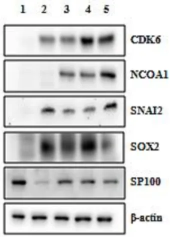
The results of the altered expression of selected proteins shown in this thesis were obtained in fulvestrant-resistant cells grown in medium containing fulvestrant and in MCF7-S0.5 grown in fulvestrant-free medium. In the future, these experiments will be conducted in different medium conditions to confirm the higher expression of the selected proteins in the resistant cell lines comparing to MCF-7/S0.5 grown in the presence of fulvestrant. To compare the expression of the selected proteins in fulvestrant-resistant cell lines Al-1461, Al-909, Al-852 and Al-448 in fulvestrant containing medium and in parental cell line MCF7/S0.5 in fulvestrant-free medium Western blotting experiments were conducted (Figure 2).
The following single bands were detected: CDK6 antibody, ~ 37 kDa; NCOA1 antibody: ~70 kDa; SNAI2 antibody: ~ 30 kDa; SOX2 antibody: ~ 34 kDa and SP100 antibody: ~ 100 kDa. The expression of CDK6, NCOA1, SNAI2 and SOX2 proteins was higher in resistant cell lines than in MCF-7/S0.5, supporting the results from microarray and RT-qPCR data at RNA level. However, SP100 protein expression showed to be higher in the parental cell line compared to the resistant cell lines. Regarding NCOA1, the bands observed were approximately at ~70 kDa and not at ~160 kDa as expected from the primary protein sequence. This can be due to degradation of the lysate by proteolysis, protein modification after translation or a splice variant. The expression of this protein should be confirmed with a different antibody however, due to time constraint that could not be performed in this work. Furthermore, one fulvestrant-resistant cell line, Al-1461, did not show a higher expression of NCOA1 protein as observed for the other resistant cell lines.
Figure 2 Comparison of expression levels of selected candidate proteins in fulvestrant-resistant cell lines and in MCF-7/S0.5 cell line with Western blotting. The bands for CDK6, NCOA1, SNAI2, SOX2 and SP100 proteins expressed in MCF-7/S0.5 cell line grown without fulvestrant are shown in lane 1. The bands for the same proteins expressed in fulvestrant-resistant cell lines Al-1461, Al-909, Al-852 and Al-448 grown with fulvestrant are shown in lanes 2, 3, 4 and 5, respectively. β-actin was used as a loading control.
28
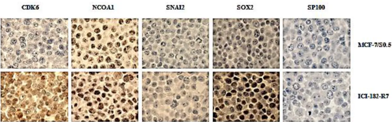
3.2.3 Validation of the altered expression of selected proteins in fulvestrant-resistant breast cancer cell lines by ICC
A FFPE cell line block containing cores of MCF-7/S0.5 and ICI-182-R7 (fulvestrant-resistant cell line) was stained with antibodies against CDK6, NCOA1, SNAI2, SOX2 and SP100 (Figure 3). The results confirmed that both CDK6 and SOX2 showed higher level of expression in ICI-182-R7 compared to MCF-7/S0.5 supporting the observations obtained by Western blotting. NCOA1, SNAI2 and SP100 did not show a clear difference in staining intensity between the sensitive and resistant cell lines although different dilutions of the antibodies against the respective proteins had been tested. The antibodies used may be not as effective in ICC as in WB or the method for antigen retrieval may interfere with the antibodies used. For these reasons, different antibodies against NCOA1, SNAI2 and SP100 or a different method or buffer for antigen retrieval should be tested to stain this cell block.
Figure 3 Comparison of expression patterns of candidate proteins in MCF-7/S0.5 and ICI-182-R7 cell lines analyzed by ICC. A FFPE cell block with cores of cell lines with different resistant profiles was stained with antibodies directed against CDK6, NCOA1, SNAI2, SOX2 and SP100. MCF-7/S0.5 corresponds to a fulvestrant-sensitive cell line and ICI-182-R7 corresponds to a fulvestrant-resistant cell line. FFPE – Formalin-fixed paraffin-embedded; ICC – Immunocytochemistry.
Although all genes showed to be over-expressed at mRNA level in fulvestrant-resistant cell lines Al-1461, Al-909, Al-852 and Al-448 compared to MCF-7/S0.5, using RT-qPCR, based on the results obtained in the validation of the expression of selected proteins by Western blotting and ICC only three genes, CDK6, SNAI2 and SOX2, were selected for further evaluation. There was not a consistent higher expression of NCOA1 protein in all resistant cell lines compared to MCF-7/S0.5 by Western blotting results. Moreover, ICC analysis for this protein did not show a clear difference in the staining pattern between the fulvestrant-sensitive and -resistant cells. Additionally, SP100 protein showed to be higher expressed in the parental cell line compared to the fulvestrant-resistant cells by Western blotting and ICC results failed to show a difference in the expression of this protein between sensitive and resistant cells. Therefore NCOA1 and SP100 were excluded from further experiments.
29 3.3 Knock down experiments
Initially, an optimization of transfection conditions was performed in order to select the best method for transfection of fulvestrant-resistant and parental MCF-7/S0.5 cell lines. Due to time constraints, knock down experiments on the genes selected for further experiments (CDK6, SNAI2 and SOX2) were conducted in only two of the four fulvestrant-resistant cell lines. Nevertheless, since the four resistant cell lines studied are clones of the same parental cell line, MCF-7/S0.5, grown in the same medium conditions, the results are expected to be comparable, as supported by the observation that the cells show similar patterns of proliferation and expression of the selected genes at both mRNA and protein levels.
After optimization of transfection conditions, transfection of MCF7/S0.5 and two fulvestrant-resistant cell lines, Al-1461 and Al-448, was conducted with siRNAs for CDK6, SNAI2 and SOX2 in different medium conditions (with and without fulvestrant). The knock down experiments were confirmed at RNA level 48 hours after transfection by RT-qPCR and at protein level 72 and 96 hours after transfection by Western blotting. Regarding functional assays, the proliferation of cells was evaluated by crystal violet staining and the death of cells by LDH measurement assay at 24, 72 and 96 hours after transfection.
As SOX2 transfection experiments did not achieve a sufficient level of knock down, ~20%, the results will not be discussed in this thesis (data not shown). The experiments have to be repeated with different siRNAs and/or different transfection method (stable transfection). 3.3.1 Optimization of transfection conditions
The transfection conditions were optimized in order to evaluate the best method for transfection of the cells selected. Chemical transfections with two different transfection reagents, OptifectTM and Transit®-2020, and electroporation were conducted in one selected fulvestrant-resistant cell line, Al-1461, with a reference gene, PKCA. Transfection by electroporation showed a higher efficiency of knock down, ~12% of PKCA expression in cells transfected with siRNA targeting PKCA compared to the expression of the gene in cells transfected with control siRNA. Therefore this method was selected to transfect fulvestrant-resistant cells and MCF-7/S0.5 (Appendix II, Figure 9).
30
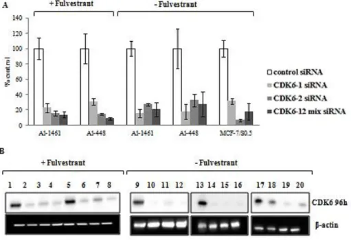
3.3.2.1 Validation of CDK6 mRNA and protein expression after CDK6 knock down in fulvestrant-resistant and MCF-7/S0.5 breast cancer cell lines
MCF-7/S0.5 in fulvestrant-free medium and fulvestrant-resistant cell lines Al-1461 and Al448 in the presence and absence of fulvestrant were transfected with two siRNAs against CDK6 by electroporation. The two siRNAs were used separately and as a mixture to reduce the risk of the effect being an off-target effect. As the proliferation of MCF-7/S0.5 cell line was significantly affected in the presence of fulvestrant (Figure 1), which could compromise the evaluation of the effect of siRNA mediated knock down in cell proliferation and death, the knock down experiments performed in the parental cell line were exclusively performed in the absence of fulvestrant. The experiments were conducted three times and comparable results were obtained. The results shown are from one representative experiment.
Figure 4 Evaluation of mRNA and protein expression after knock down of CDK6 by siRNA transfection. A) CDK6 mRNA expression analyzed by RT-qPCR 48 hours after transfection with single and combined siRNAs against CDK6. The results are shown in percentage of expression relative to cells transfected with control siRNA. The experiments were conducted in Al-1461 and Al-448 fulvestrant-resistant cell lines in the presence and absence of fulvestrant and in MCF-7/S0.5 cell line in the absence of fulvestrant. Data is shown with error bars representing mean ± SEM. SEM – Standard error of the mean. B) CDK6 protein expression levels analyzed by Western blotting 96 hours after transfection with two single siRNAs (lanes 2-3, 6-7, 10-11, 14-15 and 18-19) and combined siRNAs (lanes 4, 8, 12, 16 and 20) against CDK6 compared to transfection with control siRNA (lanes 1, 5, 9, 13 and 17). The experiments were conducted in fulvestrant-resistant cell lines Al-1461 and Al-448 in the presence of fulvestrant (lane 1-4 and 5-8, respectively) and absence of fulvestrant (lane 9-12 and 13-16, respectively). The experiments were conducted in MCF-7/S0.5 cell line in fulvestrant-free medium conditions (lane 17-20). β-actin was used as loading control.