IMMUNOSENSOR BASED ON INK PRINTED ELECTRODE FOR
STAPHYLOCOCCAL ENTEROTOXIN DETECTION
Maria Gardenny Ribeiro PIMENTA-MARTINS1
Roselayne Ferro FURTADO2;
Rosa Fireman DUTRA3;
Luiz Guilherme Dias HENEINE4;
Ricardo Souza DIAS4;
Maria de Fátima BORGES2;
Carlucio Roberto ALVES5∗
1Researcher Professor - State University of Ceara, Fortaleza, Brazil -[email protected] 2Researcher - Embrapa Tropical Agroindustry, Fortaleza, Brazil - [email protected] 3Researcher Professor - Federal University of Pernambuco, Recife, Brazil - [email protected] 4Researcher - Ezequiel Dias Foundation, Belo Horizonte, Brazil - [email protected] 5Researcher Professor - State University of Ceara, Fortaleza, Brazil - [email protected]
Recebido em: 25/05/2014 - Aprovado em: 30/06/2014 - Disponibilizado em: 30/07/2014
ABSTRACT: Staphylococcal enterotoxin is one of the more aggressive enterotoxins produced by Staphylococcus aureus strains and it is a common cause of food poisoning. Analytical methods that are sensitive, low cost and easy to use are needed to evaluate the food quality. This work describes the development of a label free immunosensor based on screen-printed AuNPs/carbon and the characterization of its analytical response for staphylococcal enterotoxin B (SEB) detection. The biosensor was constructed from self-assembled monolayer of thiols and protein A for the oriented immobilization of the polyclonal antibodies against SEB. As electrons mediator, potassium ferrocyanide was used. The electrochemical detection was direct with the parameters following: -0.2 to 0.6 V with the pulse amplitude of 0.075 V and the pulse width of 75 ms. The immunosensor showed detection and quantification limits of 0.4 µg mL-1 and 1.6 µg mL-1,respectively. The immunosensor showed quite satisfactory performance in contaminated and non-contaminated cheese samples.
Keywords: Screen-printed carbon (SPCE). Staphylococcal enterotoxin B. Biosensor. Food safety. Analysis.
RESUMO: Enterotoxina estafilocócica é uma das mais graves enterotoxinas produzidas por Staphylococcus aureus e é
uma causa comum de intoxicação alimentar. Métodos analiticos sensíveis, de baixo custo e de fácil uso são necessários para avaliar a qualidade dos alimentos. Este trabalho descreve o desenvolvimento de um imunossensor nao marcado com base eletrodo impresso de carnono/AuNPs e a caracterização de sua resposta analítica para detecção de enterotoxina estafilocócica B (SEB). O biossensor foi construído a partir de monocamada de tiol e proteína A, para a imobilização orientada dos anticorpos policlonais contra a SEB . Como mediador de elétrons foi usado ferrocianeto de potássio. A detecção eletroquímica foi direto e utilizou-se os seguintes parâmetros: -0,2 a 0,6 V com a amplitude de pulso de 0,075 V e a largura de pulso de 75 ms. O imunossensor mostrou limites de detecção e quantificação de 0,4 mg mL-1 e 1,6 mg mL-1, respectivamente. O imunossensor apresentou desempenho bastante satisfatório em amostras de queijo contaminadas e não contaminadas .
Palavras-chave: Eletrodo impresso de carbono. Enterotoxina estafilocócica B. Biossensor. Segurança alimentar.
Análises.
∗corresponding author:
1. Introduction
Staphylococcal enterotoxins (SEs) are a family of structurally related proteins, produced by Staphylococcus aureus strains (Khreich et al., 2008). SEs are low-molecular weight proteins (26-30 kDa), heat stable, resistant to gut proteases and stable in a wide range of pH (4-10) (LE LOIR; BARON; GAUTIER, 2003; Omoe et al., 2005). These characteristics make these proteins quite resistant to food processing and storage and the proper conditions of the digestive system. Staphylococcal enterotoxin B (SEB) is a cause of serious food poisoning and represents a potential agent of biological terrorism.
Traditional techniques, such as
radioimmunoassay, fluorescence-labeled
antibody assay, and enzyme-linked
immunosorbent assay (ELISA) are widely used for detecting SEB. However, these traditional immunological techniques involve
time-consuming procedures, harmful
biological markers, and expensive instruments (LANCETTE; BENNETT, 2001; TSAI; LI,
2009; Velusamy et al., 2010). An
unquestionable trend for monitoring
biological agents and food contaminants is through novel methods that present rapid analytical response, and that are sensitive,
portable and low cost (PETRENKO;
SOROKULOVA, 2004).
Electrochemical biosensors are an efficient method for detection of analytes in different types of samples including those from food (PIMENTA-MARTINS et al., 2012; LI et al., 2012). In this method, it is possible to associate the specificity of bioreceptors to analytical sensitivity of electrochemical techniques. Electrochemical
biosensors based on screen-printed
technology have been successfully used in the
analysis of contaminated food
(DOMINGUEZ-RENEDO;
ALONSO-LOMILLO; ARCOS-MARTINEZ, 2007;
YANG et al., 2010).
The technology for preparation of screen-printed electrodes (SPEs) is simple, inexpensive, versatile, and also suitable for mass production of disposable electrodes (DAI et al., 2007; GARCÍA-GONZÁLEZ et
al., 2008). SPEs are made by printing
electrically conductive inks, especially carbon on an inert support. In this case, they are
called screen-printed carbon electrodes
(SPCEs). The use of carbon inks is particularly attractive for manufacturing printed electrodes (due to low cost, lead to low background currents and a wide potential window for electrochemical devices) (WANG
et al., 1996; MORRIN; KILLARD; SMYTH,
2003). Moreover, theses disposable electrodes can be modified with nanoparticles. The large surface area-to-volume ratio of nanoparticles such as gold, silver, and carbon nanotubes are important to improve the electron transfer of
the biochemical reaction of the biosensor resulting in higher sensibility of the method and to facilitate the assembly of molecular structures on them (PINGARRON; YÁÑEZ-SEDEÑO; GONZÁLEZ-CORTÉ, 2008).
The first step for the construction of biosensors is the modification of the surface
to obtain an optimum condition of
biomolecule immobilization (RASOOLY; RASOOLY, 1999). Self-assembly has often been applied to the formation of the thin and organized film of biomolecules on a gold surface. In this paper, we used a thiol of short chain that has two functional groups (SH and
NH2), one bound to gold nanoparticles
deposited on a SPCE surface and another
group bound to Protein A from
Staphylococcus aureus. Protein A is a highly
stable receptor, capable of binding to the
crystallizable fragment (Fc) of
immunoglobulins from a large number of species. This step is important for an oriented immobilization of antibodies and it makes a more specific detection of the antigen by immobilized antibodies per portion possible (LEONARD et al., 2003; MUZZUCCHELLI
et al., 2010; VELUSAMY et al., 2010).
The possibility to miniaturize the
staphylococcal enterotoxin B (SEB)
immunosensor using a transducer element that is small in size and disposable such as the Screen Printed Electrodes (SCE) represents an important goal in the development of an amperometric staphylococcal enterotoxin B
(SEB) device. We previously showed an immunosensor for staphylococcal enterotoxin
detection based on a gold surface
(PIMENTA-MARTINS et al., 2012).
Recently, we have adapted the methodology for disposable electrodes. In this work, the results inherent to the development of the
biosensor with antibodies against
staphylococcal enterotoxin B (SEB) on screen printed carbon electrodes are reported. In specific, electrochemical characterization of the construction of biosensors and the
analytical response for staphylococcal
Enterotoxin B (SEB) were studied.
2. Experimental
2.1. Reagents and apparatus
Electrodag PF-407 C carbon ink was
acquired from the Acheson Henkel
Corporation (USA). The reagents
N-hydroxysuccinimide (NHS),
1-(3-Dimethylaminopropyl)-N'-ethylcarbodiimide hydrochloride (EDC), Cysteamine, Hydrogen
tetrachloroaurate (HAuClO4), and the
proteins: Bovine Serum Albumin (BSA) Staphylococcal enterotoxin B (SEB), anti-Staphylococcal B, and Protein A from soluble
Staphylococcus aureus were purchased from
Sigma-Aldrich, St. Louis, Mo, USA.
Unless indicated, all the antibodies and antigen solutions was prepared in 0.01
7.0. Ultra-pure water (18 MΩ cm) used to prepare all solutions was obtained from a Milli-Q water purification system (Millipore Inc., USA).
The electrochemical experiments were carried out using Autolab PGSTAT 12 potentiostat/galvanostat (Eco Chemie, The
Netherlands) with General Purpose
Electrochemical System software (GPES 4.9). In the experiments, the SPCEs with AUNPs were used as working electrodes, helical platinum wire was used as a counter electrode, and an Ag/AgCl electrode was used as reference. The electrodes were set up in a glassy electrochemical cell with 10 mL volume.
2.2. Preparation of the screen-printed
electrodes (SPCEs)
For the manufacturing of the SPCEs, adhesive plastic mold was fixed on the acetate strip substrate and it was over-coated with one
layer of carbon ink (Electrodag® PF 407-C)
using the appropriate stencils. The electrode
surface (area = 9.62 mm2) was cured for 60
min. at 60 ºC. Finally, the adhesive plastic mold was removed and the AuNPs were introduced onto the surface. Thereafter, the carbon ink thickness deposited on acetate was
measured using a digital micrometer
(Mitutoyo Corporation, Japan) and overall thickness was expressed as an average of ten
readings taken randomly on each SPCEs sample.
2.3. Construction of the biosensor
Firstly, the SPCEs were pretreated in 100 mM KCl solution for the activation of the surface according to Alonso-Lomillo et al. (2009). Screen-printed carbon electrodes with AuNPS were immersed in 10 mM cysteamine solution for 2 h at room temperature. Afterwards, the SPCEs were rinsed with ethanol and water, and dried at room
temperature (MENDES; CARVALHAL;
KUBOTA, 2008). In the next step, 2 mM EDC and 5 mM NHS solution freshly prepared in acetate buffer (pH 5.0), reacted
for 30 minutes. In sequence, 5 mg mL-1 of
protein A was added the EDC/NHS solution and left to react for 30 minutes. Finally, the modified electrode with cysteamine was incubated in protein A and EDC/NHS solution for one hour. Subsequently, the SPCEs were incubated in anti-SEB solution
(100 mg mL-1) for one hour. Then, 10 µ L 1%
BSA was dropped onto SPCEs for 1 h at room temperature. After each incubation step, the SPCEs were washed with 10 mM PBS and deionized water.
2.5. Immunosensor response
After the fabrication of immunosensor, the modified SPCEs were dropped in 10 µ L
of SEB (1 mg L-1, pH 7.4) for one hour. All
differential pulse voltammetry (DPV)
measurements were carried out in 50 mM
PBS (pH 7.4) containing 4 mM K3Fe(CN)6
redox probe system at room temperature. These measurements were obtained from -0.2 to 0.6 V with the pulse amplitude of 0.075 V and the pulse width of 75 ms. The cyclic voltammetry (CV) measurements were carried
out in 50 mM KCl and 4 mM K3Fe(CN)6
solution and scan speed of 0.050 mV s-1.
3. Results and discussion
3.1. The principle of the electrochemical immunosensor for enterotoxin
Electrochemical immunosensors are a type of biosensor that is quite popular. Immunosensors are actually a new version of
enzyme-linked immuno-sorbent assay
(ELISA), with reduced cost, improved response speed, operation convenience, and comparable or even higher sensitivity than other conventional methods of analysis. In the
development of electrochemical
immunosensors, the amount of antibodies or antigens are immobilized at the electrode. The biological molecules form the surface sensor. The interaction of the analyte-substrate is measured through electrochemical techniques. These are based on the detection of a change
in the electrical properties of the surface
transducer as a result of the immunocomplex
formation which causes a change in the transducer signal.
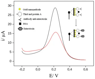
A redox probe is used in many cases, in order to detect this change (Figure 1). In the label-free immunosensors, the use of enzymes or another marker is dispensable. This fact confers the advantages of this single stage analysis, which leads to a lower cost and is easier to use than labeled-immunosensors. Direct measurements have been a success achieved in immunosensors adopting the cyclic voltammetry technique in the presence
of [Fe(CN)6]3−/4− as a redox probe for the
detection of microorganisms (CHO et al., 2008). In this work, the immunosensor was developed using the differential pulse
voltammetry (DPV) technique and
[Fe(CN)6]3−/4− as a redox probe. Generally,
the detection limit obtained from the differential pulse voltammetry is two or three orders of magnitude lower than values obtained by cyclic voltammetry, reaching the
range of 10-7 to 10-8 mol L-1 (WELCH;
COMPTON, 2006).
The K3Fe(CN)6 probe is a valuable
tool for testing the kinetic barrier of the
charge transfer between solution and
electrode interface. When antibodies against Staphylococcal enterotoxin are deposited on SPCEs, the electrode active area decreases and hinders probe ions through their pathways into the electrode. Hence, the redox reaction
of K3Fe(CN)6 decreases and consequently the
is observed when a blocking agent and the target molecule bind to a sensing surface. The approach to detection of the target analyte is illustrated in Figure 1.
Figure 1. Schematic illustration for sensing of
staphylococcal enterotoxin in 4 mM K3Fe(CN)6 and 25
mM PBS solution pH 7. -0,2 0,0 0,2 0,4 0,6 0 5 10 15 20 25 30 i/ µ A E/ V 3.2. Electrochemical characterization
The average thickness of the deposited carbon ink on acetate film was 54.5 µm (CV = 1.6%, n = 3). Analysis of the coverage of the ink printed electrode can be conducted by
cyclic voltammetric investigations
(FURTADO et al., 2012). The area of the
redox peak of the redox probe [Fe(CN)6]3−/4−
can be used in the characterization of layers with respect to their degree of coverage. Figure 2 shows that after the formation of each layer, a decreasing of the cathodic electric current and the anodic peak of the
redox probe [Fe(CN)6]3−/4− occurs. Parallel to
this, there was a noticeable separation of oxidation and the reduction peaks of the overlapping voltammograms. In this case, the separation of the redox peak is higher if there is an insulating of the electron flux. All these events are associated with the binding of the biomolecules to the surface electrode.
Figure 2. Cyclic voltammograms in 4 mM
K3[Fe(CN)6] and PBS buffer solution pH 7.4: steps
immunosensor manufacture: (____) bare surface -SPCE; (- - - -) cysteamine (10 mM); (____) protein A (0.05 mg mL-1); (……) anti-SEB (100 mg mL-1) immobilization and BSA blocking (1%). The measurements were carried out in 50 mM KCl and 4 mM K3Fe(CN)6 solution and scan speed of 0.050
mV s-1. -0.25 0.00 0.25 0.50 0.75 -80 -60 -40 -20 0 20 40 60
i
/
µ
A
E / V
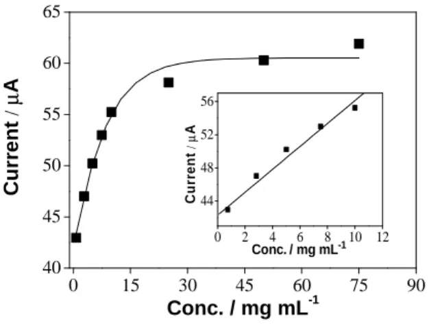
3.2.1. Calibration curveCalibration curve was obtained by using the differential pulse voltammetry (DPV) measurements. The analytical response of the AuNPS/SPCEs incubated in different concentrations of SEB prepared in PBS pH 7.4 was generated by the increasing the insulating of the electron flux of the redox probe until the surface. The calibration
equation obtained by DPV presented a good linearity with the correlation coefficient of 0.98 (P < 0.001, n = 5) according to Figure 3.
The limit of detection of 0.4 µg mL-1
was estimated considering three times the standard deviation of the measurement of the blank divided by the slope of the calibration
curve. The quantification limit of 1.6 µg mL-1
was estimated considering ten times the standard deviation of the measurement of the blank divided by the slope of the calibration curve.
Figure 3. Calibration curve of immunosensor for SEB
detection in 50 mM PBS (pH 7.4) and 4 mM K3Fe(CN)6. Measurements were obtained from -0.2 to
0.6 V with the pulse amplitude of 0.075 V and the pulse width of 75 ms. Insert: Linear curve of the calibration plot. 0 15 30 45 60 75 90 40 45 50 55 60 65 0 2 4 6 8 10 12 44 48 52 56 Conc./ mg mL-1 C u rr e n t / µ A C u rr e n t / µ A Conc. / mg mL-1
3.2.2. Detection of SEB in cheese samples
Attempting to make use of this SEB immunosensor in a complex matrix as Coalho cheese type, commercial samples were adquired for evaluating the performance of the immunosensor. Firstly, samples were
prepared according to manufacturer's
instructions from the SET-RPLA test
(Oxoid®), which uses the rapid reversed
passive latex agglutination method. The positive and negative samples thus classified
by the kit, were analyzed by the
immunosensor. According to Figure 4, the immunosensor was capable to distinguish the contaminated and non-contaminated cheese samples. These results indicate that the immunosensor can be a low cost alternative method for analytical detection of SEB.
Electrochemical biosensors are often preferred over other transducers due to the ease of the miniaturization and automation system. In this sense, the methodology developed based on AuNPS/SPCE could be adapted to miniaturized electronic systems aiming at a future commercial application for monitoring the quality of food.
Figure 4. Assessment of the immunosensor in
contaminated and non-contaminated cheese samples. Measurements were obtained from -0.2 to 0.6 V with the pulse amplitude of 0.075 V and the pulse width of 75 ms. 1 2 0.5 1.0 1.5 2.0 2.5 3.0 3.5
Samples
Negative Positivei
/
µA
4. Conclusion
A label free immunosensor for SEB detection was satisfactorily developed based on SPCE modified with AUNPs. The device
showed good performance in the
contaminated cheese samples as an alternative method of analysis. The results obtained demonstrated that by using this developed system, it is possible to detect low concentration of SEB in few minutes. Moreover, this study leads to additional exciting investigations on rapid detection of a variety of microorganisms and its toxins.
Acknowledgment
The authors would like to thank the Brazilian agencies FUNCAP, CNPq and Embrapa for their financial support.
5. References
ALONSO-LOMILLO, M. A.;
DOMÍNGUEZ-RENEDO, O.; MATOS, P. M.; ARCOS-MARTÍNEZ, J.
Electrochemical determination of levetiracetam by screen-printed based biosensors. Bioelectrochemistry, 74: 306–
309, 2009.
CHO, E. C.; CHOI, J-W; LEE, M.; KOO, K-K. Fabrication of an electrochemical
immunosensor with self-assembled peptide nanotubes. Colloids and Surfaces A:
Physicochemical and Engineering Aspects, 313: 95–99, 2008.
DAI, Z.; FANG, M.; BAO, J.; WANG, H.; LU, T. An amperometric glucose biosensor
constructed by immobilizing glucose
oxidase on titanium-containing mesoporous composite material of no. 41 modified screen-printed electrodes. Analytica
Chimica Acta, 591(2): 195–199, 2007.
DOMINGUEZ-RENEDO, O.; ALONSO-LOMILLO, M. A.; ARCOS-MARTINEZ, M. J. Recent developments in the field of
screen-printed electrodes and their related applications. Talana, 73: 202–219, 2007.
FURTADO, R. F.; ALVES, C. R.;
MOREIRA, A. C. O.; AZEVEDO, R. M.; DUTRA, R. F. A. novel xyloglucan
film-based biosensor for toxicity assessment of ricin in castor seed meal. Carbohydrate
Polymers, 20: 586–591, 2012.
GARCÍA-GONZÁLEZ, R.; FERNÁNDEZ-ABEDUL, M. T.; PERNÍA, A.; COSTA-GARCÍA, A. Electrochemical
characterization of different screen-printed gold electrodes. Electrochimica Acta, 53:
KHREICH, N.; LAMOURETTE, P.; BOUTAL, H.; DEVILIERS, K.;
CRÉMINON, C.; VOLLAND H. Detection
of Staphylococcus enterotoxin B using fluorescent immunoliposomes as label for immunochromatographic testing.
Analytical Biochemistry, 377: 182–188, 2008.
LANCETTE, G. A.; BENNETT, R. W.
Staphylococcus aureus and Staphylococcal enterotoxins, In DOWNES, F. P.; ITO, K.
(Eds), Compendium of methods for the
Microbiological examination of Foods.
Washington: American Public Health Association, 2001, pp. 387–403.
LE LOIR, Y.; BARON, F.; GAUTIER, M.
Staphylococcus aureus and food poisoning,
Genetic and Molecular Research, 2: 163–176, 2003.
LEONARD, P.; HEARTY, P. S.;
BRENNAN, J.; DUNNE, L.; QUINN, J.; CHAKRABORTY, T. Advances in
biosensors for detection of pathogens in food and water. Enzyme and Microbial
Technology, 32: 3–13, 2003.
LI, Y.; CHENG, P.; GONG, J.; FANG, L.; DENG, J.; LIANG, W. Amperometric
immunosensor for the detection of
Escherichia coli O157:H7 in food
specimens. Analytical Biochemistry, 421(1):
227–233, 2012.
MENDES, R. K.; CARVALHAL, R. F.; KUBOTA, L. T. Effects of different
self-assembled monolayers on enzyme
Immobilization procedures in peroxidase-based biosensor development. Journal of
Electroanalytical Chemistry, 612: 164–172, 2008.
MORRIN, A.; KILLARD, A. J.; SMYTH, M. R. Electrochemical characterization of
commercial and home-made screen-printed carbon electrodes. Analytical Letters, 36,
2021–2039, 2003.
MUZZUCCHELLI, S.; COLOMBO, M.; PALMA, C.; SALVADE, A.; VERDERIO, P.; COGHI, M. D. Single-domain protein
A‐engineered magnetic nanoparticles: toward a universal strategy to site-specific labeling of antibodies for targeted detection of tumor cells. ACS Nano, 4: 5693–5702,
2010.
OMOE, K.; MANISHIK, I.; HU, D.; KATO, H.; FUGANE, Y.; ABE, Y. Characterization
of novel staphylococcal enterotoxin-like toxin type P. Infection and Immunity, 73,
5540–5546, 2005.
PETRENKO, V. A.; SOROKULOVA, I. B.
Detection of biological threats. A challenge for directed molecular evolution. Journal of
PIMENTA-MARTINS, M. G. R.; ALVES, C. R.; FURTADO, R. F.; HELEINE, L. G. D.; DIAS, R. S.; BORGES, M. F. Development
of an amperometric imunosensor for detection of staphylococcal enterotoxin type A in cheese. Journal of Microbiological
Methods, 91: 138–143, 2012.
PINGARRON, J. M.; YÁÑEZ-SEDEÑO, P.; GONZÁLEZ-CORTÉ, A. Gold
nanoparticle-based electrochemical biosensors. Electrochimica Acta, 53: 5848–
5866, 2008.
RASSOLY, A.; RASSOLY, B. Real time
biosensor analysis of staphylococcal enterotoxin A in food. International Journal
of Food Microbiology, 49: 119–127, 1999.
TSAI, W.; LI, I. SPR-based immunosensor
for determining staphylococcal enterotoxin A. Sensors and Actuators B: Chemical, 136:
8–12, 2009.
VELUSAMY, V.; ARSHAK, K.;
KOROSTYNSKA, O.; OLIWA, K.; ADLEY, C. An overview of foodborne pathogen
detection: in the perspective of biosensors.
Biotechnology Advances, 28: 232–254, 2010.
WANG, J.; PEDRERO, M.; SAKSLUND, H.; HAMMERICH, O.; PINGARRON, J.
Electrochemical activation of
screen-printed carbon strips, Analyst, 121(3): 345–
350, 1996.
WELCH, C. M.; COMPTON, R. G. The use
of nanoparticles in electroanalysis: a review. Analytical and Bioanalytical
Chemistry, 384: 601–619, 2006.
YANG, M.; SUN, S.; BRUCK, H. A.; KOSTOV, Y.; ASSOLY, A. Electrical
percolation-based biosensor for real-time direct detection of staphylococcal
enterotoxin B (SEB). Biosensors and