UNIVERSIDADE DO ALGARVE
On the solvation in lipid bilayers measured by pyrene
fluorescence: Thermal and molecular composition
influence on equivalent polarity in lipidic mixtures
Dalila Lopes Arrais
Dissertação de Doutoramento em Ciências Biológicas (especialidade de Bioquímica)
Trabalho realizado sob a orientação de Prof. Doutor Jorge Manuel Martins e co-orientação de Prof. Doutor Eurico Melo
On the solvation in lipid bilayers measured by pyrene fluorescence: Thermal and molecular composition influence on equivalent polarity in lipidic mixtures.
Declaração de Autoria de Trabalho
Declaro ser a autora deste trabalho, que é original e inédito. Autores e trabalhos consultados estão devidamente citados no texto e constam da listagem de referências incluída.
Copyright © 2013
Dalila Lopes Arrais (A Universidade do Algarve tem o direito, perpétuo e sem limites geográficos, de arquivar e publicitar este trabalho através de exemplares impressos reproduzidos em papel ou de forma digital, ou por qualquer outro meio conhecido ou que venha a ser inventado, de o divulgar através de repositórios científicos e de admitir a sua cópia e distribuição com objectivos educacionais ou de investigação, não comerciais, desde que seja dado crédito ao autor e editor).
i
Acknowledgements
“I have no special talent. I am only passionately curious.” said Albert Einstein. I feel exactly the same. The need to question and find answers has driven me to these particular circumstances. The past years have been all about learning, not only in the scientific field, but also in life. I am truly thankful for the chance I have been given.
I would like to show my sincere appreciation to Prof. Jorge Martins, for all the support, friendship and wise advices throughout these (sometimes very demanding) years and for being an example in life. He once told me that fulfilling our purposes is as important as the path that leads us there and that being aware of this will make us rise above our (known) abilities. This is absolutely true! I also thank Prof. Eurico Melo for co-orienting this project.
I gratefully acknowledge FCT (Fundação para a Ciência e a Tecnologia) for the grant
SFRH/BD/41607/2007 and IBB-CBME and Universidade do Algarve, Faro – Portugal, for their financial support and for providing all the necessary conditions so that this work could be performed.
But my overall gratitude goes to every single person who has been a part of my life until the present moment. Every living creature that touches (or touched) our lives has (or had) the power to contribute to the (never ending) creation of a better version of ourselves. It is up to us to read the signs and interpret them in the best way possible as everything is a positive lesson if seen through the right perspective. This will give us the chance to live plentifully, to accept ourselves and everyone who crosses our lives. I gratefully thank my family, Bruno and all my friends and dear ones, for showing me constantly how it feels to be truly loved.
This work is dedicated to those who are always in my heart and bring light to my life!
(Este trabalho é dedicado a todos aqueles que, estando longe ou perto, numa base diária ou menos constante, me aquecem o coração e enchem a minha vida de luz!)
iii
Abstract
Phosphatidylcholine (PC), sphingomyelin (SM) and cholesterol (CHOL) are major constituents of mammalian cell membranes. DPPC/CHOL and DPPC/DMPC are well-known binary mixtures. POPC/CHOL, DOPC/CHOL, SM/CHOL, SM/POPC and egg-SM/DOPC are less studied, but also important for the comprehension of the POPC/egg-SM/CHOL mixtures. These provide complex media for which polarity is hard to access. It is mainly determined by the water penetrating the bilayer (unevenly distributed creating a polarity gradient), though the influence of the dipoles from phospholipids (e.g. –PO, –CO, – OH) and the double bond in the steroid ring of CHOL cannot be neglected. CHOL derivatives are an interesting tool to verify the influence of the double bonds in the polarization of its surroundings. Pyrene fluorescence was used to access an equivalent polarity (associated to the dielectric constant) near the lipid/water interface of lipid bilayers. POPC/CHOL and DOPC/CHOL have similar thermal behavior and variation with CHOL content, though for lower CHOL content the equivalent polarity is higher for the DOPC/CHOL mixtures. The studies with DPPC and DMPC showed that pyrene does not seem to have a marked preference for either ordered or disordered phases. For DPPC/CHOL and egg-SM/CHOL the highlight goes to the behavior of the mixtures at higher CHOL amounts, where there is a substantial change in the thermal behavior and polarity values especially for the egg-SM/CHOL mixture. Egg-SM/POPC and egg-SM/DOPC show different behavior depending on which phospholipid has a higher molar proportion. The ternary mixtures analyzed do not exhibit significant differences, though there is the indication of the existence of a more ordered environment at lower temperatures and a less ordered environment for higher temperatures. The presence of 7DHC or DCHOL in egg-SM bilayers showed a tendency for the same behavior detected upon mixing higher amounts of CHOL.
Keywords: Pyrene, Fluorescence, Equivalent polarity, Model membranes, Lipid mixtures,
v
Resumo
As fosfatidilcolinas (PC), esfingomielinas (SM) e colesterol (CHOL) são os principais constituintes das membranas celulares de mamíferos. A presença deste esterol numa bicamada reflete-se através de algumas modificações ao nível das propriedades da mesma, das quais são exemplo, alterações na permeabilidade, espessura da bicamada, empacotamento das moléculas, difusão lateral dos lípidos. Hoje sabe-se que a bicamada lipídica que as compõe é um meio bastante complexo. Além de apresentar um gradiente de fluidez (da zona da interface, na qual os segmentos das cadeias metilénicas estão mais ordenados, para a zona do interior hidrofóbico, onde estão mais desordenados), apresenta ainda um gradiente de polaridade (que depende maioritariamente da presença de moléculas de água, mais concentradas na zona da interface e em muito menor quantidade no interior hidrofóbico). No entanto, é bastante difícil medir a polaridade em sistemas lipídicos. Um método muito utilizado é o uso de sondas sensíveis a alterações no meio, que particionam para o interior da bicamada. Embora as grandes diferenças na polaridade se devam à entrada e saída de água da bicamada, há que ter em conta a existência de dipolos provenientes dos diversos grupos moleculares dos lípidos (e.g. grupos fosfato, carbonilo, hidroxilo) e ligações duplas (como a que se encontra no sistema de anéis do colesterol). Muitos estudos sobre bicamadas lipídicas são feitos em sistemas modelo, segundo determinadas condições experimentais, e apenas dão uma ideia geral sobre as propriedades dos lípidos em misturas. O foco principal, na maioria das vezes, está nas interações entre lípidos, de modo que o papel desempenhado pelos dipolos é geralmente negligenciado. Sistemas binários e ternários como os estudados neste trabalho, podem parecer demasiado simples, mas dada a complexidade da membrana, são um bom método para isolar as partes para tentar compreender o todo. As misturas DPPC/CHOL e DMPC/DPPC já foram extensivamente estudadas, mas misturas como POPC/CHOL, DOPC/CHOL, egg-SM/CHOL, egg-SM/POPC e egg-SM/DOPC são menos conhecidas. No entanto, são um bom ponto de partida para aumentar um pouco mais a complexidade do sistema em estudo e partir para misturas ternárias, como a mistura POPC/egg-SM/CHOL, que é conhecida como sendo a mistura canónica que mimetiza a formação de domínios lipídicos (ou “rafts”) em membranas biológicas. O uso de derivados de colesterol, também pode ser útil, no caso de se pretender uma melhor perceção sobre as interações lípido-esterol, como aconteceu neste trabalho, particularmente em relação à influência da ligação dupla do colesterol na polaridade sentida pela sonda.
Para aceder à polaridade nas misturas estudadas, foi utilizada a molécula de pireno, um hidrocarboneto aromático policíclico. Muito se tem especulado acerca deste tipo de sondas e sobre a consistência dos resultados que produzem, em especial, sobre o facto da inserção de uma molécula rígida poder trazer efeitos a nível estrutural, e sobre a possível existência de movimentos transversais consideráveis, que possam de certa forma alterar os resultados. O pireno inserido na bicamada localiza-se na zona mais ordenada das cadeias metilénicas. É certo que apresenta pequenos movimentos transversais, mas segundo estudos recentes em misturas POPC/CHOL, estes parecem ser negligenciáveis, bem como os possíveis efeitos estruturais provocados pela inserção desta sonda. O facto de esta sonda proporcionar uma média da polaridade nesta zona e ainda poder reportar uma média de diferentes zonas no plano da bicamada, devido à sua difusão lateral e ainda devido à difusão lateral dos lípidos,
vi
revela-se como uma vantagem na sua utilização. Geralmente, os dados são obtidos sob a forma de uma razão entre a intensidade da primeira banda (altamente sensível à polaridade do solvente) e a intensidade da terceira banda (praticamente insensível à polaridade do solvente) do espetro de emissão do pireno (normalmente representada como ⁄ ). Através da escala de polaridade do pireno (baseada no efeito de Ham) é possível relacionar esta razão diretamente com a constante dielétrica de um meio.
Com base nos diagramas de fases conhecidos para as misturas analisadas neste trabalho, foi possível obter um conjunto de resultados bastante completos, através da caracterização, em termos de polaridade (equivalente), de sistemas com diferentes composições lipídicas, a diferentes temperaturas.
No caso das misturas que envolvem fosfolípidos insaturados, verificou-se que os perfis de variação térmica de POPC/CHOL e DOPC/CHOL são equivalentes e semelhantes aos verificados para solventes homogéneos polares. No entanto, é possível detetar diferenças na polaridade para altas concentrações de colesterol (os valores de ⁄ baixam com a adição de 40 mol% de colesterol no caso da mistura POPC/CHOL e com a adição de 20 mol% do mesmo esterol na mistura DOPC/CHOL).
A mistura (quase) ideal entre DPPC e DMPC revelou que a sonda parece não ter uma preferência marcada por fases mais fluidas ou fases mais ordenadas, sendo que esta poderá depender da composição da bicamada.
Para as misturas de fosfolípidos saturados com colesterol, a análise dos resultados é um pouco mais complexa. No entanto, observa-se um comportamento semelhante para as duas misturas (embora com valores de polaridade, no geral, ligeiramente mais elevados para a mistura DPPC/CHOL): para baixas concentrações de colesterol, obtiveram-se resultados parecidos aos correspondentes às fases fluidas, e um comportamento semelhante a solventes homogéneos polares; aumentando a quantidade de esterol, os valores de ⁄ passam a não depender da temperatura, ou a depender de forma inversa (que é o caso da mistura egg-SM/CHOL).
As misturas fosfolípido-fosfolípido estudadas mostram que os perfis de variação da polaridade com a temperatura dependem de qual dos fosfolípidos está em excesso.
No caso das misturas ternárias, verificou-se que não há diferenças significativas entre os valores de polaridade para cada composição escolhida, no entanto, a variação térmica aponta no sentido da existência de uma fase mais ordenada a baixas temperaturas e uma fase menos ordenada a elevadas temperaturas.
Por sua vez, os efeitos da adição de 7DHC ou DCHOL à egg-SM, verificaram-se, de um modo geral, como sendo semelhantes aos efeitos do colesterol. Deste modo, o comportamento da mistura egg-SM para elevadas concentrações de colesterol pode não ser um efeito específico da presença da ligação dupla.
Palavras-Chave: Pireno, Fluorescência, Polaridade equivalente, Modelos de membrana,
ix
Contents
Section
PageChapter I – INTRODUCTION 1
I – 1 A general view over biological membranes... 3
I – 1.1 Lipid bilayer structure... 5
I – 1.1.1 Water... 5
I – 1.1.2 Membrane lipids... 5
I – 1.1.2.1 Glycerophospholipids... 5
I – 1.1.2.2 Sphingolipids... 7
I – 1.1.2.3 Sterols... 7
I – 1.1.2.3.1 Cholesterol is a “special” lipid... 8
I – 1.1.2.3.2 Cholesterol derivatives... 9
I – 1.1.3 How do lipids behave in aqueous solution?... 10
I – 1.1.3.1 Assembly of lipid aggregates... 11
I – 1.1.3.1.1 Experimental model membrane systems... 11
I – 1.2 Lipid bilayer physical properties... 12
I – 1.2.1 Phase transitions in single lipid bilayers... 14
I – 1.2.2 Lipidic mixtures and the observation of phase coexistence... 17
I – 1.2.2.1 Phospholipid-phospholipid mixtures... 17
I – 1.2.2.2 Phospholipid-sterol binary mixtures... 19
I – 1.2.2.2.1 Phase coexistence... 22
I – 1.2.2.3 Ternary mixtures... 23
I – 1.3 Lateral and transversal asymmetry in lipid bilayers... 25
I – 1.3.1 Membrane Proteins... 25
I – 1.3.2 Lipid domains in biological membranes... 26
I – 1.4 Lipid bilayers as permeability barriers... 28
I – 2 Polarity measurements in biological membranes... 30
I – 2.1 Polarity is a complex physicochemical property... 30
x
I – 2.2.1 General features of fluorescence spectroscopy... 33
I – 2.2.2 The use of fluorescent probes... 36
I – 2.2.2.1 Empirical scales of polarity... 37
I – 2.3 Pyrene: a well-known and widely used polarity probe... 39
I – 2.3.1 Pyrene absorption and emission characteristics... 39
I – 2.3.1.1 Pyrene fluorescence measurements must be free of experimental and instrumental artifacts... 40
I – 2.3.1.1.1 The appropriate experimental conditions... 40
I – 2.3.1.1.2 Raman scattering... 41
I – 2.3.1.1.3 The ⁄ ratio depends on temperature... 42
I – 2.3.2 Studies using pyrene... 43
Chapter II – MATERIALS AND METHODS 45 II – 1 Chemicals and solvents... 47
II – 2 Stock solutions... 47
II – 2.1 Pyrene... 47
II – 2.2 Lipids... 47
II – 3 Liposome preparation... 47
II – 3.1 Pure phospholipid liposomes... 48
II – 3.2 Phospholipid/phospholipid and phospholipid/cholesterol liposomes... 48
II – 3.3 Ternary mixtures liposomes... 49
II – 3.4 Sphingomyelin/cholesterol derivatives liposomes... 49
II - 4 Steady-state fluorescence measurements... 49
II – 5 Statistical Analysis... 50
Chapter III – RESULTS AND DISCUSSION 51 III - 1 Equivalent polarity for binary mixtures involving unsaturated phospholipids... 54
III – 1.1 POPC/Cholesterol mixtures... 54
III – 1.2 POPC/egg-Sphingomyelin mixtures... 56
xi
III – 1.4 DOPC/egg-Sphingomyelin mixtures... 62
III - 2 Equivalent polarity for binary mixtures involving saturated phospholipids... 65
III – 2.1 DMPC/DPPC mixtures... 65
III – 2.2 DPPC/Cholesterol mixtures... 67
III – 2.3 Egg-Sphingomyelin/Cholesterol mixtures... 72
III – 2.4 Egg-Sphingomyelin/POPC mixtures... 77
III – 2.5 Egg-Sphingomyelin/DOPC mixtures... 79
III – 3 Equivalent polarity for ternary mixtures of POPC, egg-Sphingomyelin and cholesterol... 82
III – 4 Equivalent polarity for binary mixtures egg-Sphingomyelin and cholesterol derivatives... 85
III – 4.1 Egg-Sphingomyelin/7-DHC mixtures... 85
III – 4.2 Egg-Sphingomyelin/Cholestanol mixtures... 88
III – 4.3 Egg-Sphingomyelin/Cholesterol derivatives vs. egg-Sphingomyelin/Cholesterol... 89
Chapter IV – CONCLUDING REMARKS 91 IV – 1 Binary mixtures involving unsaturated phospholipids... 93
IV – 2 Binary mixtures involving saturated phospholipids... 94
IV – 3 Ternary mixtures of POPC, egg-Sphingomyelin and cholesterol... 95
IV – 4 Binary mixtures of egg-Sphingomyelin and cholesterol derivatives... 95
IV – 5 Perspectives... 96
REFERENCES... 99
APPENDIX: Equivalent polarity results ( ⁄ values)... 109
POPC/Cholesterol binary mixtures... 111
POPC/Egg-Sphingomyelin binary mixtures... 112
DOPC/cholesterol binary mixtures... 113
DOPC/Egg-Sphingomyelin binary mixtures... 114
xii
DPPC/cholesterol binary mixtures... 116
Egg-Sphingomyelin/cholesterol binary mixtures... 117
Egg-Sphingomyelin/POPC binary mixtures... 118
Egg-Sphingomyelin/DOPC binary mixtures... 119
POPC/Egg-Sphingomyelin/cholesterol ternary mixtures... 120
Egg-Sphingomyelin/7-Dehydrocholesterol binary mixtures... 121
Abbreviations and Symbols
G Gibbs free energy variation
ΔH Enthalpy variation
ΔS Entropy variation
7DHC 7-Dehydrocholesterol
7DHCR 7-Dehydrocholesterol reductase
Abs Absorbance / Absorption
CAC Critical aggregation concentration
CHCl3 Chloroform
CHOL Cholesterol
CMC Critical micelle concentration
D Distance
DCHOL Cholestanol
DMPC 1,2-dimyristoyl-sn-glycero-3-phosphocholine
DOPC 1,2-dioleoyl-sn-glycero-3-phosphocholine
DPPC 1,2-dipalmitoyl-sn-glycero-3-phosphocholine
DSC Differential scanning calorimetry
E Applied electric field
egg-SM Natural egg yolk sphingomyelin
EPR Electron paramagnetic resonance
ER Endoplasmatic reticulum
ESR Electron spin resonance
EtOH Ethanol
F Fluorescence emission
F’ Fluorescence emission after solvent relaxation
G Correlation factor that characterizes the relative orientations between neighboring molecules
GPI Glycosylphosphadidylinositol
GUV Giant unilamellar vesicles
⁄ Ratio between the intensities of fluorescence of the first and third band of the pyrene emission spectrum
IC Internal conversion
IR Infrared spectroscopy
ISC Intersystem crossing
kB Boltzmann constant
Liquid crystalline or fluid lamellar phase Gel lamellar phase
Gel lamellar phase for phosphatidylcholines Sub-gel phase
Liquid disordered phase for phospholipid-cholesterol mixtures Liquid ordered phase for phospholipid-cholesterol mixtures
LAURDAN 2-dimethylamino-6-lauroylnaphthalene LUV Unilamellar vesicles
M Molar mass
MD Molecular dynamics
MeOH Methanol
MLV Multilamellar vesicles Avogadro Number
NMR Nuclear magnetic resonance 2
H-NMR Deuterium nuclear magnetic resonance
p Polarization
Ripple phase for phosphadidylcholines
PAH Polycyclic aromatic hydrocarbons
PC Phosphatidylcholine PE Phosphatidylethanolamine PI Phosphatidylinositol PG Phosphatidylglycerol Pm Molar polarizability POPC 1-palmitoyl-2-oleoyl-sn-glycero-3-phosphocholine PRODAN 6-propionyl-2-dimethylaminonaphthalene PS Phosphatidylserine PSM N-pamitoyl-sphingomyelin (N-palmitoyl-D-erythro-sphingosylphosphorylcholine) Q Charge (+q or –q) of a particle Singlet ground electronic state First singlet electronic excited state Second singlet electronic excited state
So Solid-ordered state of lipid bilayers
So1 POPC-rich solid-ordered state in PSM/POPC mixtures
So2 PSM-rich solid-ordered state in PSM/POPC mixtures
SLOS Smith-Lemli-Opitz syndrome
SM Sphingomyelin (1-ceramide-phosphorylcholine)
SUV Small unilamellar vesicles First triplet state
Second triplet state
Main phase transition temperature
T Temperature
UV-vis Ultra-violet visible range of the electromagnetic spectrum Vibrational ground state
First vibrational excited state Second vibrational excited state
Greek Letters
Molecular polarizability Permittivity in vacuum
Dielectric constant or relative permittivity (also noted as ) / molar absorption coefficient
Dielectric constant or relative permittivity (also noted as ) Excitation wavelength
Dipole moment of a molecule in the ground state Dipole moment of a molecule in the excited state Induced dipole moment
Bonding pi molecular orbital Antibonding pi molecular orbital
Mass density
Fluorescence lifetime Fluorescence quantum yield
Chapter I
I – INTRODUCTION
3
I – 1
A general view over biological membranes
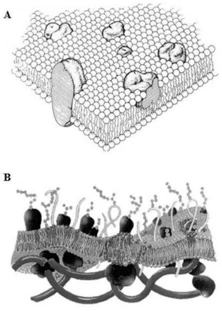
Membranes play a key role in structure and function of cells. They are complex structures that are involved in several biological events, e.g. the permeation of small molecules in and/or out of compartments, and the information transfer mostly through conformational changes induced in protein membrane components. They also provide the ideal conditions for cellular enzymes to catalyze numerous transmembrane reactions (such as molecular transport), in plane sequential reactions (electron transport chains) and to participate in the maintenance or biosynthesis of the membrane [1].
One (very) simple way to look at biological membranes is to picture them as flexible boundaries that form a permeability barrier and provide compartmentalization, i.e. sealing specific environments [2]. Essentially, it is their ability to control the nature of all communications between the inside and outside media that made them such an interesting object of study. Its barrier properties became evident in the early studies involving cells. With the advance in membrane study techniques, soon there was the notion that the membrane supporting structure was mainly of lipidic nature, it was disposed in the form of a bilayer and that there was the presence of biologically relevant proteins [3]. Singer and Nicolson [4] then collected the available information to formulate the famous “fluid mosaic model” in which biological membranes were seen as a lipid bilayer where globular proteins could diffuse freely and could be inserted into the membrane or loosely attached to it (Figure 1.1 – A). They also accounted the fact that some proteins may prefer a specific lipidic surrounding in order to be fully functional. This idea was later refined by Mouritsen and Bloom [5], giving rise to the Mattress Model: protein and lipid interactions are based in the hydrophobic matching, which leads to the accumulation of certain lipidic species around the proteins, resulting in their aggregation and clustering (Figure 1.1 – B) [3]. These views describe the basic interaction between the two major components of biological membranes: lipids and proteins. Carbohydrates account for about 10% of the weight of plasma membranes, but are invariably bound to either proteins or lipids. It is now known that membrane composition is highly variable from cell to cell and even between organelles inside the same cell [1, 3, 6].
I – INTRODUCTION
4
Figure 1.1 – (A) Singer and Nicolson fluid mosaic model: the membrane proteins are “floating in a fluid sea” of lipids and are grouped in two classes (integral and peripheral); (B) Mouritsen and Bloom mattress model: a lipid bilayer subjected to undulations that is sandwiched between polysaccharides on the outside and the cytoskeleton in the inside and displays lateral heterogeneity, lipid domains formation and thickness variation close to integral proteins (adapted from reference [3]).
Nowadays, there is much available information mostly achieved by studying the physicochemical properties of membrane components on its own or through the assembly of model membranes with varying lipidic and/or proteic compositions, under different thermodynamic parameters (e.g. temperature, pressure).
I – INTRODUCTION
5
I – 1.1 Lipid bilayer structure
I – 1.1.1
WaterWater is an important component of lipidic membranes as it stabilizes membrane structure. It plays a major role in physiological processes of great significance, such as membrane fusion, and the association with proteins and small molecules with the lipid bilayer [3, 7]. Lipids need high amounts of water to fully hydrate. The dissimilar distribution of water in biological membranes (higher amounts at the lipid/water interface and lower amounts in the hydrocarbon region) gives rise to different polarity environments (with different dielectric constants), therefore creating a polarity gradient.
Due to this, the role of water will be addressed several times throughout this work, every time it is relevant to the matter. A more detailed description on how water interacts with lipid bilayers and influences the polarity and the dielectric constant in these lipidic systems will be given in Section I – 1.4.
I – 1.1.2
Membrane lipidsLipids are responsible for the formation of the matrix which is the base for the structure of biological membranes. The membrane response to several physiological events (like high curvature regions, membrane fusion, cytokinesis, biosynthetic pathways) as well as the selective interaction with membrane proteins (assisting on the correct folding or on the achievement of optimal enzyme activity) may require the presence of different lipidic species. At the same time, as these changes in the lipidic environment are performed, the main bilayer physical properties are still maintained. This is only possible due to the existence of a great variety of lipidic chemical structures [1, 2, 8]. Lipids are small amphiphilic molecules with a polar head group and a hydrophobic hydrocarbon region [3]. Most of the information on the properties of lipid bilayers results from studies with glycerophospholipids (or just “phospholipids”), sphingolipids and sterols, so the next lines will be focused on them.
I – 1.1.2.1
GlycerophospholipidsThe glycerophospholipids are the main lipid constituents in biological membranes, usually found in most eukaryotic and prokaryotic organisms. They are glycerol-based lipids for which generally there is a phosphate group linked in the third position (sn-3) and two hydrocarbon chains attached (through ester linkages) to the first and second positions (sn-1 and sn-2,
I – INTRODUCTION
6
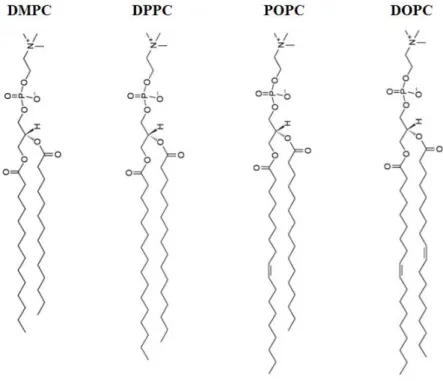
respectively). These are the so-called 1,2-diacylphosphoglycerides or phospholipids. The hydrocarbon chains vary widely in length, branching, and degree of unsaturation. Lipids containing double bonds in their methylenic chains are called unsaturated phospholipids, while the ones without double bonds are called saturated [1, 3]. The phosphate is usually linked to one functional group thus constituting the polar head group that categorizes such lipids into phosphatidylcholines (PC), phoshatidylethanolamines (PE), phosphatidylserines (PS), phosphatidylglycerol (PG) and phosphatidylinositol (PI). From these, PC are of particular interest as they are a major component in animal cell membranes. Many of these molecules have one saturated and one unsaturated chain (in animal cells, the unsaturated chain is usually esterified to the sn-2 position of glycerol). Natural occurring PC commonly display cis- double bonds [1, 8]. Figure 1.2 shows the structures of the PC used in this work: 1,2-dimyristoyl-sn-glycero-3-phosphocholine (DMPC); dipalmitoyl-sn-glycero-3-phosphocholine (DPPC); 1-palmitoyl-2-oleoyl-sn-glycero-3-dipalmitoyl-sn-glycero-3-phosphocholine (POPC); 1,2-dioleoyl-sn-glycero-3-phosphocholine (DOPC).
Figure 1.2 – Schematic representation of the structure of the phospholipids used in this work. For DMPC, the two sterified fatty acids are myristic acid (Abbreviated notation: 14:0). For DPPC, the two sterified fatty acids are palmitic acid (16:0). For POPC, the sn-1 position is sterified with palmitic acid and the sn-2 position with an oleic acid, with a cis-double bond in carbon 9 (16:0-18:1, Δ9-Cis). For DOPC, the two sterified fatty acids are oleic acid (18:1, Δ9-Cis).
I – INTRODUCTION
7
I – 1.1.2.2
SphingolipidsOne of the fundamental structures that are common to sphingolipids is the ceramide (N-acyl-sphingosine). The sphingolipids basically contain the same polar substituents as the glycerophospholipids, but they are ceramide-based. Due to their similarities regarding physicochemical properties, these two lipidic species are frequently grouped together. Sphingomyelin (SM) or ceramide 1-phosphorylcholine is one of the most important phosphosphingolipids (Figure 1.3). The terminal–OH group of ceramide is esterified with a choline group. It is widely found in animal cell membranes, generally in the outer leaflet of the plasma membrane [1, 2, 6].
Figure 1.3 – Schematic representation of PSM (N-palmitoyl-1-phosphorylcholine) (Abbreviatied notation: 16:0 SM) which is the predominant lipidic specie in the egg-SM used in this work.
I – 1.1.2.3
SterolsSterols are found in the composition of plant, animal and fungal membranes. Most have the same ring skeleton, but differ in their side chains, peripheral structure features,
I – INTRODUCTION
8
stereochemistry and number of double bonds in the ring system [6]. Cholesterol (CHOL) (Figure 1.4) is by far the most commonly found sterol in eukaryotic cell membranes.
Figure 1.4 – Schematic representation of the structure of CHOL.
It is present in animal cell plasma membranes (20 mol% - 50 mol% of its mass), lysosomes, endosomes, mitochondrial membranes (< 5 mol%), Golgi (≈ 8 mol%) and endoplasmatic reticulum (ER) (≈ 10 mol%) [9]
. Sterols are a major mean through which eukaryotic cells modulate and refine membrane properties [10]. Other important sterols are sitosterol, campesterol and stigmasterol, usually found in higher plants; ergosterol, an important component in fungal plasma membranes, and lanosterol, the sterol of some procaryotes [1, 3, 8].
I – 1.1.2.3.1
Cholesterol is a “special” lipidCHOL is a very important biomolecule with various important biological functions. It is involved in several physiological events, e.g. biogenesis, cell growth, steroid hormone and bile salt synthesis and embryonic development [10, 11, 12]. Further, CHOL acts as a precursor of the active form of vitamin D. Its synthesis is mainly confined to the ER and includes the presence of acetyl coenzyme A in a series of enzymatic steps (CHOL is indeed the regulator of some enzymes in its metabolic pathway) [2]. These are highly regulated as any deviation from its physiological concentrations may cause pathological situations. Elevated cholesterol levels have been observed in membranes of living cells and are associated with the formation of atherosclerotic plaques (in the form of CHOL monohydrate) [13], depletion of ER calcium supplies [14] and obstruction of the small bowel [15, 16]. The Alzheimer’s disease also seems to
I – INTRODUCTION
9
be related to the levels of CHOL and lipids as its distribution may be related to the formation of amyloid deposits [17].
The involvement of CHOL in membrane organization and structure characterizes it as a “membrane active sterol”. It has a major contribution in the control of membrane passive permeability to small polar molecules, by reducing average “fluidity” and free volume of the lipid bilayers. Plus, it has an important role on lateral organization, modulation of membrane thickness, enhancement of mechanical strength [18, 19] and free volume distribution (involved in controlling membrane protein activity and “raft” formation) [7, 10, 11]
. Basically, these effects in membrane structure and function are the result of the molecular structure and the interactions of this sterol with neighbor lipids (and proteins). As it is shown in Figure 1.4, CHOL is also an amphiphilic molecule, though it is not similar to a “regular” phospholipid. It is constituted by a small hydrophilic hydroxyl (–OH) group and a planar rigid hydrophobic structure (in a trans-configuration) with a short branched (isooctyl) chain segment. Its ring system has a smooth face (α-face), regular and tight, and a rough face (β-face) irregular and less tight due to the presence of the protruding methyl groups [9, 20, 21]. The smooth and rough sides of CHOL are indicated as responsible for the molecule tilt, when inserted in lipid bilayers [21]. This sterol seems to adjust its tilt angle for a better accommodation in the membrane: at low CHOL concentrations, the molecule has a large tilt, while at higher CHOL concentrations it has a smaller tilt [22, 23]. This was observed in the cases of saturated lipid bilayers and unsaturated lipid bilayers, though the molecular tilt appears to be lower in the latter case [23] [24]. This feature is believed to be essential for the ordering and condensing effect exerted by CHOL on neighbor lipids [25]. The general aspects of phospholipid-sterol interactions in lipid bilayers will be further described in Section I – 1.2.2.2.
I – 1.1.2.3.2
Cholesterol derivativesThe so-called CHOL derivatives may be a useful tool when it comes to understand certain lipid bilayer properties, especially by comparing its effects on membrane structure and function with the ones exerted by CHOL. In this work, the attention goes to 7-dehydrocholesterol (7DHC) and cholestanol (or dihydrocholesterol – DCHOL), which are represented in Figure 1.5 – A and B, respectively. Structurally, the main difference between these sterols is in the number of double bonds in the ring system: 7DHC has two double bonds, CHOL has one and DCHOL has none. 7DHC may be more rapidly oxidized than
I – INTRODUCTION
10
CHOL [26], due to its loss of planarity [27, 28], while DCHOL is similar to CHOL, in terms of planarity, condensation ability and molecular areas at air-water interface [28].
Figure 1.5 – Schematic representation of the structures of the CHOL derivatives used in this work: (A) DCHOL; (B) 7DHC.
7DHC is a biogenic intermediate in the biosynthesis of CHOL [28]. It is converted into CHOL via an enzymatic step performed by 7-dehydrocholesterol reductase (7DHCR). A defect in this enzyme leads to the accumulation of 7DHC in all tissues and this is associated with the Smith-Lemli-Opitz syndrome (SLOS) [26, 28, 29]. There are some indications that 7DHC may participate in raft formation [28, 30] and that its presence can affect the protein raft composition [31]. DCHOL also seems to promote the formation of membrane domains [28].
I – 1.1.3
How do lipids behave in aqueous solution?Above a critical concentration (referred in literature as “Critical Aggregation Concentration” – CAC, an extended notion of the classical “Critical Micelle Concentration” – CMC), lipids tend to form amphiphilic aggregates in order to shield the hydrocarbon chains of the amphiphiles from contact with water, while exposing their polar and/or charged groups to it. The hydrocarbon parts of these molecules do not interact favorably with water through dipole-dipole interactions and they are also not able to form hydrogen bonds. Due to this hydrophobic effect (liberated water molecules from the solvent cages formed around the hydrocarbon chains to stabilize them in a hydrogen-bond forming medium), the overall entropy of the amphiphile/water mixture is higher (the increase in entropy in aqueous solvent
I – INTRODUCTION
11
largely compensate the formation of amphiphilic aggregates), and the stability of the system is ensured (G < 0). So, the association of the hydrocarbon parts of the lipids will contribute to
minimize the total surface area in contact with water, while the polar domains interact with it or with other lipid head groups either through hydrogen bonding or ionic interactions, constituting an energetically stable structure in the aqueous environment [1].
I – 1.1.3.1
Assembly of lipid aggregatesThe lipid aggregation in the presence of water results in various lipid-water polymorphic arrangements for which the predominant lipidic form depends mainly on temperature, pressure, ionic strength and pH. Generally, they can be found as: micelles (which may assume different shapes); lamellar phases (extended two-dimensional sheets in the form of bilayers, at very high lipid concentrations); and, at very high temperatures, cubic phases (unilamellar structures with periodic three-dimensional order) or inverted hexagonal phases (mostly for PE) [1, 3].
From these, lamellar phases are of great interest, as the lipid bilayer is a self-aggregate “sheet” of amphiphiles (usually two molecules thick) in which the polar portions of the constituent molecules are exposed to water at the two surfaces and the apolar portions are excluded from water in the volume between these two surfaces [1, 2, 3, 8].
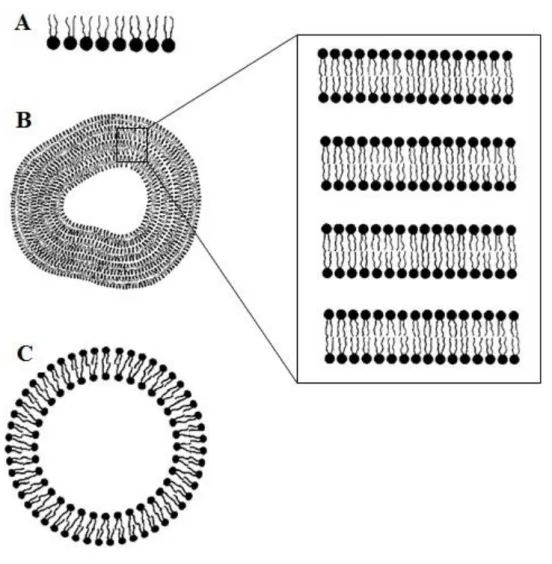
I – 1.1.3.2.1
Experimental model membrane systemsModel membrane systems are a useful tool to study the properties of pure lipids, lipid mixtures and reconstituted lipid-protein mixtures. These form spontaneously on hydration of the amphiphiles and are represented in Figure 1.6. Usually, they are studied as monolayers (using a Langmuir trough to accurately measure lipid properties like surface area and lateral pressure) or liposomes (lipid structures that usually enclose an aqueous volume). According to the already mentioned properties of lipids, when they disperse in water a heterogeneous mixture of vesicular structures is formed, with these containing concentric bilayers separated from the inside and the outside by a thin layer of water (approximately 10-20 Å) due to a strong force of repulsion (usually referred to as “hydration force”) [3, 6, 8]
. These are called multilamellar vesicles (MLV). They can be transformed into unilamellar vesicles of different diameters: small unilamellar vesicles (SUV) with about ≈ 20 nm of diameter; large
I – INTRODUCTION
12
unilamellar vesicles (LUV) with about ≈ 100 nm and giant unilamellar vesicles (GUV) in the order of 10-50 μm [1, 8].
Figure 1.6 – Schematic illustration of lipid molecular aggregates used as model membrane systems. (A) Monolayer; (B) Multilamellar vesicle or liposome (with the representation of multilamellar lipid bilayers in a
stack); (C) Unilamellar vesicle or liposome (adapted from reference [9]).
I – 1.2 Lipid bilayer physical properties
Lipid bilayers are condensed phases that with many characteristics of simple lipids though simultaneously structured. Their transbilayer profile was characterized by a number of techniques, e.g. X-ray and neutron scattering techniques, NMR (nuclear magnetic resonance),
I – INTRODUCTION
13
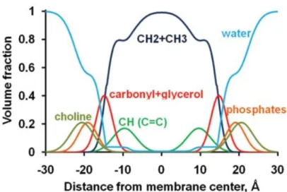
EPR (electron paramagnetic resonance) or ESR (electron spin resonance), molecular probing through optical spectroscopies and computer simulations (Figure 1.7). Roughly, lipid bilayers can be divided into four zones: (from the outside to the inside) a zone where “structured” water is deprived of forming all possible hydrogen bonds; a hydrophilic/hydrophobic region that includes the polar head groups and the upper segment of the hydrocarbon chains (where the membrane/water interface is found); a region of ordered fatty-acid segments; and a hydrophobic core with disordered methylenic chains segments [9].
Figure 1.7 – Volume fractions of lipid segments for a fluid DOPC bilayer (determined by X-ray and neutron scattering). The volume fractions of individual lipid components are represented as colored solid lines for alkyl groups ( , dark blue), double bonds (green), water (blue), carbonyls with glycerols (red), phosphates (orange), and choline (olive) groups (adapted from reference [32]).
Data from X-ray crystallography shows that lipid bilayers are closely related to liquid crystals, as the lipid molecules display a preferential orientation within the membrane [3]. The lipid polar head groups (in PC) are generally parallel to the plane of the bilayer, which seems to be the most stable arrangement as the positive and negative charges are located in a plane that is nearly parallel and electrostatically neutral [1, 6]. On the other hand, the acyl chains are usually aligned perpendicular to the membrane surface though they can have a tilt angle (the angle between the hydrocarbon chain axis and the bilayer normal). The glycerol backbone is the least flexible region of phospholipids. The acyl chains linked to the sn-1 position of
I – INTRODUCTION
14
glycerol have higher order parameters for carbons up to the middle of the chain. Then they rapidly decay toward the terminal methyl group (the region of high chain order is referred as the “order parameter plateau”) as there is the increase on the probability of carbon-carbon (C– C) trans-gauche isomerization. In contrast, chains linked to the sn-2 position have lower order parameters (due to differences in chain orientation near the glycerol) [6, 33]. The orientational and positional order of phospholipid acyl chains is the basis for the existence of a fluidity gradient [34].
Generally, the lipids that constitute lipid bilayers are polymorphic: they have different modes of hydrocarbon packing. These are mainly dependent on their chemical identity, degree of hydration, pressure, ionic strength, pH and temperature, which basically defines their final form [6]. The temperature dependent change in hydrocarbon chain order is called order or melting transition (generally addressed as a solid-to-liquid transition). These transitions occur over a very narrow temperature regime, as a consequence of the cooperativity between large clusters of n lipids (cooperative unit) that form under the influence of temperature [3]. In this particular case, we can observe different phases (i.e. fixed and well-defined physical states) [35]. In order to better understand this phenomenon, it is helpful to first consider a single lipid bilayer.
I – 1.2.1
Phase transitions in single lipid bilayersAt very low temperatures, phospholipid chains may be arranged in a rigid and highly ordered state (all-trans), easily comparable to a crystalline structure (the “sub-gel” or phase). As the temperature increases, the molecules will be arranged tightly in a two dimensional lattice in the membrane plane that corresponds to the gel phase ( ). These are phases of higher bilayer thickness due to the all-trans state of the methylenic chains (oriented perpendicular to the bilayer plane). When the temperature is raised, there is still in the existence of a two-dimensional ordered system, but the hydrocarbon chains are mostly disordered (increase in the gauche conformers) and the lattice order is lost (Figure 1.8). At this point, we are in the presence of a fluid phase, often called the liquid-crystalline phase ( ). This form is usually thought to represent the bulk of the lipids in the biological membrane [1, 3].
I – INTRODUCTION
15 Figure 1.8 – Representation of the melting transition from a solid (ordered) phase to a liquid (disordered) phase (from left to right). (A) Reduction of the hydrocarbon chain order upon increasing temperature. (B) Loss in lattice order in the polar head group region when the temperature is higher (adapted from reference [3]).
The temperature for which there is the transition from the gel to the liquid crystalline phase is called the main phase transition temperature ( ). Table 1.1 represents the for the lipids used in this work. This process is endothermic and is usually monitored through differential scanning calorimetry1 (DSC). It occurs exclusively in the plane of the membrane and is observed in all lipid bilayers regardless of the chemical identity of the lipids. At the , the lipid is partially in the gel state and partially in the liquid crystalline state. The temperature at which this phase transition occurs is mostly dependent on the chemical identity, length and degree of unsaturation of the fatty acyl chains, but the nature of the polar head group is also important. In this matter, the intermolecular forces (van der Waals interactions) play an significant role in contributing for the relative stability of the referred phases: longer chain lengths result in higher values and vice versa. The existence of cis-double bonds reduces the as this will disrupt the ability of the chains to interact optimally in the gel state [1, 3].
I – INTRODUCTION
16
Table 1.1 – Main phase transition temperatures ( ) for the phospholipids used in this work.
Phospholipid (ºC) POPC 20* POPC 2.6* DMPC 23* DPPC 41* PSM 41.3** * reference [36] ** reference [6]
It is also important to notice that phase transitions are accompanied by lateral expansion and consequent decrease in bilayer thickness, increase on the number of water molecules bound to the surface of the bilayer and nonmonotonic changes in bilayer volume. This free volume increases with the bilayer depth and is higher in the phase, due to the disorder of the hydrocarbon chains deep in the bilayer [1, 3, 6, 20].
Phospholipids with a large area requirement for the polar head group such as PC and PI, show a pretransition between the gel and liquid-crystalline states (Figure 1.9). This happens because the acyl chains are tilted with respect to the bilayer normal. This is why in PC, the gel phase is noted as . The “second” gel state is referred to as the ripple phase ( ), where the acyl chain order is lower than in the and phases. This is a consequence of the periodic one-dimensional ripples that were detected on the membrane surface (probably formed by periodic arrangements of linear gel and fluid domains in atomic microscopy studies). So, this can be seen as a partially melted lipid phase that forms prior to the melting transition. In these cases as the temperature increases we may be in the presence of a subtransition from the Lc to the phase, a pretransition from the to the and the main transition from the to the phase [1, 3].
I – INTRODUCTION
17 Figure 1.9 – Schematic representation of thermal phase behavior for an experimental system composed of phosphatidylcholines. (A) gel phase (B) ripple phase; (C) liquid crystalline phase (adapted from reference [3]).
Biological membranes seem to adapt their lipid composition such that the temperature distance between room temperature and melting transition is maintained, so they do not display phase transitions. However, the basic physical-chemistry of lipid mixtures is relevant to the understanding of biological membrane properties, especially when it regards possible lateral inhomogeneities [1, 3]. That explained some relevant lipidic mixtures will be considered.
I – 1.2.2
Lipidic mixtures and the observation of phase coexistenceI – 1.2.2.1
Phospholipid-phospholipid mixturesIn lipidic mixtures, the melting of a certain lipid is influenced by the melting behavior of neighboring lipids of different chemical nature. When the lipids randomly distribute in each of the lipid phases (i.e. exchanging lipids within the gel phase or the fluid phase will not change the free energy of the lipid matrix) one is in the presence of ideal mixing [3].
I – INTRODUCTION
18
Thermodynamic studies have clearly demonstrated that dissimilar phospholipids do not mix ideally, but many times an ideal mixing (based in the Regular Solution Theory2) is assumed in order to obtain a quantitative description of complicated experimental phase diagrams [3].
A phase diagram (exemplified in Figure 1.10) is a graphic representation of conditions at which thermodynamically distinct phases can occur. They may be calculated (theoretical) or experimentally obtained through DSC or spectroscopic techniques. Nonideal mixing of lipids is often identified by comparing the experimental phase diagram with the one predicted theoretically [1, 3]. Disaturated PC that differ only in the length of two methylenic groups in their acyl chains exhibit an almost ideal behavior in phospholipid-water dispersions, e.g. DMPC/DPPC binary mixtures [37, 38]. The phase diagram for this mixture is represented in Figure 1.10 – A, along with the one for another binary mixture used in this work, the egg-SM/DOPC mixture (Figure 1.10 – B). The latter shows an overall behavior similar to the DMPC/DPPC mixture, though with some differences.
Figure 1.10 – Phase diagrams for phospholipid-phospholipid mixtures used in this work. (A) Representation of a DMPC/DPPC phase diagram (adapted from reference [37]) where the behavior of the mixture is close to ideal; (B) Representation of a PSM/DOPC phase diagram (adapted from reference [39]), for which the overall behavior is similar to the one observed for the DMPC/DPPC mixture.
2 The components in each phase are assumed to mix randomly as in an ideal solution theory, with ΔS = 0, though neighboring lipids still contribute to the enthalpy (ΔH) of the system [3].
I – INTRODUCTION
19
So, in the gel phase, the packing requirements may prevent two lipids from being miscible, resulting in clustering or lateral phase separations, and even in the liquid crystalline phase, the two lipids may be miscible and still behave nonideally [1]. The differences in the head group constitution and mainly in chain length, may lead to lipid preferences in the nearest neighbors both in gel and liquid crystalline phases [8].
I – 1.2.2.2
Phospholipid-sterol binary mixturesCHOL has an amphiphilic structure, but it does not form bilayers of its own (it forms crystals) [3]. The miscibility of CHOL in the lipid bilayer depends both on structure of the phospholipid polar head groups and hydrocarbon chains, on temperature and phase state of the bilayers [20], but it can go as high as ≈ 67 mol % (different experimental conditions from those used in this work) [40]. When inserted in a lipid bilayer, the –OH group of CHOL is near the ester carbonyls of the phospholipids and its long axis is oriented parallel to the bilayer normal. The position of the sterol relative to the bilayer interface is mainly determined by a hydrophobic/hydrophilic balance [20, 41]. At the lipid/water interface, CHOL can lead to an even higher orientational polarization3 of the water molecules when compared to pure phospholipid bilayers [42]. On the other hand, CHOL can penetrate deep into the hydrophobic interior, which depth will depend on the sterol content of the bilayer [42]. In some cases, it can also protrude into the opposite monolayer and even cross the membrane by (passive) diffusion across the bilayer, unspecific diffusion in the presence of proteins or at the boundary of membrane domains, or through active protein-mediated transport [11]. CHOL has a highly dynamic motion parallel to the bilayer normal and displays a very rapid transbilayer motion (flip-flop) rate in liposomes [11].
Phospholipid-sterol interactions are complex. In a simple description, CHOL interacts with the phospholipids in different ways: in the polar head group zone, the –OH group of this sterol interacts mainly with the phosphate (–PO) and carbonyl (–CO) groups, participating in hydrogen bonding (it can either be a donor and an acceptor) and also in charge pairing (electrostatic interactions between the partial positive charged choline nitrogen moiety and the negatively charged CHOL oxygen) [7, 21, 24]; at the hydrocarbon region, CHOL interacts with the methylenic chains of the phospholipids mainly through van der Waals interactions and short range electrostatic effects [20].
3 Water molecules at air/water surface are highly polarized. This orientational polarization can occur in substances composed of molecules that have permanent electric dipoles. The alignment of the dipoles is temperature dependent and leads to an orientational polarizability [43], see Section I – 2.1.
I – INTRODUCTION
20
This is a consequence of the amphiphilic nature of both molecules: this way, the –OH group of CHOL and the polar head groups are in contact with water and the hydrocarbon chains and the steroid ring system are “shielded” from this contact. However, when considering phospholipid-sterol interactions the nature of CHOL as bulky rigid molecule inserted into a lipid bilayer (containing flexible phospholipid molecules) cannot be overlooked.
At this point, it is interesting to consider the phase diagram for the DPPC/CHOL mixture (Figure 1.11). This is one of the most studied and well-known phospholipid-sterol mixtures [44, 45, 46, 47]
. In the specific case of PC, the electrostatic interactions between the head groups are weak and there are only hydrogen bonding acceptor groups. The interaction with CHOL may lead to the increase of the distance between the phospholipid head groups and a consequent decrease in the electrostatic attractions or a reduction of its potential to hydrogen bond [20]. At lower CHOL (and higher temperatures) this may not be evident: the phospholipid chains are still “disordered” (translational disorder, rapid lateral diffusion, substantial degree of chain conformational disorder) as in the case of a pure phospholipid bilayer [19]. So, this is many times described as a liquid disordered phase ( ). But, as the sterol content in the mixture is gradually raised, there will be a substantial effect on the order parameters measured along the lipid hydrocarbon chain. Generally, there is an enhancement of the lipid packing increasing the molecular order of the lipid chains and a reduction of the surface area per molecule occupied by phospholipids at the air/water interface [6].
I – INTRODUCTION
21
This condensing effect is the result of CHOL steroid ring interactions with the acyl chains that force them to assume a more ordered conformation and is stronger in the case of saturated phospholipids. As a consequence, there is an increase of the conformational order and a decrease in the translational diffusion (2-3 fold up to about 10 fold) [8, 48]. These conformational constraints give rise to the formation of a fluid phase distinct from the and phases, the so-called liquid-ordered phase ( ). This is usually seen as a state of “intermediate fluidity” between the familiar gel and fluid phases formed by pure glycerophospholipids and sphingophospholipids. In the phase, the molecules can exhibit a degree of translational freedom (in a simple way viewed as lateral mobility) and translational diffusion similar to the one for the conventional fluid bilayer state, while at the same time, the configurational freedom (order) of the lipid hydrocarbon chains more closely resembles the one observed for the gel state [49, 50].
This said it is worth considering three important models that illustrate the interaction between CHOL and phospholipids: the condensed complex model, the superlattice model and the umbrella model. The condensed complex model [51, 52, 53] was proposed to explain the phospholipid-CHOL mixture properties that become highly nonideal at higher CHOL content. The phospholipid-CHOL interactions are treated as reversible chemical reactions at chemical equilibrium and it assumes that there is the formation of lipid-sterol complexes with defined stoichiometry to account for deviations from the regular solution thermodynamics. The “condensed” term indicates that complexes are formed as a consequence of the condensing effect of CHOL: the average area per phospholipid in the complex is less than would be expected for ideal mixing and the methylenic chains are in the all-trans configuration, closely packed, making this complexes, a thicker “region” in the lipid bilayer. The superlattice model [54]
affirms the existence of regularly distributed lattices (within the matrix lattice formed by membrane acyl chains and CHOL molecules) and irregularly distributed lattices that coexist in fluid sterol-containing membranes. This model states the existence of critical CHOL concentrations (20, 22.2, 25, 33.3, 40 and 50 mol %). In regular solutions, this sterol is distributed into either hexagonal or centered rectangular superlattices, whose shape and size fluctuates with time. In this case, the long range repulsive forces between the bulky steroid rings and the short range interaction between sterols and the neighboring hydrocarbon chains are invoked as being crucial for superlattice formation. The umbrella model [55] asserts that the polar head group must help to cover the nonpolar body of CHOL to avoid the unfavorable free energy that arises from the sterol’s exposition to water. The lipid head groups reorient
I – INTRODUCTION
22
and expand at the bilayer aqueous interface in order to cover CHOL (as its concentration is raised and as long as the polar head groups are capable of “shielding” the interaction of water with the steroid ring of CHOL) [48].
There was another saturated phospholipid-CHOL mixture used in this work, the PSM/CHOL mixture (for which the phase diagram will be presented in Section I – 1.2.2.3). It is interesting to notice that DPPC and SM (Figure 1.2 and Figure 1.3, respectively) display structural similarities like the identical zwitterionic hydrophilic head group, the existence of an interface section and two methylenic chains which form the hydrocarbon core. The main difference between these two species is the fact that the SM head group and interface section has hydrogen bond donor and acceptor groups (while PC only have acceptor groups), and only one variable alkyl chain [56, 57]. Though, it appears that the charge pairing interactions may be more frequent between SM and CHOL than the conventional hydrogen bonds [21, 24]. This mixture has not been extensively studied as the DPPC/CHOL mixture but its phase diagram also illustrates the existence of the already mentioned and phases as well as a large liquid-liquid coexistence regime between these two phases ranging (roughly) from 15 to about 35 mol % of CHOL, above the . [1, 3].
I – 1.2.2.2.1
Phase coexistenceThe formalisms applied for the construction of phase diagrams for mixtures of homogeneous solvents are also applied in the case of lipid mixtures. The phase coexistence is the result of lateral phase separation that seems to occur under certain conditions. In these cases, it is usual to consider one of the phases as physically continuous (or percolative) and the other as physically discontinuous or dispersed as isolated domains. An interconversion between these phases may occur as a result of changes in physicochemical properties of the bilayer (as lateral pressure, temperature, chemical composition). By crossing the critical mass ratio of phases (the percolative threshold) previously disconnected domains and their constituents can be connected as well as other can be disconnected. Systems with phase coexistence may experience interfacial or surface tension (line tension, in two-dimensional systems) between phases that drives the system toward a minimization of the free energy at the interface. Line tension (macroscopically) is the result from both hydrophobic and chain ordering mismatch at the boundary between domains [8]. Hybrid lipids are lipids that can lower this tension to zero and still have minor effects on the thermodynamics (phase diagram,
I – INTRODUCTION
23
critical temperature, etc.) of the system. This designation comes from the fact that they have a fully saturated chain and a partially unsaturated one. They are soluble in the equilibrium phases, but also interfacially active (e.g. when added to a typical saturated/unsaturated/CHOL system, they adsorb to the interface between two coexisting bulk and phases phases) [58]. Their molecular orientation is energetically favorable (e.g. POPC) [59]. Tension plays an important role in the stability of the membranes (not just tension as local pressure, but the whole distribution of local pressure determines the functioning of membranes, including functioning of proteins) [60]. A strong surface tension may lead to the separation of phases into macroscopic domains or rafts [8] (Section I – 1.3.2). In homogeneous solvents, the dielectric constant plays an important role in the miscibility, so it is possible that it will also be relevant in the case of lipidic mixtures and lipid organization into domains [61] (further considerations in this matter in Section I -2.1). Now, one has the tools to proceed to a more complex system.
I – 1.2.2.3
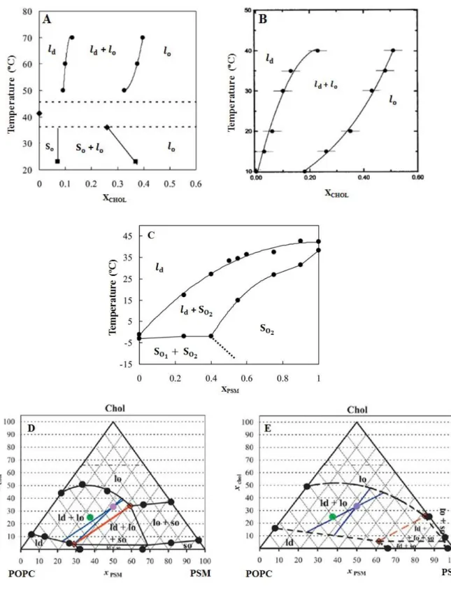
Ternary mixturesIn the recent decade, ternary mixtures of PC, SM and CHOL (three major components of the exoplasmatic leaflet of the mammalian plasma membranes) have often been investigated. Based in the studies from PSM/CHOL, POPC/CHOL and PSM/POPC (Figure 1.12 – A, B and C, respectively), the first diagram to be published for a ternary mixture (POPC/ PSM/CHOL) was the one from de Almeida et al. [62] represented in Figure 1.12 – D and E. These diagrams have a triangular representation and the compositions in mole fractions/molar proportions are given by the set of points contained within an equilateral triangle of unit side [3]
.
It is interesting to notice that CHOL seems to interact differently with saturated and unsaturated lipids [21, 24], as fully saturated PC and SM are known to have the strongest interactions with this sterol [9, 63] and seem to have a preference for its smooth side [24]. Indeed, there are experimental evidences that indicate that CHOL partitions with roughly two-fold greater affinity into vesicles prepared from saturated PC or SM than into vesicles prepared from unsaturated PC [50]. Even between saturated phospholipids, this sterol shows preferential association. It is thought to interact more strongly with sphingolipids (mainly due to the ability of charge pairing and hydrogen bonding between CHOL hydroxyl group and the neighboring SM molecules), triggering lateral separation of lipids into and domains which have been extensively characterized in model membranes [11, 64, 65, 66].
I – INTRODUCTION
24
Figure 1.12 – Phase diagrams for phospholipid-cholesterol binary and ternary mixtures used in this work. (A) PSM/CHOL (adapted from reference [62]); (B) POPC/CHOL (adapted from reference [71]); (C) PSM/POPC, is POPC-rich and is PSM-rich (adapted from reference [62]); (D) POPC/PSM/CHOL at 23 ºC (adapted from reference [62]) (E) POPC/PSM/CHOL at 37 ºC (adapted from reference [62]).
I – INTRODUCTION
25
So, phase separation can also be observed in ternary mixtures containing one low phospholipid (e.g. POPC, DOPC), CHOL and a high phospholipid (e.g. SM, DPPC) [3].
While the condensed complex model points toward that fact that these mixtures, under certain conditions, will be composed fundamentally by a SM/CHOL complex ( ) and POPC ( ) [67], other studies indicate there may be the existence of domains enriched in unsaturated lipids with low CHOL content ( ) coexisting with other domains with large amounts of saturated lipids and CHOL ( ) [68, 69, 70].
The POPC/PSM/CHOL mixture is considered the canonical raft mixture [72], as it seems to represent the essential characteristics of the lipidic components of membranes that contain rafts, as these and phases seem to coexist at concentrations that mimic the composition of the outer leaflet of the mammalian plasmatic membrane [65, 73, 74]. In compositional terms, it may be rather simple when compared to the complexity of the plasma membrane, though it is a good starting point to understand the properties for the mixing of more than two different lipids [65].
I – 1.3
Lateral and transversal asymmetry in biological membranes
Membrane lipids exhibit relatively rapid transbilayer motion (flip-flop), which is usually negligible due to the half times on the order of several days or longer. The lipid biosynthesis in the ER and Golgi complex, relatively slow membrane translocation of lipids, asymmetrical chemical composition of aqueous compartments, spontaneous curvature or the existence of “flippases” may contribute to membrane transversal asymmetry. An example of transverse lipid asymmetry is the human erythrocyte, with PC and SM on the outer surface and PE and PS in the inner half of the membrane. Membranes also exhibit lateral asymmetry (or lateral heterogeneity), as there are domains or regions within some membranes which have distinct compositions and which may separate from other portions of the membrane with respect to the diffusional exchange of components [1].
I – 1.3.1
Membrane proteinsMembranes contain between 20% and 80% (w/w) protein. These are the biochemical active membrane components thorough the form of enzymes, transporters, receptors, pores,
etc., which distinguishes each particular membrane. Nowadays, membrane proteins are


![Figure 1.11 – Phase diagram for the DPPC/CHOL mixtures (adapted from reference [45]).](https://thumb-eu.123doks.com/thumbv2/123dok_br/18808518.926445/40.892.262.540.773.1098/figure-phase-diagram-dppc-chol-mixtures-adapted-reference.webp)