UNIVERSIDADE DE LISBOA
FACULDADE DE MOTRICIDADE HUMANA
Association between newly developed body
indices with total and trunk fat mass in
older adults
Dissertação elaborada com vista à obtenção do Grau de Mestre em
Exercício e Saúde
Orientadora: Professora Doutora Diana de Aguiar Pereira dos Santos
Júris: Doutora Analiza Mónica Lopes Almeida da Silva, professora
auxiliar com agregação à Faculdade de Motricidade Humana da
Universidade de Lisboa
Doutora Maria Filomena Soares Vieira, professora auxiliar com agregação
à Faculdade de Motricidade Humana da Universidade de Lisboa
Doutora Diana de Aguiar Pereira dos Santos, Investigadora do Centro
Interdisciplinar do Estudo da Performance Humana da Faculdade de
Motricidade Humana da Universidade de Lisboa
Nuno Pedro Caldas Soares de Sousa Capilé
Lisboa, 2018
I
Acknowledgements/Agradecimentos
Este trabalho culmina num grande esforço pessoal, que não teria sido possível sem a ajuda de todas as pessoas em que nele participaram direta ou indiretamente. Devido a isso gostaria de aqui a agradecer, de uma maneira geral, a todos os eles.
Em primeiro lugar, à minha orientadora, a Professora Doutora Diana Santos, por toda a sua experiência, orientação, conhecimento transmitidos e a ajuda que me prestou, sem dúvida que foi um grande pilar para mim nesta investigação. Toda a paciência que teve para as minhas hesitações, dando-me sempre um caminho de modo a que pudesse conduzir esta investigação no sentido certo. Muito obrigado!
A todos os meus colegas de faculdade que comigo percorreram este longo caminho, os de Coimbra e os de Lisboa, como o David, o André, o Girão, o Bota, a Joana, o Chico, o Costa, o Bettencourt, o Rei, a Dani, o Rafa e de Lisboa o João, finalizando com um agradecimento especial à Susana e Juliana que tiveram comigo em ambas as cidades, e que de um modo ou do outro acabaram por me acompanhar e sofrer mais comigo nesta caminhada.
Aos meus amigos de infância que estão comigo desde sempre, um grande obrigado por todo o companheirismo e amizade que me deram durante este percurso e não só. Frederico, Rui Costa, Gonçalo, Tiago Chaves, Sara, Bruno, Ricardo Russo, Cajó e com um agradecimento especial ao Tiago Couto que para além de tudo o resto me deu um grande apoio na realização desde trabalho.
A toda a minha família que sempre me apoiou apesar de tudo e nunca me deixou desistir quando tudo parecia mais difícil. Por todo o carinho e ajuda, um muito obrigado! Um agradecimento ao meu tio e tia que me deram um grande suporte quando me mudei para Lisboa, sem esquecer a ajuda do meu irmão Bernardo. Ao meu outro irmão, o Gonçalo, por todo o apoio e oportunidades que me criou e um agradecimento especial ao meu Pai que sempre acreditou em mim e sempre me apoiou em todo o meu percurso académico.
Para finalizar, mas sem dúvida não menos importante, à minha melhor amiga, companheira, “mãe” e namorada, Adriana Sales, sem dúvida outro grande pilar em todo o meu percurso académico, onde sempre me ajudou e tentou que eu fosse pelo melhor caminho e por todo o amor que me transmitiu. Um muito obrigado a ela e à sua família.
Sem nunca esquecer a estrela mais brilhante e o meu anjo da guarda, a minha MÃE que apesar de não estar fisicamente, é a minha inspiração, quem me fez trilhar este caminho e me ensinou que tudo é possível, só temos de acreditar! Muito obrigado por toda a educação que me deste.
II
Resumo
Objetivo: Nos últimos anos foram desenvolvidos novos índices corporais, como o índice da forma do corpo (ABSI), o índice de adiposidade corporal (BAI), o índice de arredondamento do corpo (BRI) e o rácio cintura para estatura (WHtR), com vista a ter indicadores de gordura corporal melhores que o índice de massa corporal (IMC). Deste modo, a presente investigação tem como objetivo analisar a relação entre os vários índices corporais e a massa gorda (MG) total e do tronco em pessoas idosas.
Métodos: Neste estudo observacional transversal, foram avaliadas 129 pessoas (87
mulheres e 42 homens) com idades entre 60 e 84, através da densitometria radiológica de dupla energia, para obter as medidas de MG, e de medições antropométricas, para posterior cálculo dos índices. Posteriormente realizaram-se análises de correlação entre a %MG total e do tronco e os índices corporais. Foram efetuadas regressões lineares, simples e múltiplas para verificar a associação entre os índices e a percentagem de MG total. A analise de dados foi feita usando o IBM SPSS Statistics versão 25.0.
Resultados: Nas mulheres o ABSI não se associou nem com a MG total nem com a do
tronco. Relativamente aos restantes índices todos estavam associados com a adiposidade avaliada pela DXA (total: r>0.649; tronco r>0.520). O índice com um maior poder explicativo foi o IMC (r=0.798) para a MG total e o WHtR (r=0.783) para a MG do tronco. Nos homens todos os índices estavam associados quer com a MG total (r>0.427) quer com a MG do tronco (r>0.463). Quer para a MG total (r=0.742) quer para a do tronco (R=0.713) o WHtR apresentou maior explicação da MG do DXA. É contudo de realçar que não existiram diferenças de relevo entre os índices e que as associações se mantinham semelhantes quando ajustadas para o índice de massa isenta de gordura apendicular.
Conclusões: Apesar das limitações reportadas na literatura o IMC apresenta associações
semelhantes, ou até melhores, que novos índices corporais para estimar a adiposidade total e do tronco em pessoas idosas.
Palavras-chave: ABSI; BAI; BRI; IMC; WHtR; pessoas idosas; DXA; Massa gorda;
III
Abstract
Objective: In recent years, new body indexes have been developed, such as Body
Shape Index (ABSI), Body Adiposity Index (BAI), Body Roundness Index (BRI) and Waist-to-Height Ratio (WHtR), in order to have better body fat indexes than the Body Mass Index (BMI). Thus, the present research aims to analyze the relationship between the various body indexes and the total fat mass (FM) and FM of the trunk in the elderly.
Methods: In this cross-sectional observational study, 129 people (87 women and 42
men) aged 60 to 84 were evaluated through dual energy radiological densitometry, to obtain the FM measurements, and anthropometric measurements, for subsequent calculation of the indexes. Subsequently, correlation analysis were performed between the total percentage of FM and the total percentage of fat mass of the trunk and the body indexes. Linear, simple and multiple regressions were performed to verify the association between the indexes and the percentage of total FM. Data analysis was performed using the IBM SPSS Statistics version 25.0.
Results: In women ABSI was not associated with either total or trunk FM. Regarding
the remaining indexes, all of the indexes were associated with the adiposity assessed by DXA (total: r> 0.649; trunk r> 0.520). The index with a greater explanatory power was the BMI (r = 0.798) for the total FM and the WHtR (r = 0.783) for the FM of the trunk. In men, all indexes were associated with either the total FM (r> 0.427) or the FM of the trunk (r> 0.463). For the total FM (r = 0.742) and for the trunk FM (R = 0.713) the WHtR presented a greater explanation of the fat mass of DXA. It should be noted, however, that there were no significant differences between the indexes and that the associations remained similar when adjusted for the appendicular fat-free mass index.
Conclusions: BMI, despite the limitations that the index demonstrates, presents similar
or even better associations than new body indexes to estimate total and trunk adiposity in the elderly.
IV
Index
Acknowledgements/Agradecimentos ... I Resumo ... II Abstract ... III List of Tables ... V List of Figures ... V Abbreviations ... VI Introduction ... 1 Literature Review ... 3Body composition assessment in older adults ... 7
New body indices ... 9
Dual-Energy X-Ray Absorptiometry ... 14
Relevance of the study and objectives ... 16
Methodology ... 17
Type of Study ... 17
Study Conception and Design ... 17
Participants ... 17
Research Inclusion Criteria ... 18
Research Exclusion Criteria ... 18
Data Collection Instruments and Procedures ... 18
Preparation ... 18
Anthropometric data ... 18
Dual-energy X-ray absorptiometry ... 19
Statistical Analysis ... 21
Results ... 22
Discussion ... 29
Body Adiposity Index (BAI) ... 30
A body shape index (ABSI) ... 31
Waist-to-height ratio (WHtR) ... 31
Comparison between indexes ... 32
Conclusion... 33
Future research ... 33
V
List of Tables
Table 1: Descriptive characteristics of the main outcome variables ... 22
Table 2: Predictive Power of different indices in explaining total DXA fat to females. ... 27
Table 3: Predictive power of different indices in explaining total fat mass in DXA ... 28
List of Figures
Figure 1: Association between %FM and body indices, for females ... 23Figure 2: Association between %Trunk fat mass and body indices, for females ... 24
Figure 3: Association between %FM and body indices, for males ... 25
VI
Abbreviations
4C – four compartments ABSI – a body shape indexACSM – American College of Sports Medicine ALST – appendicular lean soft tissue
BAI – body adiposity index BF – body fat
BM - body mass
BMC – bone mineral content BMD – bone mineral density BMI – body mass index BRI – body roundness index
DXA – Dual Energy X-ray Absorptiometry
EWGSOP – European Working Group on Sarcopenia in Elderly People FFM – fat-free mass
FM – fat mass
GH - growth hormone HP - hip circumference
IGF - Insulin-like growth factor IL - interleukin
INE – Portuguese National Statistics
IWGS – International Working Group on Sarcopenia LM – lean mass
MM – muscle mass
TNF-α – tumor necrosis factor alpha VAP – visceral adipose tissue
VII
WC – waist circumference
WGO – World Gastroenterology Organisation WHO – World Human Organization
1
Introduction
The present dissertation is made up of six parts, with a last section for the bibliographical references that supported this investigation. In the first part, introduction, the course of the study is described. We next have the Literature Review, which describes the context and the need to study these new body indexes, this part also describes the "gold standard" method for body composition and the aging process. This chapter justifies the need to find new and more practical methodologies that will allow an approximation of body composition, and more specifically fat mass (FM) in the field. Next in the document the objectives and relevance of the research are listed followed by the applied methodology in which the selection of the sample, the instruments and procedures applied, as well as the main variables of interest in the study are explained. Further on, the results obtained are described. After this a discussion of the results is presented, where the difference between indexes and populations is perceived. The last part of the present dissertation describes the main conclusions and recommendations for future research. At the end of the entire description of the work, all the bibliographical references used in the course of this work are stated.
Aging is something common to all humans, and the increase in the elderly population is nowadays a reality on a world scale, with Portugal being no exception to this reality. According to the Portuguese National Statistics Institute (INE), out of a total of 10.562.178 Portuguese, 2.010.064 are people aged 65 and over and only 1.572.329 of the total population have between 0 and 14 years of age (INE, 2018b). The average life expectancy in Portugal is 80.6 years, with 77.6 years and 83.3 years for men and women, respectively, comparing to an average life expectancy of 67.1 years in 1960 (INE, 2017), being that the current population aging rate is registered at 128% in Portugal, which means that for every 100 children/youngsters (0-15 years old) the country has 128 elderly people (≥ 65 years); in 1960 this same index was established at 27 years (INE, 2018a). Analysing this data, it is easy to understand that the trend over the years is to have an increase in the average life expectancy and consequently an increase in the number of elderly people in Portugal. Due to the growth of this age group, and considering the deleterious effect of excess adiposity in numerous chronic diseases, it is urgent to develop new tools that can be used in routine assessment of older adults, that is at the same accurate, easy to use on the field, and low cost.
2
It is known that when it is desired to assess, in a fast and inexpensive manner, the FM, the body mass index (BMI) is the most used indicator, however, this index has some limitations (Garn, Leonard, & Hawthorne, 1986). Thus, in recent years, new indices have been developed to predict adiposity, such as a body shape index (ABSI) (Krakauer & Krakauer, 2012); the body adiposity index (BAI) (Bergman et al., 2011); the body roundness index (BRI) (Thomas et al., 2013) and the waist-to-height ratio (WHtR) (Hsieh, Yoshinaga, & Muto, 2003) which could be a great help in better understanding and evaluating body composition in older ages. It was decided that it could be useful to study these indices in this population in order to perceive their interaction and what would be the most accurate field index to give an indicator of the amount of FM and its distribution in the trunk region.
3
Literature Review
The world population is rapidly aging, studies point to an increase in the number of people aged 60 years and older from 11% in 2006 to 22% in 2050. This means that, due to this duplication, for the first time in human history, there will be more elderly population than children (0-14 years) in the total of world population (WHO, 2008). Older adults reflect the highest population growth segment in the most industrialized countries, including Portugal (Marques et al., 2014). The percentages of Portuguese adults with more than 65 years is increasing every year and is expected to increase by 33% more until 2050 (Hooyman & Kiyak, 2011). Besides the increased costs associated with this growth, Portugal, alongside with Spain and Italy, should have the highest rates of old-age dependency in relation to the other members of the European Union (Muenz, 2007). With the projected increase of older adults in most part of the world, it is critical for economic and personal reasons, that this segment of the population remains healthy and independent for as long as possible (Rikli & Jones, 2013).
However, with aging there are physiological changes that may interfere with the health status of each person. For example, the progressive increase of fat mass (FM), the reduction of lean mass (LM), bone mass (BM),or the reduction of the amount of minerals and the proportion between intra and extracellular water are some of the observed phenomes (Barbosa, Santarem, Jacob Filho, Meirelles, & Marucci, 2001).
The physiological changes related to age affect a wide range of tissues, organ systems, and functions that together may alter the preservation of physical independence (American College of Sports et al., 2009), resulting in a reduction in the quality of life, life expectancy, and high costs, related to health in the long term (Cruz-Jentoft et al., 2010).
Regarding body composition, first of all, we must keep in mind that it can be organized according to a model consisting of five levels of increasing complexity: Atomic (first level); Molecular (second level); Cellular (third level); Tissue System (fourth level); and Whole Body (fifth level) (Wang, Pierson, & Heymsfield, 1992). The fifth level characterizes body size and shape, often described by anthropometric measures such as body weight, skinfolds, circumferences, and body mass index (BMI) while the molecular level consists of six main components: water, lipids, proteins, carbohydrates, bone minerals and soft tissue minerals (Wang, et al., 1992).
4
The evaluation of body composition in older adults becomes even more important, since it allows us to understand the body and identify health risks associated with the accumulation of FM, since it has already been proven that excess body fat triggers negative effects in the development of cardiovascular diseases, bone diseases, obesity, cancers among others (Pi-Sunyer, 2009). Accordingly, assessing body composition in the elderly is crucial so that appropriate objectives can be prescribed for nutritional and/or exercise plans directed and specialized to the body needs of this group. It is possible to monitor the changes in the body composition during the disease as well as the effects of pharmacological interventions and understand the aging process with the same values in repeated measures (Aniteli, Florindo, Pereira, & Martini, 2006).
We can say that the greatest problems associated with age in most people, coupled with what was already mentioned, are physical inactivity and malnutrition, leading to two major problems of the elderly today, obesity and sarcopenia (Stoever, Heber, Eichberg, & Brixius, 2017), a term that was first proposed by Rosenberg, who described it as the loss of muscle mass age-related (Rosenberg, 1989), even though until 2009 there were no clinically accepted definition nor diagnostic criteria or treatment guidelines. The European Working Group on Sarcopenia in Elderly People (EWGSOP) (Cruz-Jentoft, et al., 2010), defined that the gradual and generalized loss of muscle mass and low muscle strength determined what was Sarcopenia (Cruz-Jentoft, et al., 2010; Cruz-Jentoft et al., 2014; Fielding et al., 2011). However, the definition of sarcopenia of EWGSOP (Cruz-Jentoft, et al., 2010), involves not only muscle mass and strength but also physical performance. On the one hand, this increases complexity, but, on the other hand, it allows the consideration of several parameters that may be important for the development of sarcopenia.
Later the International Working Group on Sarcopenia (IWGS) (Cruz-Jentoft, et al., 2010) defined that an elderly person is only considered sarcopenic if he has low muscle mass together with a slow running speed (Fielding, et al., 2011). This makes it difficult to find a general and common definition of Sarcopenia, although this problematic situation has already been discussed in several previous studies (Beaudart et al., 2015; Cooper et al., 2012; Cruz-Jentoft, et al., 2014; Volpato et al., 2014).
This pathology, which generates an age-related loss of muscle mass and strength, may arise from multiple factors that may be adjacent to the Central Nervous
5
System and result in a reduction in the number of alpha motor units in the spinal cord; it’s origin may be related to the muscular system and from a loss of muscle strength, decrease in the muscle area, decreased percentage of muscle fibers of both types and a decrease in muscle cells (aptose); or it can come from hormonal factors, resulting from insulin resistance, decreased levels of anabolic hormones, decreased GH and IGF-1, testosterone and estrogen (decreased muscle mass and strength), increased production of proinflammatory cytokines (IL-6, TNF-α and IL-1) that stimulate the loss of amino acids in muscle and slower muscle contraction; another origin may be due to nutritional factors, derived from the decline of more than 15% of basal metabolic expenditure (due to the reduction of lean tissue), reduction of food intake, malnutrition of nutrients, reduction of protein intake, carbohydrate-rich diet and tendency to consume sugar as well as vitamin D deficiency. The last source of this pathology may come from lifestyle factors, a consequence of sedentarism and physical inactivity, or even pharmacological treatments derived from other chronic diseases that can lead to very serious consequences, both for the person with the syndrome and for the country where this person lives, which may lead to morbidity, a high level of functional disability associated with a poor quality of life and a higher level of dependence on others, high costs for health care, greater probability of falls and fractures and more serious mortality. When added to all these factors is associated the increase in FM, this is a phenomenon of Sarcopenic Obesity. According to Baumgartner (2000) conclusions, about 15% of people with sarcopenia also suffer from obesity.
According to the WHO (2017), obesity is defined as an abnormal or excessive accumulation of body fat. This accumulation happens when the energy ingested is greater than the energy expended, this excess accumulates in the body in the form of fat. Obesity is classified as a chronic disease, of multifactorial origin and is associated with chronic-degenerative diseases, usually defined as having a BMI ⩾ 30 kg / m2. There are two ways to characterize obesity: android obesity, a characteristic associated with man, (trunk and abdomen) where it carries greater coronary risk, hypertension, stroke and diabetes. And gynoid obesity, which is associated with the woman (lower limbs - pelvic and thigh), where there is a lower coronary risk, hypertension, stroke and diabetes (Wallace & Ray, 2003; Wannamethee & Atkins, 2015; WHO, 2017).
6
This is a major public health problem, well recognized as a risk factor for cardiovascular morbidity and mortality in adult populations. The prevalence of obesity in the elderly continues to increase worldwide and has doubled since 1980. Data from (WHO, 2017) report that 2.8 million people worldwide die every year as a result of being overweight.
This is a complex multifaceted disease that involves, among other factors, hypothalamic, endocrine and genetic disorders, but also, the environment and the behaviour of the individual. A common misconception is that the main cause of obesity is overeating (Bayles & Ray, 2003). According to the ACSM (Wallace & Ray, 2003), the major part of evidence suggests that the energy imbalance resulting from the total consumption of excess calories, a sedentary lifestyle or a combination of this two factors is the main cause of obesity.
The major difference between the identification, definition and classification of obesity and overweight is the assessment of body fat. Besides the percentage of total body fat, the factors to be considered in determining the degree to which obesity impairs health are the location of the fat deposits and other comorbidities. The ideal scenario would be that obesity should be classified based on the assessment of body composition (in phenotype, fat cell morphology and general health), although this rarely occurs, especially in large population samples.
There are many confounding factors that contribute to underestimating the risks of obesity in the health of the elderly. These include the effect of survival, competing mortalities, relatively short life expectancy in old age, the importance of the beginning age and duration of obesity, smoking and constant weight change and involuntary weight loss (WGO, 2011).
However, there are many medical complications related to obesity in the elderly and these are mainly related to the metabolic syndrome (with glucose intolerance, hypertension, dyslipidaemia and cardiovascular disease), other disorders related to this pathology that may also appear are osteoarthritis, pulmonary dysfunction including hypoventilation obesity syndrome, obstructive sleep apnea syndrome, cancers and urinary incontinence. Obese elderly people may also have to deal with functional limitations due to decreased muscle mass and strength, increased joint dysfunction, inability to perform daily activities, frailty, and poorer quality of life, as discussed above. However, there may also be beneficial effects of obesity, such as increased bone
7
mineral density leading to a lower risk of osteoporosis and hip fracture, which results from the fat cushioning effect around the trochanter that may protect against fracture of the hip during a fall.
The percentage of obese elderly is on the rise, however, there are still many health professionals who underestimate the health consequences of obesity in the elderly, although the elderly with an excess of weight and obese do not have the same risk of morbidity and mortality as younger individuals impaired.
A slightly higher BMI value, accompanied by a lower relative mortality of the elderly compared to the younger ones, should not be interpreted as a synonym that obesity does not harm the elderly, sometimes BMI may be a less appropriate index in the elderly (WGO, 2011). Due to this, there are still many critics about the use of the same cut-off points to classify obesity in adults and older adults, since the changes that occur in body composition associated with the aging process should be taken into consideration (Silveira, Kac, & Barbosa, 2009), since older adults present a decrease in height, in the amount of water and lean body mass, and a higher percentage of fat when compared to adults (Cervi, Franceschini, & Priori, Dec. 2005).
Body composition assessment in older adults
Many methods are used to evaluate body composition, however, only a few are applicable to large groups of individuals, as in epidemiological studies, mainly due to their high cost. One of the simplest methods is the calculation of BMI (Cervi, et al., Dec. 2005).
The BMI, also known as the Quételet index, in honour of its creator Adolphe Quételet, is used to assess the nutritional status of different population types, expressed as the ratio between body mass in kg and height in m2 (kg/m2), widely used for its good correlation with body mass (r≈0.8) and low correlation with height (Cervi, et al., Dec. 2005; dos Santos & Sichieri, 2005). Applied to the risk categories the BMI intervals include underweight (<18.5 kg/m2), normal weight (18.5 – 24.9 kg/m2), overweight (25.0 – 29.9 kg/m2), grade I obesity (30.0 – 34.9 kg/m2), grade II or severe obesity (35.0 – 39.9 kg/m2
8
It can be stated that BMI has two major theoretical objectives: to easily promote comparable and interpretable estimates of body weight standardized by stature and promote estimation of fat and body composition (Silveira, et al., 2009).
However, the use of BMI in older adults is complicated due to the frequent presence of pathologies and the absence of specific cut-off points for this age group, that account for the aging process, including the loss of muscle mass, and consequently body weight. Thus, the use of BMI to classify overweight and obesity in the elderly has been widely discussed among the scientific community (dos Santos & Sichieri, 2005).
Regardless, BMI is a very valuable tool in epidemiological studies. It can be said that it is the cornerstone of the obesity classification system and its advantages are widely explored in different disciplines, from international surveillance to individual patient assessment (Sardinha et al., 2016), also having the advantage of being a practical, simple, non-invasive and low cost indicator (Prentice & Jebb, 2001), since it does not need much material other than a scale and a stadiometer, in addition to its high relation with morbidity and mortality. According to the WHO, BMI provides the most useful measure of the population level of overweight and obesity, as it is the same for both sexes and for all ages in adults. However, it should be considered an approximate guide because it may not match the degree of fat. Because a person, according to BMI, may be overweight and not overfat, or otherwise, be underweight and not excessively thin (WHO, 2000).
Therefore, to fully understand the effect of obesity on the elderly, it is also important to consider both FM and lean mass (LM) (WHO, 2000), because BMI is unlikely to provide a valid quantification of the deposit of visceral fat present in the body (Sardinha, et al., 2016), this being the great disadvantage of the use of this anthropometric measure in epidemiological studies.
Significant changes in body composition occur with age, including a relative increase in adipose tissue and a gradual decline in muscle mass, which means that total body weight and BMI may remain relatively unchanged. Since BMI does not distinguish between LM and FM, which have opposite effects on the risk of morbidity and mortality, the use of BMI in older adults may have limitations (Wannamethee & Atkins, 2015; WGO, 2011).
9
Garn, Leonard and Hawthorne (1986) listed three limitations to the use of BMI: correlation with stature (although low is still significant), correlation with fat-free mass (especially in men), and the influence of proportionality (limb/trunk size ratio), such that the individual with shorter leg length has a higher BMI value, by about five units. These limitations may jeopardize the use of BMI as an indicator of body fat (Garn, et al., 1986).
In addition to these limitations, which apply to the general population, we add the changes that occur in body composition with aging, making it even more difficult to use BMI to assess the nutritional status of this group, while agreeing with these limitations, emphasized the non-abandonment of BMI in epidemiological studies due mainly to the absence of another such simple and convenient indicator (Dhana, Kavousi, et al., 2016; Dhana, Koolhaas, et al., 2016).
New body indices
Due to the gaps that have been pointed out to the BMI over the years, the scientific community felt the need to create new indices to predict adiposity, so that the effects of this and other factors on population health could be predicted with a lower percentage of error (Santos et al., 2015).
Due to this need, several studies were carried out in the sense of finding such new indexes, with this, it was demonstrated a significant interaction between the categories of the somatotype and the body indexes, in order to explain the adiposity. This means that the accuracy of body indexes should depend on the shape of the body and the composition of the person. These conclusions reinforce that the predictions %FM that do not discriminate between the different components of the body (FM and LM) can lead to erroneous conclusions, particularly due to morphological characteristics related to greater muscle development (Santos, et al., 2015).
Research has been conducted to identify four new indexes that include a body adiposity index (BAI) (Bergman, et al., 2011; Lam et al., 2013), a body shape index (ABSI) (Krakauer & Krakauer, 2012), the body roundness index (BRI) (Thomas, et al., 2013) and the waist-height ratio (WHtR) (Ashwell, Gunn, & Gibson, 2012; Hsieh, et al., 2003), which allows a regional indicator of adiposity in the abdomen region.
Accordingly, and to address the limitations of BMI, Bergman et al. (2011) proposed the body adiposity index (BAI). This was developed in the BetaGene study in
10
Mexican-Americans and was validated in the study "Triglyceride and Cardiovascular Risk in African Americans (TARA)" (Bergman et al., 2011; Santos et al., 2015).
Body roundness index (BAI) is defined as a way of directly estimating %FM in men and women from different ethnicities and can be calculated only from the hip circumference and height, and therefore it does not require body weight for its calculation, unlike BMI. Due to these characteristics, it can be used in clinical settings even in remote locations with very limited access to reliable scales (Bergman et al., 2011). This was designed to solve the problem of over categorization created by the BMI, but although it was developed to mitigate this issue, it was proved in a recent study that this new index was the least effective in the FM forecast by the 4C model, in athletes (Santos et al., 2015). Although no statistical corrections were required, body roundness index (BAI) was already established as lower than BMI in the prediction of % FM (Bergman et al., 2011; Lam et al., 2013), however, it has never been tested in elderly populations.
However, the validation of the relationship between body roundness index (BAI) and % FM does not extend by sex and race (Santos et al., 2015), for body adiposity index (BAI) to be used as widely as BMI, it is necessary to validate it in other ethnic and age groups and to examine its utility as a predictor of health outcomes (Lam et al., 2013).
The a body shape index (ABSI) is a new anthropometric measure introduced by Krakauer & Krakauer (2012), proposed as a predictor of premature mortality related to obesity, independent of BMI in the US population and more recently in the European population (Krakauer & Krakauer, 2014). ABSI is considered more comprehensive than other traditional anthropometric measures. Being based on WC, a body shape index (ABSI) is independent of height, weight and BMI, this may be important to elucidate on the predictive ability of abdominal obesity that cannot be attributed to BMI alone. However, the use of a body shape index (ABSI) as a predictor of total and specific mortality has not yet been validated in older adults.
The body shape, as measured by ABSI, appears to be a substantial risk factor for premature mortality in the general population derived from basic clinical measures. ABSI expresses the excess risk derived from an elevated WC in a convenient way that is complementary to BMI and other known risk factors (Krakauer & Krakauer, 2012).
11
The association of a body shape index (ABSI) with mortality risk can be addressed through its components. At a certain height and weight, the high ABSI index may correspond to a higher fraction of visceral fat compared to peripheral tissue, a lower fraction of muscle mass (MM) in the limbs, and a lower fat-free mass index (Dhana et al., 2016).
With this, it is noted that there are some conceptual advantages of introducing a body shape index (ABSI) in scientific studies, such as the sublinear addition of the WC compared to the BMI, together with the non-linear association of the WC with height and that, instead of WC, avoids regression uncertainty associated with WC and BMI co-linearity (Krakauer & Krakauer, 2012). Another advantage to be considered is that a high index of ABSI may correspond to a higher fraction of visceral (abdominal) fat in relation to the peripheral tissue, this physiological aspect may explain the association with the mortality rate, being that excess fat has been associated with a variety of potentially adverse metabolic changes. Equally important is to note that individuals with a high ABSI have a lower fraction of limb muscle mass (lean mass and limb circumference have shown strong negative correlations with mortality risk) (Krakauer & Krakauer, 2012).
As a body shape index (ABSI) has a stronger and bigger association with mortality than BMI, it can be used as a selection criteria for clinical studies, thus running a lower risk in the conclusions, since the ABSI varies less, which makes it more sensitive to the accuracy of the biometric measurements on which it is based (Krakauer & Krakauer, 2012).
A lifestyle change that reduces ABSI, such as a training program, diet or both, for example, can generate health benefits regardless of whether or not there is weight loss.
However, the use of ABSI as a predictor of total and specific mortality has not yet been investigated in older adults where the predictive capacity of traditional risk factors decreases (Grundy, Cleeman, Rifkind, & Kuller, 1999; Stevens et al., 1998).
Another recently developed index by Thomas et al. (2013) was the body roundness index (BRI), which combines height and waist circumference to predict body fat percentage and assess the patient's health status. It comes from three large databases
12
and is based on geometric theories. This was derived to quantify body shape independent of stature, as a predictor of %FM and visceral adipose tissue (VAT).
To develop this index, the authors modelled the human body shape as an ellipse (closed curved line) or an oval shape, which captures the body's circumference in relation to height (roundness of the body) (Santos, et al., 2015). This new measurement is geometrically derived from body artillery and provides consistent predictions of %FM and VAT prediction, being comparable in accuracy to the best existing predictors of WC, hip circumference, and BMI. This geometric derivation not only serves as a proxy for the % FM and VAT as well as it can be applied as a visual tool to compare body types and to identify the location of the body type in relation to a reference range of healthy body roundness (Thomas, et al., 2013). This also serves for assessments of health status (Thomas, et al., 2013). This new index combines height, waist circumference (WC) and hip circumference (HC), and is calculated from the waist eccentricity.
These geometrical "new measures" applied to the body were introduced many years ago, more precisely in 1609 by the German astronomer Johannes Kepler, to quantify the circularity of the planetary orbits. For this, the degree of roundness of an ellipse was used, being characterized by a non-dimensional value called eccentricity. This eccentricity was applied to develop a simple numerical quantifier of the roundness of the body: the roundness index of the body (BRI). Eccentricity was also applied to predict the % FM and %VAT. While the notion of roundness of the body improves body shape quantification and provides a more accurate estimate of total % FM and % VAT, the BRI calculation sacrifices simplicity compared to BMI (Thomas, et al., 2013). Body indexes such as BAI or BRI were developed to predict the % FM and not the FM distribution (Thomas et al., 2013), in this case body roundness index (BRI) brings together two main concepts in obesity phenotypes and risk assessment that were not adequately addressed by the BMI: predicting total %FM and the %VAT using body anthropometric measures.
One advantage, known from the BRI, in relation to BMI, is the improvement in the predictions of VAT in the patients, which is the most dangerous for health. Thus, the newly developed BRI index is being shown to be positively related to mortality and other health markers (such as cardiovascular and the development of type 2 diabetes) (Thomas, et al., 2013).
13
It is concluded that the proposed model and developed tool (software platform) acquired by these existing models, including measurements of body circumferences through well-established geometric methodology and providing these measures in an easily usable software platform, have improved the information and guided the treatment to improve obesity-related health (Thomas, et al., 2013).
Finally, we have an index that has been studied and developed longer than those previously mentioned, which is the waist-to-height ratio (WHtR), which is recognized as an effective index to identify central obesity, being related to cardiometabolic risk factors (Hsieh et al., 2003), and with no relation to body weight and fat distribution. A meta-analysis has shown that WHtR is statistically superior to waist circumference (WC) and BMI in cardiometabolic risk assessment (Ashwell et al., 2012).
Waist-to-height ratio (WHtR) is a simple and effective anthropometric index to identify central fat distribution in individuals, normal or overweight with a high metabolic risk, equally applicable to men or women (Hsieh et al., 2003), it also doesn´t take into consideration body weight and fat distribution (Dhana et al., 2016).
According to a study carried out with a part of the Japanese population, only WHtR correlated positively for both sexes, while the remaining anthropometric indices studied, except height, were positively correlated with the morbidity index for coronary risk factors. In all the indices investigated, it was the best to signal the cardiometabolic risk, independently of the height of the individuals studied (Hsieh et al., 2003).
It was the first time that robust statistical evidence from studies involving more than 300.000 adults from various ethnic groups shows the superiority of WHtR compared to WC and BMI to detect cardiometabolic risk factors in both sexes (Ashwell et al., 2012 ).
To sum up, the development and the search for new body indexes that seek to improve and deepen the knowledge regarding body composition, has been of extreme importance for the scientific community and for those who investigate this area, accordingly, exercise prescription can be more detailed and individualized, so that better results can be achieved every time. It is true that not all methods are available and are as simple and practical to use as BMI, however, all these new indexes and methods of body composition assessment may provide us with new, more detailed, and important information concerning different populations, ethnicities, sex, and morphologies.
14
Dual-Energy X-Ray Absorptiometry
Dual-energy X-Ray absorptiometry (DXA) was introduced in the clinical area in the 1980s, with the main objective of measuring bone mineral density (BMD) to diagnose osteoporosis and other bone diseases (Toombs et al., 2012). But, with the advance of the technology it also allowed to assess the body composition, that is, the measurement of lean mass, fat mass, and bone mass (Roubenoff, Kehayias, Dawson-Hughes, & Heymsfield, 1993).
Originally, pencil beam densitometers were used, being replaced 10 years after their creation by fan beams, which allowed a faster and better resolution scan (Tothill, Hannan, & Wilkinson, 2001). More recently, new scanners were introduced, which provided better resolution and image quality, as well as a wider platform and a greater weight limit, thus allowing the scan of obese individuals (Cole & McClung, 2006; O'Connor & Enright, 2006), the development of the platform also allowed a reduction in scan time without compromising accuracy and without substantially increasing the radiation dosage (Toombs et al., 2012). Some authors even state DXA as a reference technique for assessing body composition at the molecular level and is based on a 3-compartment model (FM, LST, and BMC) (Bazzocchi, Ponti, Albisinni, Battista, & Guglielmi, 2016).
Using a 3-compartment model approach, DXA can estimate fat mass (FM), lean soft tissue, and bone mineral content (BMC) regionally and in the whole body (Bazzocchi, et al., 2016). That being said, we can state that DXA is a widely accepted method for assessing total or regional body composition (Kohrt, 1998; Prior et al., 1997; Shen, St-Onge, Wang, & Heymsfield, 2005).
The past decades in the history of the DXA have been characterized by technological advances that made possible to reach a method of corporal evaluation quite competent in terms of precision and truthfulness (Bazzocchi et al., 2016).
These factors are reinforced by the fact that this machine is fast, easy to operate, reproducible, non-invasive because of its low radiation to the patient, safe, reliable and still relatively inexpensive if compared to CT or MRI (Haarbo, Gotfredsen, Hassager, & Christiansen, 1991; Kohrt, 1998; Tothill, 1995). All these advantages make this method ideal for clinical use and longitudinal studies (Bazzocchi et al., 2016). And make it a
15
diagnostic tool very helpful and convenient for the evaluation of body composition (Toombs, et al., 2012).
Despite all the advantages that the DXA technology presents it also presents limitations (Toombs et al., 2012). The fact that a whole body scan can only be performed on individuals smaller than the scan area, which varies between 185 and 197 cm, depending on the equipment (Wang et al., 1992). This limitation particularly affects tall people, but it can also affect obese participants (from 147 kg to 204 kg, depending on how recent the equipment) or persons broader than the scan area (typically obese participants), that is of 76 cm. The thickness and depth of the tissues may be another limiting factor, as X-rays may not penetrate them the way they should (Bazzocchi et al., 2016). DXA is also known to underestimate visceral or central fat.
16
Relevance of the study and objectives
Nowadays, we face an increasing and prolonged aging of the world population, making it necessary to improve our knowledge in the evaluation of body composition in this age group in order to be able to prescribe exercise in a more individualized manner, with the objective to increase the quality of life of the elderly.
The body indexes that will be studied and analysed appear to be more than just good predictors of body composition, in different population types. This research will raise questions about the use of ABSI, BAI, BRI and WHtR in adult individuals over 60 years old and apparently healthy, as an indicator of total and regional (trunk) FM, in order to analyse if the new indexes are more effective, or not, than other anthropometric measures (BMI).
Thus, this research aims to analyse the association of newly developed body indexes as indicators of total and trunk adiposity, in order to perceive which is the best predictor of fat mass, using a voluntary sample of Portuguese older adults, in the Lisbon area. To achieve this objective, sample data was collected through DXA and anthropometric variables at the FMH Exercise and Health laboratory. Through the data collected, statistical tests were conducted to understand which of the indexes is actually better in predicting total and trunk FM. Factors that may influence this relationship, such as gender, age and the appendicular lean soft tissue index (ALSTI), were considered through statistical analysis of this relationship.
Accordingly, the present study intends to contribute to a greater knowledge about the predictors of adiposity and by doing this, contribute to an improved health and quality of life of Portuguese older adults, especially in terms of body composition, promoting an alternative approach to evaluate it in the field.
17
Methodology
This chapter contains a description of the approach taken in this research, including the type of study, the design of the study and its target population, the sample, and also the methods and procedures for data collection.
Type of Study
This is an observational, quantitative, cross-sectional, analytical study.
Study Conception and Design
For the accomplishment of the present study, the body composition through the DXA was evaluated and anthropometric measurements were done in a sample of healthy older adults who volunteered to participate.
Participants
The study population consists of 129 older adults (87 women and 42 men), aged between 60 and 84 years, who volunteered to participate in data collection at the Exercise and Health Laboratory of the Faculty of Human Kinetics of the Lisbon University.
All participants were previously informed of the voluntary character of their participation and confidentiality of the collected data.
The Ethics committee of the Faculty of Human Kinetics approved the investigation and all participants signed an informed consent form in accordance with the regulations of this committee.
18
After we analysed the database every participants should have:
Research Inclusion Criteria
Age older or equal to 60 years of age;
Assessment of body composition by DXA;
Anthropometric measurements;
Healthy participants.
Research Exclusion Criteria
Younger than 60 years of age;
Nor performing all assessments;
Unhealthy participants.
Data Collection Instruments and Procedures
Preparation
After twelve hours of fasting, the participants were evaluated. This evaluation included anthropometric evaluation (weight, height and circumferences) and evaluation of body composition through DXA. The evaluations were thus performed in the morning in the exercise and health laboratory by the same professional.
Anthropometric data
Participants were weighed to the nearest 0.01 kg minimal clothes on a scale and without shoes (SECA, Hamburg, Germany). Height was measured to the nearest 0.1 cm with a stadiometer (Seca, Hamburg, Germany), according to standardized procedures (Lohman, Roche, & Martorell, 1988).
Circumferences were measured according to standardized procedures (Lohman, et al., 1988; NIH, 1998) with an anthropometric tape (Lufkin W606PM, Apex Tool Group, Sparks, Maryland U.S.A.) and reported to the nearest 0.1 cm.
Waist circumference was measured at minimal respiration by positioning an inelastic tape parallel to the floor and immediately above the iliac crest, according to the
19
NIH procedures (NIH, 1998). To assess the hip circumference the participant stood straight with arms at the sides and feet together. The measurer squatted at the side of the subject so that the level of maximum extension of the buttocks could be seen. An inelastic tape was placed around the buttocks in a horizontal plane without compressing the skin (Lohman, et al., 1988).
Body indices calculations
Body mass index, body adiposity index (Bergman, et al., 2011), body roundness index (Thomas, et al., 2013), a body shape index (Krakauer & Krakauer, 2012), and waist-to-height ratio (Hsieh, et al., 2003) were calculated as described in equation 1 to 5: 𝐵𝑜𝑑𝑦 𝑚𝑎𝑠𝑠 𝑖𝑛𝑑𝑒𝑥 = 𝑏𝑜𝑑𝑦 𝑚𝑎𝑠𝑠 (𝑘𝑔) ℎ𝑒𝑖𝑔ℎ𝑡 (𝑚)2 (1) 𝐵𝑜𝑑𝑦 𝑎𝑑𝑖𝑝𝑜𝑠𝑖𝑡𝑦 𝑖𝑛𝑑𝑒𝑥 = ℎ𝑖𝑝 𝑐𝑖𝑟𝑐𝑢𝑚𝑓𝑒𝑟𝑒𝑛𝑐𝑒 (𝑐𝑚) ℎ𝑒𝑖𝑔ℎ𝑡 (𝑚)1.5 (2) 𝐵𝑜𝑑𝑦 𝑟𝑜𝑢𝑛𝑑𝑛𝑒𝑠𝑠 𝑖𝑛𝑑𝑒𝑥 = 364.2 − 365.5 × 𝑤𝑎𝑖𝑠𝑡 𝑒𝑐𝑐𝑒𝑛𝑡𝑟𝑖𝑐𝑖𝑡𝑦 (3) 𝐴 𝑏𝑜𝑑𝑦 𝑠ℎ𝑎𝑝𝑒 𝑖𝑛𝑑𝑒𝑥 = 𝑤𝑎𝑖𝑠𝑡 𝑐𝑖𝑟𝑐𝑢𝑚𝑓𝑒𝑟𝑒𝑛𝑐𝑒 (𝑚) 𝑏𝑜𝑑𝑦 𝑚𝑎𝑠𝑠 𝑖𝑛𝑑𝑒𝑥 (𝑘𝑔/𝑚2) 2/3 × ℎ𝑒𝑖𝑔ℎ𝑡 (𝑚) 1/2 (4) 𝑊𝑎𝑖𝑠𝑡 − 𝑡𝑜 − ℎ𝑒𝑖𝑔ℎ𝑡 𝑟𝑎𝑡𝑖𝑜 =𝑤𝑎𝑖𝑠𝑡 𝑐𝑖𝑟𝑐𝑢𝑚𝑓𝑒𝑟𝑒𝑛𝑐𝑒 (𝑚) ℎ𝑒𝑖𝑔ℎ𝑡 (𝑚) (5)
Dual-energy X-ray absorptiometry
Participants total and regional body composition was assessed by DXA utilizing a whole body (QDR-1500, Hologic, Waltham, USA, pencil beam mode, software version 5.67 enhanced whole-body analyses) system. Prior to testing, the system was
20
calibrated according to the manufactures recommendations. Following the protocol for DXA described by the manufacturer, a step phantom with six fields of acrylic and aluminium of varying thickness and known absorptive properties was scanned to serve as an external standard for the analysis of different tissue composition. The same lab technician positioned the subjects, performed the scans and executed the analysis according to the operator's manual using the standard analysis protocol. Variables included in this study were total fat mass, trunk fat mass and appendicular lean soft tissue.
21
Statistical Analysis
Data analysis was performed using IBM SPSS Statistics version 25.0 (IBM, Chicago, Illinois, U.S.A.). Because significant interactions were found between ABSI with sex in the association between ABSI with total fat mass (p=0.013) all analyses were conducted separately for males and females.
Normality was tested using the Shapiro-Wilk test. Descriptive statistics including means ± SD were calculated for all outcome measurements. Comparisons between sexes were conducted using the independent sample t-Test or the non-parametric Mann-Whitney U test when normality was not verified.
Partial correlation analyses (adjusted for age) were conducted to verify the association between body indices with both total and trunk percent fat mass.
Simple and multiple linear regression analyses were used to verify the association between BMI, BRI, BAI, ABSI, and WHtR with total percent fat mass assessed by DXA.
Three models were developed, first testing only the association between each body indices with total fat mass, model 2 was additionally adjusted for age and model 3 was conducted to verify if body indices remained significant after adjusting for ALST index.
22
Results
This chapter was organized with the aim of providing important data for the development of this thesis.
Table 1 summarizes the participants’ demographic data, anthropometric measurements and body composition.
Table 1: Descriptive characteristics of the main outcome variables
Variables Females n=87 Mean ± SD Males n=42 Mean ± SD Total n=129 Mean ± SD Age (years) 70.24 ± 6.79 70.48 ± 5.71 70.32 ± 6.43 Body mass (kg) 64.57 ± 11.93** 74.64 ± 10.09 67.85 ± 12.28 Height (cm) 153.12 ± 6.01** 166.63 ± 5.15 157.52 ± 8.55 Waist circumference (cm) 88.38 ± 10.14** 97.12 ± 8.07 91.22 ± 10.34 Hip circumference (cm) 100.85 ± 9.15 98.36 ± 5.57 100.04 ± 8.22 Fat mass (%) 40.81 ± 5.68** 28.60 ± 4.98 36.83 ± 7.91
Trunk fat mass (%) 42.19 ± 7.51** 33.53 ± 6.55 39.37 ± 8.26
ALST index (kg/m2) 5.79 ± 0.72** 7.45 ± 0.78 6.33 ± 1.07 BMI (kg/m²) 27.54 ± 4.79** 26.85 ± 3.14 27.32 ± 4.32 BRI 4.97 ± 1.55 5.04 ± 1.06 5.00 ± 1.41 BAI (%) 35.35 ± 5.52** 27.77 ± 2.77 32.88 ± 5.97 ABSI (m11/6 kg-2/3) 0.079 ± 0.004** 0.084 ± 0.003 0.080 ± 0.004 WHtR 0.578 ± 0.068 0.583 ± 0.048 0.580 ± 0.062
** significantly different from males at p<0.01 * significantly different from males at p<0.05
Abbreviations: ALST, appendicular lean soft tissue; BMI, body mass index; BRI, body roundness index; BAI, body adiposity index; ABSI, a body shape index; WHtR, waist-to-height ratio.
In terms of body mass and height, males demonstrated higher values than females. Regarding the body composition evaluated through the DXA, it is verified that the female sample presents a total FM significantly higher than males. It is also noted that females present a BMI and BAI significantly higher than the observed in the male sample, whereas ABSI shows the opposite, with the male sample presenting significantly higher results. In the remaining indices (BRI and WHtR) no statistically significant differences between sexes were found. Regarding age, there was no significant difference between genders. The average BMI observed (27.32 ± 4.32 kg / m2) is representative of overweight.
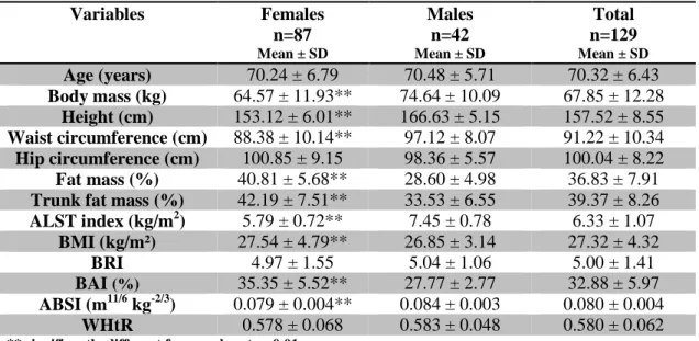
Partial correlation analyses, adjusting for age, were conducted to analyse the association between all body indices with total and trunk fat mass. In figures 1 to 4 are
23
illustrated these associations for females (figure 1 and 2 for total and trunk fat mass, respectively) and males (figure 3 and 4 for total and trunk fat mass, respectively).
Abbreviations: BMI, body mass index; BRI, body roundness index; BAI, body adiposity index; ABSI, a body shape index; WHtR, waist-to-height ratio.
*Regression coefficients are adjusted for age
24
Figure 1 demonstrates that the association between %FM and ABSI was not significant (p=0.238). All the other body indices were associated with adiposity, explaining between 42% (BAI) and 63% (BMI) of % FM from DXA in females.
Abbreviations: BMI, body mass index; BRI, body roundness index; BAI, body adiposity index; ABSI, a body shape index; WHtR, waist-to-height ratio.
*Regression coefficients are adjusted for age
25
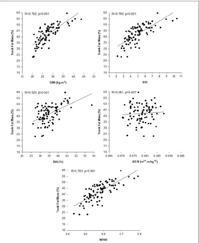
Also for Trunk %FM, ABSI was not associated with values from DXA (Figure 2). The other body indices explained between 27% (BAI) and 61% (WHtR) of Trunk %FM variability for females.
Abbreviations: BMI, body mass index; BRI, body roundness index; BAI, body adiposity index; ABSI, a body shape index; WHtR, waist-to-height ratio.
*Regression coefficients are adjusted for age
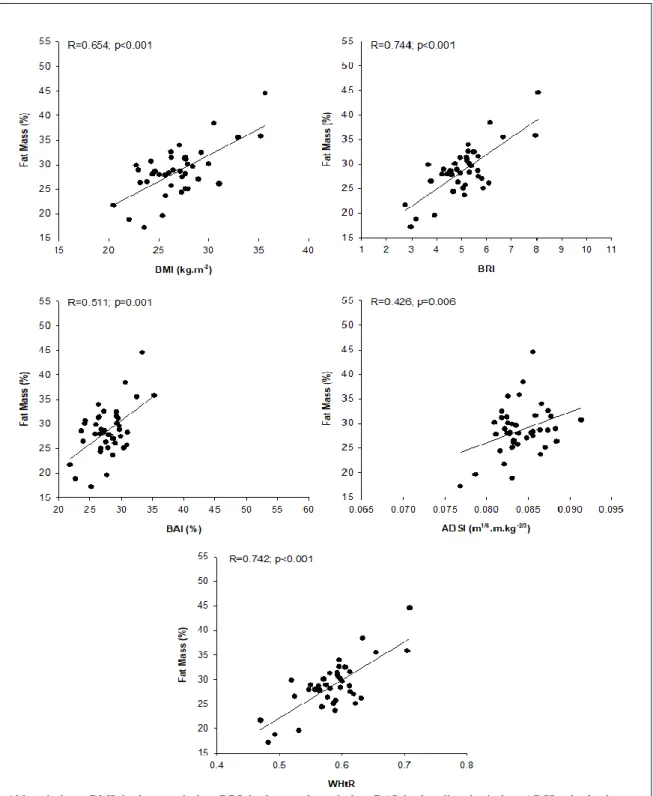
Figure 3: Association between %FM and body indices, for males
Figure 3 demonstrates that body indices explained between 18% (ABSI) and 55% (BRI) %FM variability for males.
26
Abbreviations: BMI, body mass index; BRI, body roundness index; BAI, body adiposity index; ABSI, a body shape index; WHtR, waist-to-height ratio.
Regression coefficients are adjusted for age
27
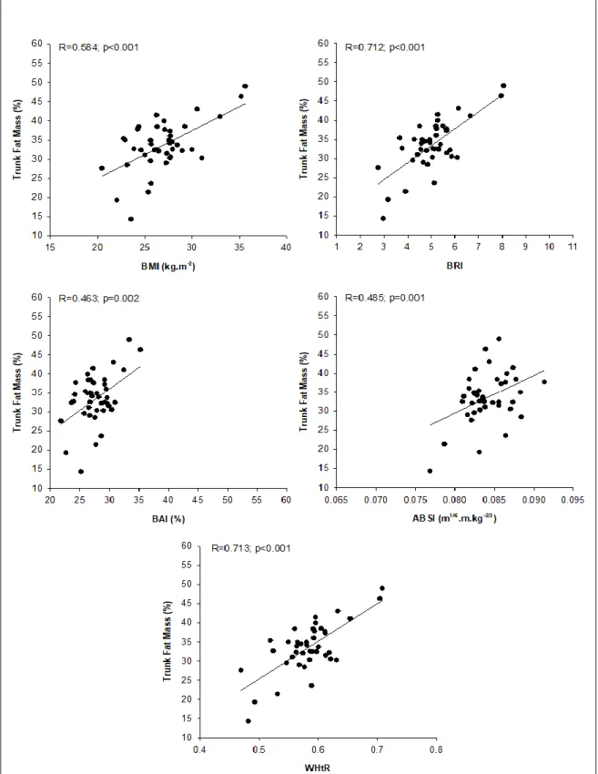
From Figure 4 it is possible to verify that all body indices were related to trunk %FM from DXA. Overall, body indices explained between 21% (BAI) and 51% (WHtR) of trunk %FM variability for males.
In table 2 are represented the females’ results of simple and multiple linear regression analysis for body indices with %FM from DXA. Model 1 represents simple associations, model 2 was adjusted for age, and model 3 was additionally adjusted for ALST index.
Table 2: Predictive Power of different indices in explaining total DXA fat to females. Females
Model 1 Model 2 Model 3
BMI Age ALST Index R 0.944** --- --- r=0.796 (p<0.001) 0.941** 0.068 --- r=0.800 (p<0.001) 1.480** -0.050 -4.754** r=0.893(p<0.001) BRI Age ALST Index R 2.560** --- --- r=0.699 (p<0.001) 2.704** -0.191* --- r=0.735 (p<0.001) 3.700** -0.0225* -2.951** r=0.780 (p<0.001) BAI Age ALST Index R 0.659** --- --- r=0.641 (p<0.001) 0.665** -0.107 --- r=0.654 (p<0.001) 0.671** -0.107 -0.099 r=0.654 (p<0.001) ABSI Age ALST Index R -235.3 --- --- r=0.156 |p=0.148 -208.0 -0.040 --- r=0.163 |p=0.325 -175.5 -0.051 2.596* r=0.367 |p=0.007 WHtR Age ALST Index R 59.1** --- --- r=0.709 (p<0.001) 62.8* 0.200* --- r=0.747 (p<0.001) 87.7** -0.240** -3.210** r=0.799 (p<0.001) ** significant at p<0.001 *significant at p<0.005
Abbreviations: ALST, appendicular lean soft tissue; BMI, body mass index; BRI, body roundness index; BAI, body adiposity index; ABSI, a body shape index; WHtR, waist-to-height ratio.
For females, with the exception of ABSI, all body indices were positively associated with %FM. These associations were independent of age and also ALST index. Also, independent of each body index and age, ALST index was negatively related to adiposity.
28
Table 3 represents the results from simple and multiple linear regression analysis for body indices with %FM from DXA in males. Similarly to women, model 1 represents simple associations, model 2 was adjusted for age, and model 3 was also adjusted for ALST index.
Table 3: Predictive power of different indices in explaining total fat mass in DXA Males
Model 1 Model 2 Model 3
BMI Age ALST Index R 1.065** --- --- r=0.672 (p<0.001) 1.064** -0.002 --- r=0.672 (p<0.001) 1.906** -0.050 -5.250** r=980 (p<0.001) BRI Age ALST Index R 3.509** --- --- r=0.749 (p<0.001) 3.442** -0.096 --- r=0.540 (p<0.001) 4.441** -0.185* -3.163 r=0.869 (p<0.001) BAI Age ALST Index R 0.952** --- --- r=0.530 (p<0.001) 0.915* -0.095 --- r=540 (p<0.001) 1.194** -0.151 -2.302* r=0.624 (p<0.001) ABSI Age ALST Index R 626.7* --- --- r=0.344 | p=0.026 790.0* -0.283* --- r=0.464 | p=0.009 865.8* -0.268 0.714 r=0.475 | p=0.020 WHtR Age ALST Index R 77.4** --- --- r=0.746 (p<0.001) 76.0** -0.103 --- r=0.755 (p<0.001) 97.4** -0.193* -3.107* r=0.865 (p<0.001) ** significant at p<0.001 *significant at p<0.005
Abbreviations: ALST, appendicular lean soft tissue; BMI, body mass index; BRI, body roundness index; BAI, body adiposity index; ABSI, a body shape index; WHtR, waist-to-height ratio.
In males, all body indices were positively associated to %FM from DXA. These associations were independent of age and ALST index. Independently of BMI, BRI, BAI, and WHtR, but not ABSI. ALST index was negatively related to adiposity.
29
Discussion
Due to the need to evaluate the body composition in older adults and to promote their well-being and quality of life, the opportunity to study new body indexes as indicators of total and regional adiposity lead to the current investigation. The current study investigated the association between newly developed body indices with total FM and trunk FM, assessed by DXA in older adults (age≥60 years old).
To the present date, there are not many studies carried out with samples similar to this one. Thus, the discussion of results will focus on the existing ones, together with studies performed in samples with different characteristics.
In the present investigation males and females were similar in age, but the same can not be verified regarding other variables, it is noteworthy that the men who make up this sample have a higher height and body mass, although the women have a greater BMI. The male sample also presents a higher average, %FM and trunk FM, however, a greater WC is observed in the male sample.
Our results demonstrated that BMI explained 45% and 63% of the variation of %FM derivate by DXA, in males and females, respectively.
Body Roundness Index (BRI)
BRI was created (Thomas, et al., 2013) in order to estimate body shape individually as a predictor of percentage of FM and VAT. Thomas et al. (2013) modelled the body shape as an ellipse or an oval shape that could capture the circumference of the body and relate it to the stature (roundness of the body) and by doing this provided a simple calculator based on the user interface (https: //www.pbrc.edu/bodyroundness). Yet there are few or no studies conducted in that direction using this index.
The sample here presented has an average BRI 5.00 ± 1.41, explaining 55% of fat mass for men and 49% for women, these values are higher than those observed by Santos et al. (2015) in a population of athletes with an average BRI of 2.43 ± 0.65 and with an explanatory power of 36% and 25% for men and women, respectively. In our sample of older adults the BRI correlation with adiposity was higher than the observed in Satntos et al (2015) study in both males and females (r2>0.51).
30
Body Adiposity Index (BAI)
According to the results presented, BAI explained FM from DXA by 28% in males and 42% in females. The average BAI in the current investigation was 32.88 ± 5.97%, this value is higher than the observed by Santos et al. (2015) in athletes (23.0 ± 3.3%). In the same study the author verified that BAI explained 7% and 14% of FM whereas in our study it was 28% and 41% for men and women respectively. Although in our study the BAI was positively associated, in both sexes with the fat mass, the same did not happen in the female population of the study developed by Lam et al. (2013), these researchers studied the comparison between BAI and BMI in estimating adiposity in a Chinese population in Singapore that mixed healthy volunteers and patients from the Department of Family and Community Medicine at Khoo Teck Puat Hospital (KTPH) with an average age of 39.3 ± 11.6 and had an average BAI of 31.2 ± 5.4, also lower than that found in our sample and in the present investigation (r = 0.654 (BMI) vs. 0.511 (BAI), for males and r = 0.798 (BMI) vs. 0.649 (BAI), for females) in Lam et al. (2013) study the BAI, when stratified by gender, obtained a lower R than BMI (r = 0.81 (BMI) vs. 0.74 (BAI), for males and r = 0.87 (BMI) vs. 0.82 (BAI) for females). In another research conducted with obese women, it has also been shown that BAI does not outperform BMI to predict FM (Geliebter, Atalayer, Flancbaum, & Gibson, 2013). Also Ramirez-Velez et al. (2016) observed BAI to be unable to explain the FM mass deliberated by the DXA, since it underestimated it, when adjusted for gender, in a study with obese adult population of Colombia, where it was observed that individuals with less fat, have a higher correlation between BAI and FM. In another study conducted by Sung, Oh, & Lee (2014) in Korean women, the average BAI was 26.9 ± 3.3, being lower than the average BAI of our sample, which shows a coefficient of correlation of 0.758, which is higher than the one observed in the females of our study with an R = 0.649, both adjusted for age. However, they observed that BMI remains a better predictor of percentage of FM.
Although the results described above point to the contrary, Bergman et al., the original creators of BAI, were able to validate it in a study of African Americans, after having developed it with a population of Mexican Americans. However, the results have been inconsistent as demonstrated previously. Albeit, the reasons for this discrepancy are unclear, but since BAI quantifies adiposity based on height-adjusted hip
31
circumference, different distributions of body fat between populations may be reflected in different BAI's (Bennasar-Veny et al., 2013).
A body shape index (ABSI)
In the study presented, it can be noted that ABSI shows no association with the percentage of fat mass in females.
According to the results, our sample presents an average ABSI of 0.080 ± 0.004. This value is higher than that observed by Santos et al. (2015) in an investigation where they tried to verify the association of ABSI and other body indexes in a sample of athletes (0.074 ± 0.003), with an average age of 22.6 ± 4.6. In Santos et al. investigation it was verified that ABSI explained 22% of FM in men but no significant associations in women. In our study ABSI explained only 18% of %FM in males and no significant associations were verified in females. However, contrary results were found by Dhana et al. (2016) in an investigation analysing the association of ABSI, BMI and WC with fat mass and fat-free mass in 3612 older adults, where the mean ABSI was 0.083 ± 0.0040 and 0.077 ± 0.005, for men and women, respectively. In Dhana et al, (2016) study multivariate models for confounding factors showed that a higher ABSI was associated with a higher fat mass index (β 1.01, 95% CI 0.85, 1.17) and lower fat free mass index (β -0.28, 95% CI -0.38, -0.17) in men. On the other hand, in the female group, the highest ABSI value was not associated with the FM.
Another study carried out by Biolo et al. (2015) in 200 overweight or obese individuals, where ABSI and BMI did not correlate, concluded that ABSI may be a good predictor of sarcopenic obesity in individuals of this stature.
Waist-to-height ratio (WHtR)
According to the presented results, this sample presents an average WHtR of 0.580 ± 0.062. Swainson, Batterham, Tsakirides, Rutherford, & Hind (2017) that compared 5 anthropometric indices (BMI, WHR, WC, WHtR and WHT.5R) in a sample of 81 adults of different ethnicities, in order to analyse the best predictor of %FM derived by DXA, defined WHtR as the best index, with R2 = 0.76 and R2 = 0.60 for men and women, respectively. In contrast, our research obtained a lower R2 for both sexes (R2 = 0.50 and R2 = 0.56, for men and women, respectively). A meta-analysis developed by Ashwell, et al. (2012) sustains the results of Swainson et al. (2017), since