José Manuel Rocha Pereira
Bachelor Degree in Molecular and Cellular Biology
Improvement of a photoautotrophic
chassis robustness for Synthetic
Biology applications
Dissertation to obtain the Master of Science Degree in
Biotechnology
Supervisor: Paula Tamagnini, Associated Professor,
Faculty of Sciences of University of Porto
Co-supervisor: Catarina Pacheco, Postdoctoral researcher
Institute for Molecular and Cell Biology,
Porto
December 2015
Jury:
President: Prof. Doutor Carlos Alberto Gomes Salgueiro
Examiner: Prof. Doutor Adriano José Alves de Oliveira Henriques
III
José Manuel Rocha Pereira
Improvement of a photoautotrophic
chassis robustness for Synthetic
Biology applications
Dissertation to obtain the Master of Science Degree in Biotechnology
V
Improvement of a photoautotrophic chassis
robustness for Synthetic Biology applications
Copyright reserved to José Manuel Rocha Pereira, FCT-UNL and UNL.
The Faculty of Science and Technology and the NOVA University of Lisbon have the perpetual right, and without geographical limits, to archive and publish this dissertation through press copies in paper or digital form, or by other known form or any other that will be invented, and to divulgate it through scientific repositories and to admit its copy and distribution with educational or research objectives, non-commercial, as long as it is given credit to the author and editor.
VII
Acknowledgements
The last year has been very fulfilling to me, but very challenging as well. I had the opportunity to work in the exciting field of synthetic biology and deepen my knowledge in this area. However, it wasn’t always easy and for that I want to thank to innumerous people for their support, positivism and contribution.
First I would to thank my supervisor Prof. Paula Tamagnini for kindly receiving me in her research group (Bioengineering and Synthetic Microbiology), supervising me with the best advice and vision and giving me the opportunity to tackle issues involving the sustainability of our sweet “home”, the Earth.
To Catarina Pacheco, my awesome co-supervisor, I thank you very deeply for your support, advice, patience and guidance. The path to this moment wasn’t always easy and you truly helped me to always see Science in a positive way, since “it is what it is and we just have to move on”.
For the Cyanofactory project members a huge thank you for all your support and input: To Filipe Pinto for your tremendous help and advice; To Paulo Oliveira for your constant input, help and generosity; To Meri for the contagious happiness and your help in everything I needed and finally to Eunice Ferreira for your constant support, advice and of course the
“photosynthesis” moments during coffee breaks.
Then I would like to acknowledge my “Comrade-in-arms”, Steeve Lima, for the support, laughable moments and thoughtful conversations. We weren’t always in agreement, but it is with great pleasure that I got to meet you.
For the remaining members of our group, thank you very much for your hospitality, great spirit and help.
To Prof. Isabel de Sá Nogueira, thank you so much for your advice, guidance and everything you taught me from the very beginning of my journey in university. Additionally, I express my gratitude for introducing me to the exciting field of synthetic biology and help me find new paths for my short career. A special word for the members of the Microbial Genetics group (Lab 327) for your contribution as well.
For all my friends, including my Bachelor and Master degree’s colleagues, a big “thank you” for accompanying me along this path since my first day at university.
To Cláudia, for your absolute and strong friendship. Without you, this journey would have been much harder. Thank you so much for your support, caring and the laughable moments of pure happiness. To your family, a special thank you as well.
To my family, which is excitingly getting bigger and bigger, for all the love and support, I thank you deeply.
Finally, to my parents, there are no words to describe how I feel about you. All the
IX
Abstract
Cyanobacteria are photoautotrophic microorganisms with great potential for the biotechnological industry due to their low nutrient requirements, photosynthetic capacities and metabolic plasticity. In biotechnology, the energy sector is one of the main targets for their utilization, especially to produce the so called third generation biofuels, which are regarded as one of the best replacements for petroleum-based fuels. Although, several issues could be solved, others arise from the use of cyanobacteria, namely the need for high amounts of freshwater and contamination/predation by other microorganisms that affect cultivation efficiencies. The cultivation of cyanobacteria in seawater could solve this issue, since it has a very stable and rich chemical composition. Among cyanobacteria, the model microorganism
Synechocystis sp. PCC 6803 is one of the most studied with its genome fully sequenced and genomic, transcriptomic and proteomic data available to better predict its phenotypic behaviors/characteristics. Despite suitable for genetic engineering and implementation as a microbial cell factory, Synechocystis’ growth rate is negatively affected by increasing salinity levels. Therefore, it is important to improve. To achieve this, several strategies involving the constitutive overexpression of the native genes encoding the proteins involved in the production of the compatible solute glucosylglycerol were implemented, following synthetic biology principles. A preliminary transcription analysis of selected mutants revealed that the assembled synthetic devices are functional at the transcriptional level. However, under different salinities, the mutants did not show improved robustness to salinity in terms of growth, compared with the wild-type. Nevertheless, some mutants carrying synthetic devices appear to have a better
physiological response under seawater’s NaCl concentration than in 0% (w/v) NaCl.
Keywords:
XI
Resumo
As cianobactérias são microrganismos fotoautotróficos com elevado potencial na indústria biotecnológica, devido aos seus simples requisitos nutricionais, capacidade fotossintética e plasticidade metabólica. O sector da energia é considerado um dos principais alvos para a sua utilização, particularmente, na produção de biocombustíveis de terceira geração como substitutos dos combustíveis fósseis. Contudo, a utilização eficaz de cianobactérias apresenta alguns problemas como a necessidade de elevadas quantidades de água doce e contaminações/predação por outros microrganismos. Desta forma, o cultivo de cianobactérias utilizando água do mar pode ser uma das soluções, uma vez que esta possui uma composição química bastante rica e estável. Entre as cianobactérias, o microrganismo modelo Synechocystis sp. PCC 6803 é um dos mais estudados, tendo o seu genoma sido totalmente sequenciado e com informação ao nível da genómica, transcritómica e proteómica disponível para melhor prever determinados comportamentos fisiológicos. Apesar de ser geneticamente manipulável e útil em biotecnologia, a taxa de crescimento de Synechocystis é afetada negativamente por níveis elevados de salinidade. Assim, a halotolerância deste microrganismo necessita de ser melhorada. Para isso algumas estratégias, baseadas na sobre expressão constitutiva dos genes nativos de Synechocystis que codificam proteínas envolvidas na produção do soluto compatível glicosilglicerol, foram implementadas seguindo os princípios da biologia sintética. Uma análise dos mutantes obtidos revela funcionalidade dos módulos sintéticos ao nível transcricional. Contudo, analisando o crescimento dos mutantes de
Synechocystis, em diferentes salinidades, verifica-se que estes não apresentam um melhoramento da robustez à salinididade comparado com a estirpe selvagem. No entanto, alguns mutantes com módulos sintéticos parecem responder melhor a uma concentração de NaCl idêntica à da água do mar, em vez de 0% NaCl.
Palavras-chave:
XIII
Table of Contents
Acknowledgements ... VII
Abstract ... IX
Resumo ... XI
List of figures ... XV
List of tables ... XVII
List of abbreviations, acronymes and symbols ... XIX
1. Introduction ... 1
1.1 A worldwide problem ... 1
1.2 Biofuels as an alternative to petroleum-based fuels ... 1
1.2.1 First and second generation biofuels... 1
1.2.2 Third generation biofuels as the most promising alternative ... 2
1.3 Cyanobacteria ... 2
1.3.1 Cyanobacteria in biotechnology ... 3
1.4 Synthetic Biology ... 4
1.4.1 Synthetic Biology of cyanobacteria ... 6
1.5 Halotolerance in cyanobacteria ... 6
1.5.1 Compatible solutes ... 7
1.6 Synechocystis sp. PCC 6803 as a model organism ... 8
1.6.1 Glucosylglycerol in Synechocystis sp. PCC 6803 ... 9
1.7 Objectives ... 11
2. Materials and methods ... 13
2.1 Bacterial strains and standard growth conditions ... 13
2.2 Synthetic devices assembly... 13
2.3 Agarose gel electrophoresis ... 14
2.4 DNA purification and quantification ... 14
2.5 Polymerase chain reaction (PCR) ... 15
2.5.1 Colony PCR ... 16
2.6 DNA ligation, E. coli
DH5α transfor
mation and plasmid DNA purification ... 16
2.7 Cyanobacterial DNA extraction ... 16
2.7.1 Phenol-Chloroform DNA extraction protocol ... 16
2.8 Southern blot of the Δ
ggpS knock-out mutants ... 17
2.9 Synechocystis transformation by electroporation ... 17
XIV
2.11 Total RNA extraction and transcription analysis by quantitative real-time PCR
(RT-qPCR) ... 18
3. Results and discussion ... 21
3.1 Synechocystis tolerance to salinity ... 21
3.2 Strategies to improve Synechocystis halotolerance ... 22
3.2.1 Design and assembly of synthetic devices based on
Synechocystis’
native
genes involved in GG production ... 22
3.2.2 Generation of mutants with the synthetic devices for GG production ... 24
3.3 Functional characterization of selected mutants carrying synthetic devices for GG
production ... 26
3.3.1 Growth analysis of Synechocystis mutants under different salinities ... 26
3.3.2 Transcriptional analysis by quantitative real-time PCR (RT-qPCR) of the
relative fold expression of ggpS and ggpP genes ... 30
4. Conclusion ... 33
5. Future perspectives ... 35
XV
List of figures
Figure 1.1 Biobrick Assembly Standard RFC[10] overview.. ... 5
Figure 1.2. Representation of the three halotolerance groups of bacteria and their common compatible solutes.. ... 8
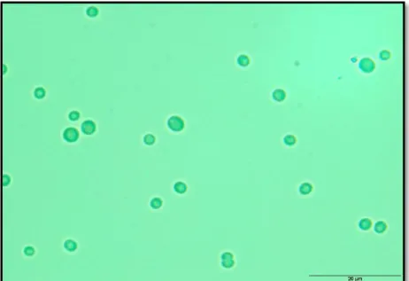
Figure 1.3. Microscopic view of Synechocystis sp. PCC 6803. ... 9
Figure 1.4 Schematic overview of the biosynthetic pathway of glucosylglycerol in
Synechocystis... 10
Figure 3.1 Salt stress effect on the growth of Synechocystis sp. PCC 6803 wild-type under different NaCl concentrations in BG11 medium... ... 21
Figure 3.2 Schematic representation of the synthetic devices designed and generated in this work. ... 23
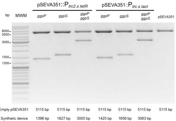
Figure 3.3 DNA electrophoresis of the plasmids with synthetic devices digested with XbaI
and PstI confirming the correct assembly of the synthetic devices into pSEVA351.. ... 24
Figure 3.4 Confirmation of segregation of Synechocystis ggpS knock-out mutants by Southern blot. ... 25
Figure 3.5 DNA electrophoresis of PCR products confirming the Synechocystis mutants carrying the pSEVA351 and pSEVA351 with synthetic devices specified by the type of promoter (Ptrc2.x.tetR and Ptrc.x.lacI) and respective ggpP and/or ggpS ORFs.. ... 25
Figure 3.6 Salt stress effect on the growth of Synechocystis sp. PCC 6803 mutants under different NaCl concentrations (% w/v) in BG11 medium.. ... 27
Figure 3.7 Transcriptional analysis by RT-qPCR of ggpS and ggpP transcripts for the wt,
XVII
List of tables
Table 2.1. List of plasmids used in this work. ... 14
Table 2.2. List of synthetic devices generated in this work. ... 14
Table 2.3. List of oligonucleotides used in this work. ... 15
XIX
List of abbreviations, acronymes and symbols
Amp - Ampicillin
AmpR - Resistance to ampicillin
bp - Base pair
cDNA - Complementary DNA Cm - Chloramphenicol
CmR - Resistance to chloramphenicol
dH2O - Deionized water
DHAP - Dihydroxyacetone phosphate DNA - Deoxyribonucleic acid
dNTP - Deoxyribonucleoside triphosphate EDTA - Ethylenediaminetetraacetic acid G3P - Glycerol-3-phosphate
gDNA - Genomic DNA GG - Glucosylglycerol GGA - Glucosylglicerate GB - Glycine betaine
GGPP - Glucosylglycerol-phosphate phosphatase GGPS - Glucosylglycerol-phosphate synthase
HEPES - 4-(2-hydroxyethyl)-1-piperazineethanesulfonic acid buffer Km - Kanamycin
KmR - Resistance to kanamycin
LB - Lysogeny broth
mRNA - Messenger ribonucleic acid OD - Optical density
ON - Overnight
ORF - Open reading frame PBR - Photobioreactor
PCC - Pasteur culture collection PCR - Polymerase chain reaction RBS - Ribosome binding site RNA - Ribonucleic acid r.p.m. - Revolutions per min RT - Room temperature
RT-qPCR - Reverse-transcriptase quantitative polymerase chain reaction S.D. - Standard deviation
XX WF - Water footprint
1
1.1
A worldwide problem
According to the United Nations, the World’s population is expected to grow from a current 7.3 to 9.5 billion people by 2050. From this total, the main rise will come from the urban population for 6.3 billion, where a two-fold increase is estimated1. Consequently, several issues
arise to be solved as the demand for food, freshwater and energy increases2. The latter needs
special attention, since its global demand is estimated, by the International Energy Agency (IEA), to be 46% more than the observed energy consumed in 2010 by 2035, within the current energy policies in vigor3. In the same way, it also reports the main energy supply would still
come from fossil fuel for about 80% of the total need. As a result, the carbon dioxide (CO2)
emission would rise up by 46% leading to negative effects in the environment, including global temperature rise3. Additionally, fossil fuels are finite sources of energy with estimated
exhaustion periods of 50-100 years and 100-200 years for oil/gas and coal, respectively, leading to a possible energy and economic insecurity as their demand rises. Particularly, due to limited alternatives to liquid transportation fuels from petroleum compared with electric power generation alternatives, such as wind, hydro and solar renewables4.
1.2
Biofuels as an alternative to petroleum-based fuels
Biofuels are nearly carbon neutral renewable liquid fuels produced from biomass, which might be organic/biological waste or plant and microbial based5,6. Considered to be the
renewable solution for transportation fuels, biofuels have shown a clear production increase over the last decade, where their production has increased by more than fivefold over it7. This is
particularly true, due to their possible use in the current transportation infrastructure to some extent, apart from being a cleaner renewable energy supply6,8,9.
Biofuels comprise three different types depending on their original feedstock.
1.2.1 First and second generation biofuels
First generation biofuels are the most abundant and derive from cultivated crop feedstock4. The main commercial available biofuels are bioethanol from microbial fermentation
of sugar compounds and biodiesel from vegetable oil transesterification4,8. Despite great
promise, however, several ethical and environmental issues arisen, namely, the competition with the food sector for arable land and food crops, such as sugarcane and corn2,4,10. Mainly
with a growing world population. Additionally, soil degradation and desertification are among other drawbacks faced with first generation biofuels2,6,11.
As an alternative, second generation biofuels emerged, since they don’t impose such a problem regarding food security as first generation’s6,12. These result primarily from the
1. Introduction
2
for microbial fermentation13. This accounts for the non-food and cheap portion of it, about 98.5%
of the total global plant biomass6. Nevertheless, despite the cheap and abundant feedstock, the
technology employed is still very costly. As a result, this type is non-commercially viable yet, being about two to three times more expensive than petroleum based fuels14.
1.2.2 Third generation biofuels as the most promising alternative
Third generation biofuels are receiving special attention as a solution for the problems imposed by first generation’s15. These are microalgae and cyanobacteria based, which are
photoautotrophic microorganisms by performing photosynthesis16. The most striking advantage
relies on microalgae/cyanobacteria requirement for lesser land, arable land is not necessary, because these microorganisms provide higher net energy yields and have higher growth rates than plant crops16,17. In this way, there would not exist a competition with the food sector.
Despite these common advantages, between microalgae and cyanobacteria there are several differences that make the latter more suitable for complex manipulations and applications18.
Hence, cyanobacteria are being pushed forward as an ideal organism for biotechnological applications in the bulk chemicals sector19,20.
1.3
Cyanobacteria
Cyanobacteria compose a vast group of Gram-negative autotrophic prokaryotes capable of using photosynthesis to produce biomass21,22. Morphologically, they range
remarkably from unicellular to colonial and filamentous with a varying size of up to two orders of magnitude23. Thereby, in conjunction with their diverse physiology, these bacteria are capable
of withstand extreme environmental conditions, from high/low temperatures, pH and salinities21.
Regarded as the first photosynthetic organisms originated on Earth, they are thought to be the main contributors, later with algae/plants, of the actual oxygenic atmosphere19. In addition, they
are estimated to contribute to about 25% of the current global carbon fixation24. These bacteria
can fix atmospheric CO2, while harvesting solar electromagnetic radiation, in the visible range25.
Photosynthetically, cyanobacteria have higher yields for solar energy conversion than algae and plants, for 10% against 5% and 1%, respectively16. Despite, being superior photosynthetic
organisms, with chlorophyll a as the main photosynthetic pigment, cyanobacteria are also
capable of grow photoheterotrophically or chemoheterotrophically21. The ability of some
cyanobacteria to also perform atmospheric molecular nitrogen fixation shows their very diverse metabolic plasticity21. In this way, differentiating them again from plants regarding the need for
minimal nutrients to thrive.
In the last decades, molecular biology, modification and characterization tools allowed for a deeper understanding of these microorganisms26. In fact, to date, there are at least 265
3
1.3.1 Cyanobacteria in biotechnology
In the last years, cyanobacteria have been receiving a huge attention in the biotechnological sector. Mainly, due to their interesting photosynthetic features, biologically active compounds and their possible genetic manipulation to produce several chemicals of interest27,29. Currently, these prokaryotes are tested, with scaled-up processes being or already
implemented, in a wide range of applications from biofuel, polyesters, fertilizers and commodity chemicals production to biorremediation22,27–29. Even though their multidisciplinary use is
evident, cyanobacteria are principally seen as the future’s most promising tool for biofuel production, as mentioned above. This is true, since they can be potentially used in an economically and environmentally effective sustainable way, in order to replace most of the current global use of fossil fuels16. Although, they share most of the advantages also associated
with algae, several others arise which make them more suitable in the long run. Some of these are a higher photosynthetic efficiency, a simpler genetic background which makes it easier to genetically manipulate and the capacity for natural transformation18.
Despite great promise, cyanobacteria, as well as algae, utilization in biotechnological applications still pose several challenges29. These comprise processes such as in cell disruption
(mechanical, enzymatic or chemical) to access the intracellular biomolecules, harvesting and cultivation16,19. Cyanobacteria and algae are usually cultivated in open and closed ponds or in
photobioreactors (PBRs)16. One of the biggest issues relates with the high water evaporation
rates associated. Although the water evaporation is greatly addressed by a PBR system, its water footprint (WF), which is the freshwater use/expenditure in a certain activity, is still very high. This could still pose a huge pressure over the world’s freshwater reserves, even though the employment of microalgae in biotechnology, notably in biodiesel production, is comparably less impactful than the use of most plant crops30. Such holds important meaning since for first
generation biofuels, the WF can range considerably. Indeed, the WF to produce 1 Kg of biodiesel can range from an estimated 2168 to 15331 L with plant crops, such as sugar beet and sorghum, respectively. In contrast, for microalgae based biodiesel, it is estimated to be up to 3650 L depending of the cultivation system30. However, as an example, according to P.
Gerbens-Leenes et al31, if all transportation fuels in Europe by 2030 were to be third generation
based, according to the IEA projection for transportation fuels needs, then the Europe’s blue
WF (freshwater from surface and groundwater reserves) would increase up to four fold from the current value. As a result, the use of microalgae, as well as cyanobacteria, would still be very severe when the proper system and/or improvements are not employed.
The solutions thought to be ideal include the improvement of current technology to avoid water loss between all downstream processes, especially for cell cultivation and harvest16.
1. Introduction
4
wastewater treatment by reducing its nutrient content. As a drawback, this is specially directed for wild-algae, i.e. microalgae that naturally inhabit these type of sewage waters16,32. As a result,
if engineered model cyanobacteria to produce different chemicals were to be used, this could lead to competition with other adapted microorganisms. Additionally, wastewater has a diverse inconstant composition turning the cultivation of cyanobacteria unstable16,33. Seawater, in
contrast, has a composition more constant and regular with a wide range of nutrients essential to cyanobacterial growth, except for phosphorous16. Its use is also estimated to reduce the
biofuels production life-cycle need for freshwater by up to 90%30. In addition, its use could also
prevent growth of more halointolerant competing and predator organisms that would affect cyanobacterial growth19. Remarkably, cyanobacteria can withstand a wide range of salinity
(concentration of dissolved inorganic ions) levels. Nonetheless, the growth rate or even survival of some main genetically engineerable cyanobacteria are affected by high levels of osmotic stress34. Hence, it could be important to tackle this by implementing a synthetic biology
approach in order to improve the robustness to salinity.
1.4
Synthetic Biology
Synthetic biology is a new field within biology originated in the 21st century35. It is
characterized, fundamentally, as the rational design of new molecules and genetic/metabolic networks or the re-design of existing ones in ways not observable in nature (therefore synthetic). Additionally, through engineering principles and/or some modelling/predictive tools from systems biology, synthetic biology practitioners aim to understand and apply biology, to attain new functionalities and biologic systems, at levels not possible with genetic engineering itself26,36–39.
Synthetic biology basilar foundations which contribute to biology’s engineerability are standardization, abstraction and decoupling36. Standardization, as the name implies, refers to
the use of globally accepted and reference standards in an interchangeable way. As for abstraction, biological parts also called BiobricksTM, such as promoters, ribosome binding sites
(RBS) and transcriptional terminators are used as building blocks which, through standardized measurements and consequent predicted behaviors for most of them, help manage biological complexity. These can be assembled into devices which will be transformed into a certain biological organism (chassis). As a result, more complex systems are formed in order to perform a desired function. Such represents a decoupling process where a complex and difficult problem is divided into smaller and simpler ones, which can be combined to possibly solve it36,40.
Every day, new BiobricksTM are generated and uploaded into online open access
databases, such as the Registry of Standard Biological Parts database, from the Biobrick’s
5
Figure 1.1 Biobrick Assembly Standard RFC[10] overview. Each part is flanked by a prefix (upstream) and suffix (downstream) with recognition sites for four different restriction enzymes (E –EcoRI, X –XbaI, S –
SpeI, P –PstI) and are assembled through molecular cloning techniques into complexer devices. The feasability of this system relies on the compatibility and ligation of the S and X overhangs which forms a
“scar” sequence between both parts without restoring any of these recognition sites. The resulting device
is also flanked by both prefix and suffix allowing further assemblies until the final and desired synthetic device is obtained41. Adapted from http://parts.igem.org/Help:Standard_Assembly_%28zoom%29; accessed in June 2015.
The former is the most common way of access biological parts. Here, the usual cloning techniques in molecular biology are used in order to assemble the parts according to a standardized system, such as the Biobrick Assembly Standard RFC10. Accordingly, every biological part is preceded by a Biobrick prefix which has two restriction enzyme sites for EcoRI
(E) and XbaI (X). Concomitantly, it is also followed by a Biobrick suffix which contains the restriction sites for SpeI (S) and PstI (P) (to note that biobricks cannot have any of these restriction sites in their sequence)41. In this way, different biological parts can be assembled by
cleaving a vector containing “part #1” (donor) with E and S and a vector with E and X containing “part #2” (recipient) for upstream cloning, see Figure 1.1, or digesting the “part #1” containing vector (recipient) with S and P and the “part #2” containing vector (donor) with X and P for downstream cloning. This system takes advantage of the X and S compatible overhangs,
1. Introduction
6
where several parts can be assembled in an intuitive mode42. Alternatively, a digitally designed
device can be obtained by a DNA synthesis process, where the time consuming steps associated with DNA cloning techniques can be avoided. In fact, this practice is becoming more popular due to a continuous fall in DNA synthesis costs as seen for DNA sequencing years ago, despite its still relative high cost35,36. Altogether, these aspects in conjunction with a thriving
community are pushing synthetic biology forward to be developed and implemented in a global, interactive and educational way.
1.4.1 Synthetic Biology of cyanobacteria
The main development regarding synthetic biology has been done, essentially, in heterotrophic bacteria. The majority of parts and synthetic devices created are targeted to the Gram-negative bacterium Escherichia coli, the Gram-positive bacterium Bacillus subtilis or the eukaryotic yeast Saccharomyces cerevisiae (http://parts.igem.org/Catalog#Browse_chassis; accessed in June 2015). Despite having some orthogonality, many of these parts do not have the same predicted behaviors in other hosts. This is true, notably, for cyanobacteria where many of the well characterized promoters and RBSs strengths are not the same as in E. coli, for example26,43. Consequently, cyanobacterial synthetic biology is still lagging behind compared
with other chassis.
Despite lacking many functional well characterized parts, several efforts are being done in order to fill this gap concerning cyanobacterial engineering. Indeed, in the last five years many tools and parts have been created and tested, while others are currently being so26,44,45.
At the same time, many established synthetic biology projects in cyanobacteria research
contribute for its growth e.g., the Cyanofactory’s European (http://www.cyanofactory.eu/) and Japanese (http://www.tuat.ac.jp/~cyano/) projects
The growth in cyanobacterial synthetic biology research is clearly derived from these bacteria capacities. As said before, the ability to thrive autotrophically, with a low nutrient requirement, in conjunction with the available molecular biology tools make them excellent chassis for biotechnological applications. As a result, cyanobacteria are being deeply studied, in
order to fulfill its promise as the so called “green E. coli”46.
1.5
Halotolerance in cyanobacteria
Cyanobacteria, just as other bacteria, are classified into three different groups according to their tolerance, i.e. halotolerance. Basically, these comprise freshwater (tolerance up to 3.5% (w/v) sodium chloride (NaCl)), moderately-halotolerant (tolerance up to ~10% (w/v) NaCl) and halophilic bacteria (tolerance up to 17.5% (w/v) NaCl), see Figure 1.234,47. Normally,
cyanobacteria thrive by maintaining a constant osmotic and ionic concentration, intracellularly, in their more hyperosmotic cytoplasm. Thereby achieved to regulate external water uptake and consequently maintaining an adjustable turgor pressure in order to grow47. When an external
7
the water availability is reduced since a higher ionic concentration leads to less free water available34,47. Actually, less free water availability implies a lower enzymatic activity within the
microorganism, possibly affecting its growth49. To face these issues, bacteria have developed
two different strategies, namely, the “salt-in” and “salt-out” strategies to acclimate against high salt stressing conditions34,47,50. The “salt-in” strategy is characteristic of very halophilic bacteria,
such as some archaea orders, it consists on a high inorganic ion uptake into the cell (up to ~22.5% (w/v), primarily KCl). Additionally, In order to resist high ionic stress, halophilic bacteria have also a proteome and consequently metabolism highly resistant to elevated ionic concentrations47. The “salt-out” strategy is the most widely used mechanism by bacteria to face
osmotic stress. The objective is now to achieve a low ionic concentration within the cell, since enzymatic activity would be affected by higher levels of sodium, for example. To maintain an osmotic equilibrium, the cells synthesize small molecules called compatible solutes, which act as osmotic regulators. Compatible solutes allow the cell to adjust the osmotic concentration, while extruding small inorganic ions, mainly sodium. This way, bacteria acclimate and can recover their former state34,47,50.
1.5.1 Compatible solutes
Compatible solutes are low-molecular mass organic molecules, usually with no charge, ranging from sugars to aminoacids and their derivatives. These are extremely useful compounds due to their osmotic and protective properties against dissecation and high/low temperatures, and the possibility of being biosynthesized in high amounts without having a negative effect on the cell’s metabolism34,47,50. Interestingly, the type of compounds produced by
different organisms is intrinsically correlated to the organisms’ halotolerance group. For freshwater bacteria, the sugars sucrose and trehalose are the main ones. As for moderately halotolerant, these are glucosylglycerol (GG) and glucosylglicerate (GGA). While for halophilic bacteria, the main compatible solutes produced are glycine betaine (GB) and glutamate betaine, as shown in Figure 1.2.
Cyanobacteria, as autotrophic microorganisms synthesize their compatible solutes de novo. However, cyanobacteria possess transporters for compatible solutes uptake47,51. They
use this mechanism to avoid a constant leakage of de novo synthesized compatible solutes, in order to prevent energy and carbon waste. In this way, the type of transporters encoded in a cyanobacterial genome is tightly related with the type of compatible solute they produce34,47. On
1. Introduction
8
Figure 1.2. Representation of the three halotolerance groups of bacteria and their common compatible solutes. Additionally, NaCl tolerance limits for each group is shown, as well as the molecular structure of each compatible solute. Adapted from Hagemann (2011).
1.6
Synechocystis
sp. PCC 6803 as a model organism
Among the vast group of cyanobacteria, the model freshwater cyanobacterium
Synechocystis sp. PCC 6803 (hereafter Synechocystis) was the first photosynthetic organism to have its genome fully sequenced and annotated52. Additionally, the vast data available allowed
the implementation of genome-wide metabolic models (e.g, iSyn811), which help to predict its cellular phenomena to some extent53–55. Besides, being a photosynthetic bacterium, as well as
naturally transformable (homologous recombination), with other transformation techniques also applicable, such as electroporation, make it of high scientific and biotechnological interest26. Synechocystis has been deeply studied since its discovery. Thus allowed a better understanding of the many aspects surrounding photosynthesis, circadian rhythms and several other mechanisms from gene regulation to environmental stress. Some of these studies have been useful to research and understand other organisms, such as higher plants, due to its
similarity with plant’s chloroplasts29.
Morphologically, this unicellular spherically shaped bacterium, as shown in Figure 1.3, is polyploid with about 12 copies of its 3.6 Mbase pair (bp) sized chromosome, as well as having seven different endogenous plasmids26. Physiologically, Synechocystis has a doubling time of 8
to 12 hours (h) when growing phototrophically on a minimum nutrient medium26,29. All these
9
Figure 1.3. Microscopic view of Synechocystis sp. PCC 6803.
1.6.1 Glucosylglycerol in
Synechocystis
sp
.
PCC 6803
Sucrose and glucosylglycerol (GG) are the compatible solutes biosynthesized by the cyanobacterial microorganism Synechocystis sp. PCC 6803 naturally. Sucrose is utilized, mainly, under low osmotic concentrations. On the other hand, GG is of special interest since its responsible for Synechocystis tolerance to salinities up to 6% (w/v) NaCl, when not acclimated47,56,57. GG is produced in a two-step biosynthetic pathway where adenosine-5’
-diphosphoglucose (ADP-glucose) and glycerol-3-phosphate (G3P) are the precursors, as shown in Figure 1.4. G3P originates from the biochemical transformation of dihydroxyacetone phosphate (DHAP) derived from the Calvin cycle, oxidative pentose phosphate and/or glycolysis pathways. The first biochemical reaction is catalyzed by the glucosylglycerol-phosphate synthase (GGPS), generating an intermediate called glucosylglycerol-phosphate (GGP) which is not protective against osmotic stress58. However, when dephosphorylated by the second step
enzyme, glucosylglycerol-phosphate phosphatase (GGPP), the compatible solute GG is then obtained conferring its osmotic protective properties to allow Synechocystis survival at higher salinity levels47,50.
These two enzymes present full activity only in a hyperosmotic medium59. For example,
according to Hagemann et al60, who tested the in vitro activity of the GGPS enzyme in a crude
protein extract from Synechocystis, the maximum activity is achieved when in the presence of
~0.6% (w/v) NaCl, which is about one fifth of seawater’s average NaCl concentration (~3%
1. Introduction
10
Figure 1.4 Schematic overview of the biosynthetic pathway of glucosylglycerol in Synechocystis. The first catalytic step is performed by glucosylglycerol-phosphate synthase (GGPS), where the intermediate glycerol-3-phosphate is formed. Afterwards, this intermediate is desphosphorylated by glucosylglycerol-phosphate phosphatase (GGPP) to form the final compatible solute glucosylglycerol. Molecular structures from chEBI and Chembase Databases.
Indeed, when growing in low-salt conditions, this cyanobacterium does not show any meaningful traces of GG, intracellularly. But, when a salt shock occurs, the GG synthesis is rapidly started due to these electrostatic interactions disturbance, without any lag-phase, while transmembrane transporters also extrude ions, mostly sodium50. Afterwards, when most of the
toxic sodium is extruded, there is an upregulation of the genes encoding the GG production enzymes, depending on the salt concentration. At the same time, the cell’s metabolism is restored and photosynthesis resumed to produce the necessary energy, as more GG is biosynthesized until a certain steady-state is reached up to 24h later34,60,62. Additionally, GGPS
and GGPP were also tested in the presence of high levels of NaCl (up to 6% (w/v)) showing that the activity is maintained and how important this characteristic is physiologically34. This allows Synechocystis, when has its housekeeping proteins suddenly affected and inhibited by salt (NaCl), to recover faster from a bacteriostatic effect, as stated above. Despite being involved in the same biosynthetic pathway, both GGPS and GGPP are encoded by two genome far located genes, the ggpS (bp position 1948824 to 1947325 - sll1566, cyanobase) and ggpP/stpA (bp position 3041493 to 3042407 - slr0746, cyanobase), respectively34,58,63. Interestingly, some Synechocystis ggpS and ggpP knock-out mutants have been generated and studied. According to Marin et al59 and Hagemann et al58, mutants carrying these mutations were unable to grow on
medium supplemented with more than 3.2% (w/v) NaCl, suffering a consequent cells lysis after salt shock. Both ggpS and ggpP transcription is salt regulated. Indeed, they are upregulated in higher salt conditions, although they are also transcribed, but at a lower extent, under isotonic conditions. Little is known about the regulation mechanism for ggpP. Nonetheless, for ggpS, the proposed regulation process involves the presence of a repressor protein (GgpR), encoded by a small gene (ggpR) which overlaps the promoter and transcriptional start point of ggpS. This repressor binds to the ggpS promotor, under low salt conditions, repressing its transcription, which is resumed after GgpR inactivation by NaCl64. All these interligated elements contribute
11
1.7
Objectives
The main goal of this study was to identify and implement strategies to improve
Synechocystis halotolerance using a synthetic biology approach.
For this purpose we:
(I) Start by establishing Synechocystis tolerance limits to different salinity levels; (II) Identify candidate genes to improve Synechocystis tolerance to salinity, in the
particular case of this work the native ones: ggpS and ggpP;
(III) Design and assemble several synthetic devices with these genes, following synthetic biology standards;
13
2.1
Bacterial strains and standard growth conditions
The cyanobacteria Synechocystis sp. PCC 6803 (obtained from the Pasteur Culture Collection of cyanobacteria, Paris, France) and a ggpS knock-out mutant (ΔggpS) strains, were kept in BG11 medium65 at 30 ºC and a 12 h light (25 μE m-2 sec-1) /12 h dark regimen.
Cosine-corrected irradiance was measured with a quantum meter (Dual Solar/Electric Quantum Meter, Spectrum Technologies, Inc.). When cultured in solid medium, BG11 supplemented with 1.5% (w/v) Difco® Agar Noble, 0.3% sodium thiosulfate and 10 mM TES–KOH buffer (pH 8.2) was
used. The strains E. coli DH5α (Stratagene) and One Shot® TOP10 chemically competent E. coli (Invitrogen) were used for molecular cloning purposes and cultured at 37 ºC in selective Lysogeny Broth (LB)66 medium. For solid medium, 1.5% (w/v) Bacteriological Agar was added.
When necessary, BG11 and LB media were supplemented with the appropriate antibiotic, chloramphenicol (Cm, 10 or 25 μg mL-1), ampicillin (Amp, 100 μg mL-1), or kanamycin (Km, 50
μg mL-1in LB; 25 to 500 μg mL-1 in BG11).
2.2
Synthetic devices assembly
In order to improve Synechocystis halotolerance, the coding sequences of the native
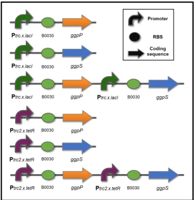
ggpS and ggpP were amplified by PCR, from Synechocystis gDNA, and used in the assembly of the synthetic devices. The devices enable the constitutive overexpression of these genes and two sets of three different devices were designed. Two have ggpS or ggpP, while the third carries both genes with ggpS downstream of ggpP. The difference between the sets relies on the promoter used, see Table 2.2. One was assembled with the synthetic Ptrc2.x.tetR (medium
strength), while the other with the synthetic Ptrc.x.lacI (high strength). The BioBrickTM RBS
(BBa_B0030) was retrieved from the Registry of Standard Biological Parts (http://parts.igem.org/) in the pSB1A2 (Table 2.1). The cloning process was performed according to the Biobrick Assembly Standard RFC10 (Figure 1.4), as follows: for upstream assembly, (I) the vector containing the promoter was digested with EcoRI and SpeI: and the fragment was ligated to the recipient vector (pSB1A2) RBS digested with EcoRI and XbaI. For downstream assembly, (II) the coding sequences digested with PstI and XbaI were ligated to the recipient vector digested with PstI and SpeI. Finally, (III) all the synthetic devices, digested with PstI and XbaI, were cloned into the shuttle vector pSEVA351 (Table 2.1), cut with PstI and
2. Materials and methods
14
Table 2.1. List of plasmids used in this work.
Plasmid Resistance
marker Purpose/ Description Source
pJ201:: P
trc2.x.tetR
KmR Plasmids with synthetic promoters DNA 2.0, Inc. pJ201:: P
trc.x.lacI
pSB1A2 AmpR
High-copy number BioBrickTM plasmid for E. coli cloning;
Plasmid containing the BioBrickTM RBS B0030
Repository of standard biological
parts (MIT)
pGEM-T AmpR TA-cloning of PCR products Promega
pGDggpS.KS AmpR/ KmR Plasmid used to generate DNA probe for Southern blot
Our lab (unpublished data)
pSEVA351 CmR Replicative shuttle vector for Synechocystis
transformation SEVA-DB
The DNA digestions were carried out using the FastDigestTM Restriction Enzymes
(ThermoScientific) according to the manufacturer’s specifications. Additionally, the assembled plasmids were confirmed by restriction with the appropriate enzymes and/or PCR followed by DNA sequencing (STABVIDA).
Table 2.2. List of synthetic devices generated in this work.
Plasmid (backbone) Synthetic devices
pSEVA351
P
trc2.x.tetR::RBS(B0030)::ggpP
P
trc2.x.tetR::RBS(B0030)::ggpS
P
trc2.x.tetR::RBS(B0030)::ggpP_Ptrc2.x.tetR:: RBS(B0030)::ggpS
P
trc.x.lacI:: RBS(B0030)::ggpP
P
trc.x.lacI:: RBS(B0030)::ggpS
P
trc.x.lacI:: RBS(B0030)::ggpP_Ptrc.x.lacI:: RBS(B0030)::ggpS
2.3
Agarose gel electrophoresis
Nucleic acids electrophoresis analysis was performed in 1% (w/v) agarose (NZYTech) gels, with 1 x TAE buffer67 supplemented with 0.5 μg mL-1 of ethidium bromide. Bands were
visualized under ultra-violet (UV) light with a Gel DocTM XR+ Imager (Bio-Rad). The
GeneRuler™ DNA Ladder Mix (ThermoScientific) was used as molecular weight marker.
2.4
DNA purification and quantification
15
The PCR assays were performed using the GoTaq® DNA polymerase (Promega) for
confirmation purposes and the Phusion® High-Fidelity DNA polymerase (ThermoScientific) for
ORF amplification from Synechocystis genomic DNA (gDNA), following the manufacturer’s
instructions. In each PCR reaction (20 μL), 1U of DNA polymerase was used and the
magnesium chloride (MgCl2) and deoxyribonucleoside triphosphate (dNTP) concentrations were
1.5 mM and 0.2 mM, respectively. As for oligonucleotides, see Table 2.3, the final concentration
was 0.5 μM. For confirmation purposes, PCRs were performed in a thermocycler (Bio-Rad) using the following profile: 3 min denaturation step at 95 ºC; followed by 25 cycles of 30 sec at 95 ºC, 30 sec at annealing temperature (see Table 2.3. List of oligonucleotides used in this work.) and 72 ºC for extension (1 min for every Kbp of the target DNA was used); a final extension step at 72 ºC for 7 min. As for the reactions employing the Phusion® High-Fidelity DNA polymerase
(ThermoScientific) the PCR profile was: 30 sec denaturation step at 98 ºC; followed by 35 cycles of 10 sec at 98 ºC, 30 sec at 60ºC and 45 sec at 72 ºC; a final extension step at 72 ºC for 7 min.
Table 2.3. List of oligonucleotides used in this work.
Restriction enzyme recognition sites are underlined
Primers Sequence 5’ → 3’ (ºC) Ta Purpose
VF2 TGCCACCTGACGTCTAAGAA
50 Confirmation of constructs in pSB1A2; DNA sequencing
VR ATTACCGCCTTTGAGTGAGC
PS1 AGGGCGGCGGATTTGTCC
58 Confirmation of constructs in pSEVA351; DNA sequencing PS2 GCGGCAACCGAGCGTTC
ggpS.5O GCTGGCTCGAGACCGTAGGGCAG
58 Southern blot DNA probe ggpS.5I GATTACAACCGGTTGTAATCACGGCTA
BBa_ggpP.F GTTTCTTCGAATTCGCGGCCGCTTCTAGATG GTATTACACCAACAACGTTTCTCC
60 ggpP ORF amplification BBa_ggpP.R GTTTCTTCCTGCAGCGGCCGCTACTAGTATTATTACTGGGAAAAATGGACTCTTCG
BBa_ggpS.F GTTTCTTCGAATTCGCGGCCGCTTCTAGATGAACTCATCCCTTGTGATCCTTTAC
60 ggpS ORF amplification BBa_ggpS.R GTTTCTTCCTGCAGCGGCCGCTACTAGTATT
ATTACATTTGGGGGGGCTCTCCCAGTACC
ggpP.FI ATTACAAACGGGCATTGAAGC
56
RT-qPCR
ggpP.RI TGTCCGATTGTGATAGTAACG
ggpS.FI CGTGGGCACCAATCCGGCAAATATC
56 ggpS.RI GGTTAGTCAACACCGCATCGGGTAG
rnpBF1 CGTTAGGATAGTGCCACAG
56
rnpBR1 CGCTCTTACCGCACCTTTG
S.petB1F CCTTCGCCTCTGTCCAATAC
56
2. Materials and methods
16
2.5.1 Colony PCR
For confirmation of Synechocystis or E. coli transformation, a colony PCR was performed. Cells from each colony were transferred to 20 μL of deionized water (0.2 mL PCR tube) and incubated at 95 ºC for 5 min followed by a short spin. Finally, 2 μL of the supernatant were used in the PCR reaction, as described in section 2.5.
2.6
DNA ligation,
E. coli
DH5α transformation
and plasmid DNA
purification
DNA ligations were performed with the T4 DNA Ligase (ThermoScientific) according to
the manufacturer’s instructions. The vector:insert ratio used was 1:3 or 1:5 and the ligation reactions were incubated ON at 25 ºC. Ligations using the pGEM®-T-Easyvector (Promega)
were carried out as described in the manufacturer’s instructions.
The assembled plasmids were then transformed into chemically competent E. coli
DH5α or One Shot® TOP10 chemically competent E. coli (Invitrogen) cells. For E. coli DH5α,
200 μL of cells were mixed with the DNA ligation and incubated on ice for 20 min. Afterwards, the mixture was heat shocked at 42 ºC for 90 sec in a water-bath, followed by an incubation on ice for 2 min. Then, 800 μL of LB medium were added to the cells that were left to recover for 45-90 min, in an orbital shaker at 37 ºC. As for the One Shot® TOP10 chemically competent E. coli (Invitrogen) the transformation process was performed according to the manufacturer’s instructions. For both strains, 100 μL of the cell suspension were plated onto LB-agar supplemented with the appropriate antibiotic and then incubated ON at 37 ºC.
To isolate plasmid DNA, cells from isolated colonies were inoculated in 5 mL of LB medium supplemented with the appropriate antibiotic and incubated ON at 37 ºC with vigorous shaking (200 r.p.m). Plasmid DNA was prepared with the GenEluteTM Plasmid miniprep Kit
(Sigma) from 4 mL of culture and following the manufacturer's instructions.
2.7
Cyanobacterial DNA extraction
For confirmation of Synechocystis transformants by PCR, DNA extraction was performed using 2 mL of culture centrifuged at 14100 xg for 1 min and washed with 500 μL of dH2O. Then, the cells were centrifuged again at 14100 xg for 1 min, resuspended in 150 μL of
dH2O and 1 μL of RNase solution (20 mg mL-1, Sigma) and 0.1 g of 425-600 nm glass beads
(acid washed, Sigma) were added. Cells were disrupted by two cycles of vigorous vortexing for 1 min followed by incubation on ice for 1 min. Finally, the cells were centrifuged at 14100 xg for 1 min and 100 μL of the supernatant was kept. For the PCR reactions, 5 μL of supernatant.
2.7.1 Phenol-Chloroform DNA extraction protocol
17
glass beads (acid washed, Sigma), 25 μl of 10% (w/v) SDS, 250 μl of phenol (pH 7.0) and 250
μl chloroform (for a 1:1 (v/v) ratio) were added and cells were disrupted by five cycles of vigorous vortexing for 30 sec followed by incubation on ice for 1 min. The aqueous/organic phases were separated by centrifugation at 13000 xg for 10 min at 6 ºC and the upper aqueous phase was extracted twice with an equal volume of chloroform (500 μl). The DNA was precipitated with 1/10 volumes of 3 M sodium acetate (pH 5.2) and 2.5 volumes of ice cold 100% (v/v) ethanol at -20 °C for 1 hour. Afterwards, samples were centrifuged at 13000 xg for 20 min at 6 ºC. Then, the resulting pellet was washed with ice cold 70% (v/v) ethanol, dried, and resuspended in water and kept ON at 4 ºC for full hydration. Finally, for Southern blot only, 1 μL of RNase solution (20 mg mL-1, Sigma) was added to samples for 1h at 37 ºC and the gDNA
integrity checked by agarose gel electrophoresis.
2.8
Southern blot
of the Δ
ggpS
knock-out mutants
The DNA probe (1223 bp) for the Southern blot assay was generated by PCR with the primers ggpS.5I and ggpS.5O (Table 2.3) covering the 5’ flanking region of the ggpS gene using the pGDggpS.KS as template. Then, 300 ng of PCR product was labelled with digoxigenin using the DIG High Prime DNA Labelling kit (Roche Molecular Biochemicals). The DNA probe labelling and efficiency testing were performed according to the manufacturer’s instructions.
The Southern blot was carried out using the Synechocystis strains gDNA (4 μg) that was digested with AvaII Fast-Digest® (ThermoScientific) for 45 min at 37 ºC, followed by an
agarose gel electrophoresis. The remaining protocol was performed according to the DIG High Prime DNA Detection Starter kit (Roche Molecular Biochemicals) instructions. The final results were observed with a Chemi DocTM XRS+ Imager (Bio-Rad).
2.9
Synechocystis
transformation by electroporation
The transformation of the assembled plasmids into Synechocystis was performed by electroporation, based on the Chiaramonte et al69 and Ludwig et al70 optimization protocols. Synechocystis cultures of a wt and ΔggpS strains were cultured at 25 ºC and continuous light regimen to an OD730~0.5. Cells were harvested by centrifugation at 4190 xg, for 10 min and
washed three times with 10 mL of 4-(2-hydroxyethyl)-1-piperazineethanesulfonic (HEPES) acid buffer 1 mM, pH 7.5. The cells were then resuspended in 1 mL of HEPES and 60 μL of this suspension were mixed with 2 μg of plasmid DNA and electroporated with a Bio-Rad Gene PulserTM (Bio-Rad), at a capacitance of 25 μF. The resistance used was 400 Ω for a constant
time of 9 msec with an electric field of 12 kV cm-1. Immediately after the electric pulse, the cells
were transferred to 50 mL of fresh BG11 medium (100 mL Erlenmeyer flask) and incubated for 24 h at 25 ºC in a continuous light regimen (20 μE m-2 sec-1). Next, the 50 mL of culture was
2. Materials and methods
18
spread onto Immobilon-NC membranes (0.45 μm pore size, 82 mm, Millipore) resting on solid BG11 petri-dishes supplemented with 10 μg mL-1 of chloramphenicol, at 25 ºC in a 16 h light / 8
h dark regimen. Colonies were observed after 1-2 weeks and were transferred to liquid BG11 medium with the same antibiotic concentration.
2.10
Halotolerance growth experiments
Pre-cultures of Synechocystis strains were grown in an orbital shaker at 150 r.p.m, at 30 ºC and under a 12 h light (25 μE m-2 sec-1) / 12 h dark regimen, until an OD730 of ~2 was
reached. When necessary the medium was supplemented with chloramphenicol (Cm, 10 μg mL -1) and/or kanamycin (Km, 25 μg mL-1). Then, the cultures were diluted, in fresh BG11 medium
without antibiotic, to a final OD730~0.5. Afterwards, 50 mL of the dilution were transferred to 100
mL Erlenmeyer flasks (previously sterilized) containing NaCl, providing the cultures with the following final NaCl concentrations: 0%, 3%, 5% and 7% (w/v). These cultures were maintained in the same conditions as the pre-culture and their growth was monitored measuring the OD730,
using a Shimadzu UVmini-1240 spectrophotometer. Each experiment was performed in duplicate and under aseptic conditions for 16 days.
2.11
Total RNA extraction and transcription analysis by
quantitative real-time PCR (RT-qPCR)
Synechocystis cultures were prepared and cultured as described in section 2.10. Cells were grown until an OD730~1 in 100 mL of BG11 medium (without antibiotic), in the presence or
absence of NaCl: 0, 3 and 5% for wt; 0 and 3% for ΔggpS mutant; 0 and 5% for the remaining mutants with synthetic devices. Cells were collected by centrifugation at 4190 xg for 10 min and the pellet was resuspended in 1 mL of fresh BG11 medium and transferred to screw-cap 2 mL tubes. Cells were centrifuged at 4190 xg and the pellet was resuspended in 500 μL of medium and 2 volumes (1 mL) of RNAprotect® Bacteria Reagent (Qiagen) was added and the mixture
was vortexed for 5 sec, then incubated for 5 min at RT and centrifuged at 5000 xg for 10 min. The cell pellets were stored at -80 ºC.
For RNA extraction, the TRIzol® Reagent (Ambion) was used in combination with the
PurelinkTM RNA Mini Kit (Ambion). Briefly, the cells were disrupted in 1 mL TRIzol containing 0.2
g of 425-600 nm glass beads (acid washed, Sigma) using a FastPrep®-24 (MP Biomedicals) (2
× 60 sec at a setting of 4.0 m sec-1), and the following extraction steps were performed
according to the manufacturer's instructions. The RNA samples were treated with On-column PureLink® DNase for 1.5 hours at 25 ºC, following the manufacturer's instructions. RNA was
quantified on a NanoDrop ND-1000 (NanoDrop Technologies, Inc.), the integrity/quality was checked using the ExperionTM RNA StdSens Analysis Kit (Bio-Rad). The absence of gDNA
19
primers and following the manufacturer’s instructions. cDNA synthesis was confirmed by PCR with the rnpB primers, using 1 μL of cDNA.
For relative gene expression quantification, RT-qPCRs were performed for the ggpP
(ggpP.RI and ggpP.FI primers), ggpS (ggpS.RI and ggpS.FI primers) and the reference genes
rnpB and petB (Table 2.3)71. Five-fold standard dilutions of cDNA were made (1/5; 1/25; 1/125;
1/625) and used to check the relative efficiency and quality of the primers. The RT-qPCRs were carried out on iQTM 96-well PCR plates covered with Optical Sealing Tape (Bio-Rad). The
reaction mixtures were manually assembled and contained 0.25 μM of each primer, 10 μL of iQTM SYBR® Green supermix (Bio-Rad) and 2 μL of template cDNA (dilution 1/25). The PCR
profile was: 3 min at 95 ºC; followed by 35 cycles of 30 sec at 95 ºC, 30 sec at 56 ºC and 30 sec at 72 ºC. Negative controls (no template cDNA) were included and a melting curve analysis was performed in all assays. RT-qPCRs were performed with one biological replicate and technical triplicates/duplicates of each cDNA sample in the iCycler iQTM5 Real-Time PCR Detection
System (Bio-Rad). The obtained data were analyzed using the iQTM5 Optical System Software
21
3.1
Synechocystis
tolerance to salinity
The model photosynthetic cyanobacterium Synechocystis sp. PCC 6803 has huge potential to be used as a synthetic biology chassis. Due to its singular characteristics, this bacterium is being widely studied in order to fulfill its place in the biotechnology field. Indeed, many applications originated with its utilization ranging from bioremediation to biologically active biomolecules and biofuels production22,29. However, the downstream processes involved in
cyanobacteria cultivation, as for microalgae, require large amounts of freshwater. Despite less severe than the plant crops water usage for biofuel production, the estimated water needs still
pose risk to the World’s freshwater reserves, if cyanobacteria utilization is to be intensive31.
Therefore, the use of seawater in Synechocystis cultivation is one of the solutions to overcome this issue. Besides its abundance, it has a very stable chemical composition with almost all the nutrients essential for cyanobacterial growth16. Additionally, it would be also useful to avoid
contamination from undesired organisms that could compete with or predate cyanobacteria, including Synechocystis19.
Synechocystis is a moderately halotolerant bacterium. According to Pandhal et al57 and
Ferreira56, this microorganism grows in salinities up to 6% (w/v) NaCl, which is about two-fold
higher than seawater’s NaCl concentration. However, even though it withstands such high salinities, its growth is severely affected with increasing levels of NaCl.
3. Results and discussion
22
As shown in Figure 3.1, a Synechocystis wild-type strain was cultured under 0, 3, 5 and 7% (w/v) NaCl for 16 days. From the results obtained, in 3% (w/v) NaCl there is a breakdown in growth of about 20%, while for the 5% condition it is around 50%, compared with the same strain growing in 0% NaCl. Finally, at 7%, the non-acclimated cells of Synechocystis are unable to grow and therefore end up dying after a few days. These results are in agreement with the ones obtained by Ferreira56. Other studies report a NaCl tolerance limit between 5.9 - 7% (w/v)
which is within the range of the results obtained here51,72. This drawback makes Synechocystis’
cultivation in seawater unattractive for the biotechnological industry. Therefore, strategies to improve its robustness to salinity are desirable.
3.2
Strategies to improve
Synechocystis
halotolerance
To date, some authors tested different strategies to improve the robustness of several organisms against salt stress. Some of these are essentially based in the transport or biosynthesis of osmotic regulators e.g. compatible solutes. As an example, Klähn et al73
transformed the gene ggpPS from the heterotrophic bacterium Azobacter vinelandii, encoding a combined GG-phosphate synthase/phosphatase enzyme (GGPPS) for glucosylglycerol (GG) production, into Arabidopsis thaliana. Unlike the wild-type without GG production, three independent Arabidopsis lines of transformants had accumulation of high amounts of GG at different levels. Interestingly, the line with lower GG amount acquired tolerance against salt stress. The strains with higher GG concentrations showed a slow growth under control conditions and no improvement in halotolerance. A different approach was utilized by Waditee
et al74, in this case, the freshwater cyanobacterium Synechococcus sp. PCC 7942 was
transformed with heterologous genes encoding: a Na+/H+ antiporter, a catalase, enzymes from
the biosynthetic pathway of betaine (compatible solute) and/or a chaperone. Unexpectedly, only the strain expressing the Na+/H+ antiporter was able to grow in NaCl concentrations up to 3%
(w/v) and seawater. In other studies, the same type of strategy actually had opposite results: the expression of a different Na+/H+ antiporter conferred Na+ sensitivity, while the presence of
betaine biosynthesis and catalase genes resulted in higher halotolerance for the same microorganism75–77. These results clearly show that an improvement in an organism
halotolerance is achievable. However, the complexity and unpredictability of a biological system still poses difficulties in its engineering. Therefore, an iterative approach will be used. In this work, the strategies to improve Synechocystis robustness to salinity are based on the overexpression of the native genes involved in GG production.
3.2.1 Design and assembly of synthetic devices based on
Synechocystis
’
native genes involved in GG production
23
which are transcribed under a tight regulatory mechanism depending on the ionic intracellular concentrations47,50.
To improve Synechocystis robustness to salinity, this work strategies rely on the constitutive overexpression of both ggpS and ggpP to increase the available pool of GGPS and/or GGPP enzymes and the intracellular concentration of GG to confer improved halotolerance. Additionally, codon optimization of both ORFs was not necessary and there was certainty in their functionality in this microorganism. In order to implement these strategies, synthetic devices employing these genes separately or together were designed. As shown in Figure 3.2, two different synthetic promoters were utilized, the Ptrc2.x.tetRand Ptrc.x.lacI with relative
strengths 30 and 59 times higher than the reference promoter PrnpB, respectively78. The RBS
(B0030) was obtained from the Registry of Standard Biological Parts (http://parts.igem.org/Part:BBa_B0030) and no transcriptional terminator was added since there is one already present in the recipient vector26. As a result, two identical sets of synthetic
devices in which only the promoter differs were assembled, in this work, for a total of six devices (Figure 3.2).
Figure 3.2 Schematic representation of the synthetic devices designed and generated in this work.
![Figure 1.1 Biobrick Assembly Standard RFC[10] overview. Each part is flanked by a prefix (upstream) and suffix (downstream) with recognition sites for four different restriction enzymes (E – EcoRI, X – XbaI, S – SpeI, P – PstI) and are assembled t](https://thumb-eu.123doks.com/thumbv2/123dok_br/16671936.742752/25.892.229.670.183.577/biobrick-assembly-standard-downstream-recognition-different-restriction-assembled.webp)