October, 2012
Escola de Engenharia
Patrícia Maria Alves
Isolation and characterization of vaginal
microorganisms and its association with
Bacterial Vaginosis
UMinho|20
12
P
atrícia Maria Alv
es
Isolation and characterization of v
aginal microorganisms
and its association wit
h Bacterial V
Dissertation for MsH degree
Master thesis in Bioengineering
Supervisor:
Dr. Nuno Cerca
Co-Supervisor:
Dr. Ana Henriques
October, 2012Escola de Engenharia
Patrícia Maria Alves
Isolation and characterization of vaginal
microorganisms and its association with
Bacterial Vaginosis
Nome: Patrícia Maria Alves
Endereço electrónico: [email protected] Bilhete de identidade: 13594954
Título Dissertação de Mestrado:
Título em PT : Isolamento e caracterização de microrganismos vaginais e a sua associação com a Vaginose Bacteriana
Título em EN : Isolation and characterization of vaginal microorganisms and its association with Bacterial Vaginosis
Orientador: Doutor Nuno Cerca
Co-orientadora: Doutora Ana Henriques
Ano de conclusão: 2012
Mestrado em:
Mestrado Bioengenharia
É autorizada a reprodução integral desta dissertação apenas para efeitos de investigação, mediante declaração escrita do interessado, que a tal se compromete.
Universidade do Minho, ___ / ___ / ___
i
AKNOWLEDGEMENTS
Quero expressar o meu sincero agradecimento a todos aqueles que por qualquer motivo, bom ou mau, me acompanharam e ajudaram ao longo desta minha caminhada. O meu sincero obrigado pela ―exigência, apoio, disponibilidade, orientação, oportunidade e amizade‖ (Nuno Cerca e Ana Henriques)
Obrigado pelo apoio e ajuda, à maravilhosa equipa da qual fiz parte ―Grupo NC‖ em especial, à Virgínia, Luís, António, Ana Isabel, Cármen, Tatiana e Joana!
Obrigado aos colegas do LMA 1 por me terem recebido tão bem e por toda a disponibilidade prestada.
Ao Departamento de Engenharia Biológica da Universidade do Minho, o meu sincero agradecimento por ter disponibilizado as instalações indispensáveis à execução de todo o trabalho experimental.
Como não podia deixar de ser, quero agradecer às pessoas que fizeram este trabalho acontecer, mesmo que indiretamente, foram as pessoas chave para que nunca desistisse e ultrapassasse qualquer obstáculo.
Aos meus queridos pais, que são os melhores do mundo, o meu OBRIGADO por toda a paciência, carinho, preocupação e apoio! Sem vocês tudo o que sou hoje seria impossível! "O prazer do amor é amar e sentirmo-nos mais felizes pela paixão que
sentimos do que pela que inspiramos" La Rochefoucauld
À minha irmãzinha, Carmen, por todo o carinho e apoio que mesmo sem saber me fortalece! ―A amizade não se força...Mas tem uma força que se intensifica a cada
instante‖
Ao Hélder Borges, por todo o amor, carinho, segurança, confiança, demonstrando-me sempre que era capaz! Pela paciência com que aturou as minhas ―crises existenciais‖ ao longo desta minha caminhada. Obrigado por me fazeres sorrir quando não havia vontade e por me mostrares que para tudo existe sempre uma solução! Tudo se resume numa única palavra... ―afinidade‖!
ii
A todos os meus amigos por todo o apoio, companheirismo e amizade que sempre demonstraram. Em especial aos ―Amigos de sempre e para sempre‖! (Frederico Magalhães, Cláudia Moreira, Tânia Brito, Joana Castro e Ângela Mucha)
This work was supported by European Union funds (FEDER/COMPETE) and by national funds (FCT) under the project with reference FCOMP-01-0124-FEDER-008991 (PTDC/BIA-MIC/098228/2008).
iii
Isolamento e caracterização de microrganismos vaginais e a sua
associação com a Vaginose Bacteriana
RESUMO
A vaginose bacteriana (VB) é uma das condições ginecológicas mais comuns que afetam as mulheres em idade fértil em todo o mundo, tendo sido associada a graves consequências para a saúde pública. Devido à sua complexidade e à diversidade dos microorganismos presentes, a sua etiologia é desconhecida. Dados recentes associaram a presença de biofilmes anaeróbios, tanto na vagina saudável como em VB, levando à teoria de que os microorganismos que formam biofilme, como Gardnerella vaginalis, podem ser relevantes para a etiologia da VB. Biofilmes são estruturas complexas que protegem os microorganismos podendo conferir resistência aos antibióticos e às defesas naturais do hospedeiro. Assim, o objetivo deste trabalho foi isolar e caracterizar a população microbiana presente na vagina de mulheres portuguesas. Para isso, foram recolhidas 54 amostras de exsudado vaginal de mulheres saudáveis ou diagnosticadas com BV. Após a caracterização inicial, 15 estirpes foram isoladas e identificadas. O passo seguinte consistiu na caracterização fenotípica das estirpes, como a capacidade intrínseca para formar biofilme e a tolerância a antibióticos, através da determinação das concentrações mínimas inibitórias (CMI´s). A capacidade intrínseca de formação de biofilme de cada um dos isolados foi avaliada em condições anaeróbias durante 48 horas utilizando diferentes meios de crescimento. Foi possível observar que todas as estirpes isoladas apresentaram elevada capacidade para formar biofilme, dependendo dos meios de crescimento utilizados. As estirpes que demonstraram maior capacidade para formar biofilme foram G. vaginalis, Enterococcus faecalis, Streptococcus spp.,
Bifidobacterium breve e Propionibacterium acnes. Além disso, os resultados da
susceptibilidade antimicrobiana demonstraram que a maior parte dos microrganismos apresentaram CMI´s semelhantes às previamente descritas na literatura.
Este trabalho permitiu fazer uma caracterização da flora microbiana vaginal de mulheres portuguesas e é o primeiro estudo deste tipo em Portugal. Estes resultados devem ser tidos em conta na investigação da epidemiologia e patogénese da VB.
v
Isolation and characterization of vaginal microorganisms and its
association with Bacterial Vaginosis
ABSTRACT
Bacterial vaginosis (BV) is one of the most common gynaecological conditions affecting women in reproductive age worldwide, being linked to serious public health consequences. Due to their complexity and to the diversity of microorganisms involved in this condition, the exact aetiology is inconclusive, but recent reports referring to the presence of anaerobic biofilms led to the theory that the microorganisms which form the biofilm, like Gardnerella vaginalis, may be relevant for the aetiology of BV. Biofilms are complex structures that protect the microorganisms involved increasing their resistance to antibiotics and natural defences of the body. As such, the aim of this work was to isolate and characterise the microbial population present in the vagina of Portuguese women. In order to achieve this, 54 samples of vaginal exudate were collected in clinical settings from Portuguese women that were either healthy or had been diagnosed with BV. After an initial characterization, 15 unique strains were isolated and identified. The next step was the phenotypic characterization of the isolated strains, which included the determination of their intrinsic capacity to form biofilm and tolerance to some antibiotics typically prescribed by physicians around the world, by determining the minimum inhibitory concentrations (MIC). The intrinsic capacity to grow as biofilm of each one of the isolate was assessed under anaerobic conditions for 48 hours and using different media. It was observed that all isolates had the capacity to form biofilm, but this was depending on the growth media used. The strains that showed a higher capacity for biofilm formation were G. vaginalis, Enterococcus faecalis,
Streptococcus spp., Bifidobacterium breve and Propionibacterium acnes. Furthermore,
results of antimicrobial susceptibility assays showed that for the most part of microorganisms had an MIC similar to those previously reported in the literature.
This work allowed the characterization of the vaginal microbial flora in Portuguese woman and is the first study of this kind in Portugal. These results should be taken into account when researching the epidemiology and pathogenesis of BV.
vii
PAGE INDEX
AKNOWLEDGEMENTS ... i
RESUMO ... iii
ABSTRACT ... v
PAGE INDEX ... vii
LIST OF FIGURES ... xi
LIST OF TABLES ... xv
NOMENCLATURE ... xvii
LIST OF PUBLICATIONS ... xix
CHAPTER 1. General Introduction ... 1
1.1. Vaginal flora microenvironment ... 3
1.2. Bacterial Vaginosis ... 4
1.2.1. Clinical features and diagnosis of BV ... 5
1.2.2. Treatment of BV ... 7
1.3. The microbiology of BV and role of recently defined BV-associated bacteria ... 9
1.3.1. The etiology of BV ... 9
1.3.2. BV-associated bacteria ... 9
1.4. Vaginal microbial diversity ... 13
1.4.1. Isolation - Culture perspective ... 13
1.4.2. Isolation - Molecular Perspective ... 14
1.4.3. Virulence factors of G. vaginalis ... 15
1.4.3.1. Initial adhesion to epithelium ... 15
1.4.3.2. Cytotoxicity of G. vaginalis ... 16
1.4.3.3. Formation of biofilm ... 17
1.5. Outline and objectives of this thesis ... 21
CHAPTER 2. Isolation and identification of microorganisms from clinical vaginal samples ... 23
2.1. Introduction ... 25
viii
2.3. Materials and Methods ... 25
2.3.1. Study population and sample collection ... 25
2.3.2. Treatment of the clinical samples ... 26
2.3.3. Isolation and identification methods ... 26
2.3.3.1. Culture methods ... 26
2.3.3.2. Isolation and identification of bacterial isolates ... 27
2.3.3.3. Molecular methods ... 30
2.4. Results and discussion ... 32
2.4.1. The importance of the vaginal microflora characterization ... 32
2.4.2. Isolation and identification of vaginal isolates ... 33
2.4.2.1. Sequencing results ... 33
2.4.3. Associations between vaginal bacterial communities in women with and without BV ... 41
2.4.3.1. Bacteria isolated from BV- diagnosed vaginal clinical samples ... 42
2.4.3.2. Bacteria isolated from healthy vaginal clinical samples ... 45
2.4.3.3. New strains isolated in BV- diagnosed and healthy vaginal samples ... 47
CHAPTER 3. Characterization of microorganisms isolated from clinical vaginal samples ... 49
3.1. Introduction ... 51
3.2. Objectives ... 52
3.3. Materials and Methods ... 52
3.3.1. Biofilm formation method ... 52
3.3.1.1. Media and strains used for biofilm forming assays... 52
3.3.1.2. Biofilm formation assays ... 53
3.3.1.3. Statistical analysis ... 54
3.3.2. Antibiotic susceptibility determination ... 54
3.4. Results and discussion ... 56
3.4.1. Analysis of biofilm formation ... 56
3.4.2. Determination of susceptibility to antibiotics ... 64
3.4.2.1. Characterization of treatment with antibiotics in BV... 64
ix
SUPPLEMENTS ... 75 REFERENCE LIST ... 87
xi
LIST OF FIGURES
Figure 1.1. Gram staining of vaginal fluid smear from clinical sample of healthy women that we prepared (original magnification, x1000). There is a predominance of
Lactobacillus spp. which are responsible for suppressing the growth of other species. .. 3
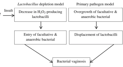
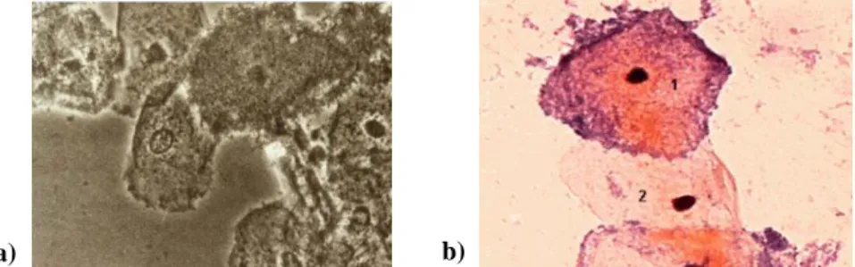
Figure 1.2. Two models explaining the pathogenesis of BV, the loss of lactobacilli model suggests that there is a decrease in the production of H2O2 by lactobacilli as the main event, which allows the proliferation of anaerobic bacteria, resulting in BV; while the primary pathogenic model suggests that entry of facultative anaerobic bacteria will cause loss of lactobacilli resulted in BV. Adapted from Srinivasan and Fredericks, (2008). ... 5 Figure 1.3. "Clue cells" in both images (original magnification, x400). a) some cells have irregular edges that define the "clue cells", those that do not have this characteristic are not "clue cells"; b) represents the normal vaginal bacterial microscopy image bacterioscopy, in which a cell 1 is a "clue cell" in which the G. vaginalis bacterium is adhered to the surface of cell, while in the cell 2 does not. Adapted from Livengood, (2009). ... 6 Figure 1.4. a) Epithelial cell totally covered by Gram-positive bacteria (clue cell), G.
vaginalis. Source: http://farm3.static.flickr.com/2056/2369779554_f4d9b48760_z.jpg,
accessed August 1, (2012); b) Gram staining of Gram-variable G. vaginalis AMD bacterium that we prepared (original magnification, x1000). ... 10 Figure 1.5. Scanning electron microscope (SEM) of G. vaginalis biofilm. Source: http://www.gardnerella.mic.vcu.edu/images/gv_em.jpg, accessed August 1, (2012). ... 10 Figure 1.6. Gram staining shows Gram-positive bacteria, with A. vaginae visible as single cells, in pair or short chains. Adapted from Geißdörfer et al., (2003). ... 12 Figure 1.7. Gram staining of vaginal smear of clinical sample that we received shows curved rods, morphotypes associated with Mobilluncus spp. (original magnification, x1000). ... 12 Figure 1.8. Growth of the bacterium G. vaginalis AMD in CBA selective medium. ... 14 Figure 1.9. Proposed mechanisms for antibiotic resistance of biofilms. Yellow region: is characterized by low penetration, the antibiotic (yellow) may fail to penetrate the
xii
surface layers of the biofilm. Green region: some of the bacteria can differentiate themselves to protect the phenotypic state (green). Red region is in regions of lack of nutrients or accumulation of waste products (red), the action of the antibiotic can be antagonized. Adapted from Stewart and Costerton, (2001). ... 17 Figure 1.10. Schematic representation of the stages of microbial biofilm forming multiple bacteria development. The progression of biofilm, initiated with bacterial contamination, followed by colonization, leading to critical colonization and systemic infection. Adapted from Phillips et al., (2008). ... 19 Figure 1.11. Biofilms formation of strains of G. vaginalis and anaerobic bacteria associated with BV. a) Bacteria were grown anaerobically in Brain Heart Infusion Broth supplemented with glucose (sBHIG) at 37°C for 24 h. The adhered cells were stained with safranin; b) Quantitative assessment of the capability of formation of the biofilm formation was made by dissolving safranin stain in 33% acetic acid and measuring optical density (OD) OD562. Adapted from Patterson et al., (2010). ... 20 Figure 2.1. Method of making a streak plate to obtain pure cultures. a) Loop is flame-sterilized, and then a loopful of one colony is removed from plate. Streak is made over a sterile agar plate, spreading out the microorganisms. Following the initial streak, subsequent streaks are made at angles to it, the loop being re-sterilized with heat between streaks. Adapted from Seeley, Seeley et al., (1991); b) Colonies of the bacterium G. vaginalis 5-1 grown on CBA plates. This bacterium was isolated by Patterson et al., (2010) from women without BV as diagnosed by the Nugent gram stain scoring system. ... 28 Figure 2.2. Gram staining of isolates that we prepared (original magnification, x1000). Gram stain showing a) Gram-positive (purple) Staphylococcus epidermidis bacterium; b) Gram-negative (pink) Escherichia coli bacterium. ... 29 Figure 3.1. Structure of the antibiotics used in the study described in this chapter: (A) metronidazole, (B) tinidazole, (C) clindamycin, (D) vancomycin and (E) rifampicin. . 55 Figure 3.2a. Intrinsic ability of biofilm formation of each strain in sBHI. This assay was performed at 37ºC, during 48 h. After this time the suspension was removed, and biofilms were subjected to one wash with 1x PBS to analyze the strength of attachment. The percent growth was calculated as OD 600 nm biofilm/ (OD 600 nm biofilm +OD 600 nm planktonic). Error bars represent the standard deviation of 3 independent experiments.
xiii
Statistical differences are marked with* (P < 0.05) from biofilm formation of these strains with washing or no washing of the cells with PBS buffer after removed planktonic cells were analyzed using independent-samples T-test. ... 59 Figure 3.2b. Intrinsic ability of biofilm formation of each strain in different media. This assay was performed at 37ºC, during 48 h. After this time the suspension was removed, and biofilms were subjected to one wash with 1x PBS to analyze the resistance thereof. The percentage growth was calculated as OD 600 nm biofilm/ (OD 600 nm biofilm + OD 600 nm planktonic). Error bars represent the standard deviation of 3 independent experiments. Statistical differences are marked with* (P < 0.05) from biofilm formation of these strains with washing or no washing of the cells with PBS buffer after removed planktonic cells were analyzed using independent-samples T-test. ... 60
xv
LIST OF TABLES
Table 1.1. Scheme for grading Gram stained vaginal contents. Adapted from Livengood, (2009). ... 7 Table 1.2. In vitro susceptibility of 93 strains of G. vaginalis to 25 antimicrobial agents. Adapted from Kharsany et al., (1993). ... 8 Table 1.3. Koch´s postulates. Adapted from Evans, (1993). ... 11 Table 1.4. Detailed composition of the vaginal microflora of 515 vaginal swab samples, as determined by culture and tDNA intergenic spacer PCR (tDNA-PCR) based identification. Adapted from Verhelst et al., (2005). ... 13 Table 2.1. Primer sequences used for the PCR assays used in this study. ... 31 Table 2.2. Microorganisms isolated from 3 clinical vaginal swabs from women with BV (UM054, UM027, UM034 and UM035). ... 35 Table 2.3. Microorganisms isolated from 4 clinical vaginal swabs from women without BV (UM016, UM022, UM031 and UM042). ... 38 Table 2.4. Results of sequencing of microorganisms isolated of vaginal clinical samples using BLAST. ... 41 Table 3.1. Characteristics of the antibiotics used in the study described in this chapter. ... 55 Table 3.2. Qualitative analysis of biofilm formed for each isolated bacterium in 9 different media, based on the Patterson et al., (2010) method. ... 58 Table 3.3. In vitro susceptibilities of microorganisms isolated from clinical samples to 5 antimicrobial agents: Metronidazole (MD), Tinidazole (TZ), Clindamycin (CM), Rifampin (RF) and Vancomycin (VM) using method described by Harwich et al., (2010) in 3.3.2 section of Materials and Methods. ... 65
xvii
NOMENCLATURE
Symbols
P Significance value ºC Temperature min Time (minutes)
s Time (seconds)
ABBREVIATIONS
BAP Biofilm-associated protein BHI Brain Heart Infusion
BLAST Basic Local Alignment Search Tool
BV Bacterial Vaginosis CBA Columbia Blood Agar
CM Clindamycin
DGGE Denaturing Gradient Gel Electrophoresis DNA Deoxyribonucleic acid
EDTA Ethylenediaminetetraacetic acid
ELISA Enzyme-Linked Immunosorbent Assay EPS Extracellular Polysaccharides
FBS Fetal Bovine Serum HBT Human bilayer Tween
HIV Human Immunodeficiency Virus IgA Immunoglobulin A
LAM 1 Laboratory of Applied Microbiology 1 LB Luria Broth
LBG Luria Broth supplemented with Glucose MD Metronidazole
MIC Minimum Inhibitory Concentrations MRS De Man-Rogosa and Sharpe agar
MRSG De Man-Rugosa and Sharpe agar supplemented with Glucose NCBI National Center for Biotechnology Information
xviii
PBS Phosphate Buffered Saline PCR Polymerase Chain Reaction rDNA ribosomal DNA
RF Rifampin
rRNA Ribosomal Ribonucleic acid sBHI Brain Heart Infusion supplemented
sBHIG Brain Heart Infusion supplemented Glucose SEM Scanning electron microscope
SPSS Statistical package for the social sciences TAE Tris-acetate-EDTA
tDNA-PCR tDNA intergenic spacer PCR
TMPD NNN'N'-tetramethyl-p-phenylenediamine TSA Tryptic Soy Agar
TSB Tryptic Soy Broth
TSBG Tryptic Soy Broth supplemented with Glucose TZ Tinidazole
VLY Vaginolysin VM Vancomycin
xix
LIST OF PUBLICATIONS
Abstracts and Posters
Alves P, Castro J, Sousa C, Cereija T, Henriques A, Cerca N, (2012) Biofilm
formation potential of clinical isolates associated with bacterial vaginosis. In Biofilms 5, 10-12 December 2012, Paris, France.
Salgueiro D, Machado A, Alves P, Martinez J, Henriques A, Cerca N, (2011) Presence of Gardnerella vaginalis in healthy Portuguese women – a pilot study. In Microbiotec, 1-3 December 2011, Braga, Portugal.
xx
xxi
I dedicate this thesis to my parents, sister and boyfriend
"Nós nunca nos realizamos. Somos dois abismos - um poço fitando o céu." Fernando Pessoa
CHAPTER 1
General Introduction
Chapter 1 | 3
1.1. Vaginal flora microenvironment
Microbial communities have a strong influence in human health and quality of life. Therefore, the bacterial community of the human vagina can have a profound impact on women's health, since microorganisms play a critical role in determining the biochemical profile and inflammation of the vaginal environment (Srinivasan and Fredericks, 2008).
Despite decades of investigations, based on growth technologies of human vaginal flora, recent studies using culture independent methods significantly increased the knowledge of bacterial diversity in this important niche (Donachie et al., 2007). Diverse microorganisms can be found in healthy women that are mostly colonized with lactobacilli such as Lactobacillus crispatus, Lactobacillus jensenii and Lactobacillus
gasseri (Zhou et al., 2004) though a variety of other bacteria may be present (Vásquez et al., 2002) (Figure 1.1).
Figure 1.1. Gram staining of vaginal fluid smear from clinical sample of healthy woman that we prepared
(original magnification, x1000). There is a predominance of Lactobacillus spp. which are responsible for suppressing the growth of other species.
The definition of a healthy vaginal environment is more complex than originally thought due to bacterial diversity observed among individuals (Kim et al., 2009). Thus, the identity and diversity of these populations remain largely unknown and the complex interaction between the various members of the vaginal flora is still poorly understood (Zhou et al., 2004). This means that it is essential to have accurate knowledge about the microbial ecosystem of the human vagina of healthy women, because there is still much to understand about how bacterial communities in this niche promote health and facilitate disease (Srinivasan and Fredericks, 2008).
4 | Chapter 1
1.2. Bacterial Vaginosis
Bacterial vaginosis (BV) is the main vaginal disorder of women in reproductive age worldwide (Harwich et al., 2010) and, although not life threatening, leads to increased risk of preterm delivery (Hillier et al., 1995), and more severe gynecological infections such as pelvic inflammatory disease (Haggerty et al., 2004) and increase risk of Human Immunodeficiency Virus (HIV) infection acquisition (Schmid et al., 2000).
The etiology of this condition has been long debated, and despite the impact on women's health, little is known about its cause and pathogenesis. This microecologic disorder is characterized by not being associated with a specific etiologic agent (Aroutcheva et al., 2001).
Women without BV typically show Gram-positive rods, revealing a predominance of lactobacilli, particularly L. crispatus and L. jensenii (Livengood, 2009). However, it is likely that over time, microbial communities in the human vagina may be affected in a negative way due to various factors, such as woman's age, hormonal fluctuations (menses, or contraception), pregnancy, sexual activity (frequency of sex and numbers of sexual partners), health status (such as diabetes, infections) and as well as various lifestyle habits and hygiene practices (such as douching) (Srinivasan and Fredericks, 2008).
During BV, most of the beneficial bacteria (like L. crispatus, L. jensenii, L.
gasseri) (Srinivasan and Fredericks, 2008) (Figure 1.1) are replaced, by concomitant
overgrowth of anaerobic or facultative bacteria (Fredricks et al., 2005) generally Gram-negative or Gram-variables cocci and rods, normally associated with the gastrointestinal tract. Cultures of vaginal fluids from individuals with BV microorganisms typically present bacteria such as Gardnerella vaginalis and a combination of other bacteria such as, Prevotella, Porphyromonas, Mobiluncus spp., Atopobium vaginae and Mycoplasma spp. (Livengood, 2009).
Lactobacilli are responsible for promoting a healthy ecosystem, producing lactic acid, hydrogen peroxide (H2O2) and bacteriocins, which have antimicrobial properties that inhibit microorganisms that are pathogens of this niche, maintaining a healthy ecosystem (Srinivasan and Fredericks, 2008). The growth of anaerobic bacteria is associated with increased production of proteolytic enzymes, which break the vaginal epithelial peptides into a variety of amines that at, an elevated pH, becomes volatile causing bad odor. Amines are associated with an increase in transudation and exfoliation
Chapter 1 | 5
of squamous epithelial cells, resulting in a white fluid that is expelled from the vagina, creating an imbalance in the vaginal flora (Sobel, 2000).
It is unknown if the primary event of BV is the loss of lactobacilli or the acquisition of facultative anaerobic bacterial communities found in this infection, or if these two events are simultaneous processes (Figure 1.2). It is also possible that the primary etiologic agent is another unknown factor, that changes in vaginal flora reflect a downstream event in the pathogenesis of BV (Srinivasan and Fredericks, 2008).
Figure 1.2. Two models explaining the pathogenesis of BV, the loss of lactobacilli model suggests that
there is a decrease in the production of H2O2 by lactobacilli as the main event, which allows the
proliferation of anaerobic bacteria, resulting in BV; while the primary pathogenic model suggests that entry of facultative anaerobic bacteria will cause loss of lactobacilli resulted in BV. Adapted from Srinivasan and Fredericks, (2008).
1.2.1. Clinical features and diagnosis of BV
The diagnosis of BV is generally done through a series of clinical criteria from pelvic examination and microscopic examination of the exudate (Amsel's criteria), or by microscopic interpretation of vaginal fluid by Gram staining technique (Nugent criteria) (Srinivasan and Fredericks, 2008). The Amsel criteria are the most frequently method used to diagnose BV in most countries, including Portugal (Henriques et al., 2012). At least 3 of 4 Amsel criteria must be present to establish the diagnosis of BV, including the increase of pH of vaginal fluid > 4.5; positive "wiff test" in the detection of an odor similar to rotten fish (with the addition of 10% potassium hydroxide (KOH ) in a sample containing vaginal fluid); presence of ―clue cells‖ (> 20%) in vaginal fluid where the
Decrease in H2O2-producing
lactobacilli
Overgrowth of facultative & anaerobic bacterial
opportunists
Lactobacillus depletion model Primary pathogen model
Entry of facultative & anaerobic bacterial
Displacement of lactobacilli
Bacterial vaginosis Insult
6 | Chapter 1
vaginal epithelial cells are coated with bacteria creating indistinct borders (Figure 1.3) and white vaginal discharge, with homogeneous characteristics (Srinivasan and Fredericks, 2008).
Figure 1.3. "Clue cells" in both images (original magnification, x400). a) some cells have irregular edges
that define the "clue cells", those that do not have this characteristic are not "clue cells"; b) represents the normal vaginal bacterial microscopy image bacterioscopy, in which a cell 1 is a "clue cell" in which the G.
vaginalis bacterium is adhered to thesurface of cell, while in the cell 2 does not. Adapted from Livengood, (2009).
For the clinical diagnosis of BV, Nugent’s criteria form a standardized method of Gram stain interpretation of vaginal samples to detect the shift of normal vaginal flora to other microorganism allowing the scoring system of vaginal smears (Delaney and Andrew, 2001). The Nugent score is calculated by assessing the presence of some microorganisms in vaginal smears that are graded on a scale based on the presence or absence of some microorganisms, such as large Gram-positive rods - Lactobacillus morphotypes (decrease in Lactobacillus - scored as 0 to 4), small Gram-variable rods - G.
vaginalis morphotypes (scored as 0 to 4), curved Gram-variable rods - Mobiluncus spp.
morphotypes (scored as 0 to 2). Therefore, the score can range from 0 to 10 and score between 7 to 10 are classified as BV (Sha et al., 2005) (Table 1.1).
For these two evaluation criteria of vaginal flora samples, Nugent scoring allows the evaluation of a change in vaginal flora continuously. Because Amsel criteria are dependent on the accuracy of the clinician, the Nugent score was favored for diagnosing BV due to its higher sensitivity and reproducibility (Onderdonk et al., 1977). Nevertheless, assessment of from vaginal smear slides samples can also be subjective and thus requires the experience of a reader (Chaijareenont et al., 2004).
b) a)
Chapter 1 | 7
Table 1.1. Scheme for grading Gram stained vaginal contents. Adapted from Livengood, (2009).
Score
Lactobacillus Morphotypes
Gardnerella and Bacteroides spp. Morphotypes Curved Gram-Variable Rods 0 4 + 0 0 1 3 + 1 + 1 + or 2 + 2 2 + 2 + 3 + or 4 + 3 1 + 3 + 4 0 4 + Total Interpretation 0 – 3 Normal 4 – 6 Intermediate 7 - 10 Bacterial vaginosis
Morphotypes are scored as the average number seen per oil immersion field. Note that less weight is given to curved Gram-variable rods. Total score = lactobacilli + G. vaginalis and bacteroides spp. + curved rods. Quantitation: 0, No morphotypes present; 1 +, < 1 morphotype present; 2 +, 1 to 4 morphotypes present; 3 +, 5 to 30 morphotypes present; 4 +, 30 or more morphotypes present.
1.2.2. Treatment of BV
Current research has revealed more detailed aspects for the treatment of BV. Aims of the treatments of BV have two main goals: to eradicate the anaerobic microorganisms, and to allow growth of lactobacilli producing H2O2.
Many studies have been performed in order to find antibiotics that can specifically eradicate the anaerobic microorganisms that cause the symptoms of BV, trying to overcome the resistance that these organisms sometimes show to multiple antibiotics. Results of several experiments demonstrate that the fact that in BV the vaginal flora is comprised of several Gram-variable bacteria, like G. vaginalis, antimicrobials specifically active against Gram-positive or Gram-negative bacteria, have little or no action against Gram-variables microorganisms, explaining the common resistance to them (Gilbert et
al., 1997).
In an attempt to find antimicrobial agents active against microorganisms normally associated with BV, including the bacteria G. vaginalis (Kharsany et al., 1993), various concentrations of antimicrobials were tested to find the minimum concentration capable of inhibiting the growth of pathogenic microorganisms, called a Minimum Inhibitory Concentration (MIC), as shown on table 1.2 (Kharsany et al., 1993).
8 | Chapter 1
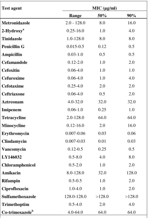
Table 1.2. In vitro susceptibility of 93 strains of G. vaginalis to 25 antimicrobial agents. Adapted from
Kharsany et al., (1993).
Test agent MIC (µg/ml)
Range 50% 90% Metronidazole 2.0 - 128.0 8.0 16.0 2-Hydroxya 0.25-16.0 1.0 4.0 Tinidazole 1.0-128.0 8.0 8.0 Penicillin G 0.015-0.5 0.12 0.5 Ampicillin 0.03-1.0 0.5 0.5 Cefamandole 0.12-2.0 1.0 2.0 Cefoxitin 0.06-4.0 1.0 1.0 Cefuroxime 0.06-4.0 1.0 4.0 Cefotaxime 0.25-4.0 2.0 2.0 Ceftriaxone 0.06-4.0 0.5 2.0 Aztreonam 4.0-32.0 32.0 32.0 Imipenem 0.06-1.0 0.25 1.0 Tetracycline 2.0-128.0 64.0 64.0 Minocycline 0.12-16.0 2.0 16.0 Erythromycin 0.007-0.06 0.03 0.06 Clindamycin 0.007-0.03 0.01 0.03 Vancomycin 0.12-0.5 0.25 0.5 LY146032 0.5-8.0 4.0 8.0 Chloramphenicol 0.5-2.0 1.0 2.0 Amikacin 8.0-128.0 32.0 128.0 Rifampin 0.5-0.5 1.0 2.0 Ciprofloxacin 1.0-4.0 1.0 2.0 Sulfamethoxazole 128.0-128.0 >128.0 >128.0 Trimethoptim 0.5-4.0 2.0 4.0 Co-trimoxazoleb 4.0-64.0 64.0 64.0
a 2-Hydroxymetabolite of metronidazole [1-(2-hydroxyethyl)-2 hydroxymethyl- 1,5-nitroimidazole]
b sulfamethoxazole-trimethoprim in a 19:1 ratio.
Recent investigations indicate that following intravaginal treatment with antibiotics such as metronidazole or clindamycin (Santiago et al., 2012) the cure rates were between 80 - 90% at the end of treatment (Livengood, 2009). However, a study suggests that three months after the end of therapy, there was a possible recurrence of BV
Chapter 1 | 9
(Polatti, 2012). A study in Portugal on the perception of specialist doctors regarding to BV, also reported that the recurrence of these microorganisms after antimicrobial chemotherapy is high (Henriques et al., 2012). This relapse can occur due to inadequate treatment of microorganisms associated with BV, due to the resistance to metronidazole and others antibiotics or due to the fact that these microorganisms have recently been recognized as biofilm-forming (described in section 1.4.3). Because of these complications, with relapse of BV, new solutions have been proposed and Nyijesy and his collaborators demonstrated that the regeneration of lactobacilli using preparations of probiotic lactobacilli, which are known to be specialized organisms which dominate the healthy vagina, is likely to happen (Nyirjesy et al., 2006). However, this research has been hampered by the complications involving both the application of probiotic
Lactobacillus spp., such as dominance by these organisms, such as L. crispatus, L. rhamnosus, and L. reuteri (Livengood, 2009) over other existing microorganisms.
1.3. The microbiology of BV and role of recently defined BV-associated
bacteria
1.3.1. The etiology of BV
BV is considered a polymicrobial condition, and recently several new fastidious bacteria have been found to be associated with BV.
An attempt to find a single etiological agent to explain the pathogenesis of BV has been inconclusive (Ferris et al., 2007). Particular attention has been attributed to G.
vaginalis, as this bacterium can be found in the vaginal flora of a majority of women with
BV (95% of cases); however is also present in 50% of healthy vaginal flora (Livengood, 2009). The bacteria Mobiluncus spp., curved bacilli highly mobile, is found only when BV is detected, but only 50% of cases of BV (Nyirjesy et al., 2007). A. vaginae is an anaerobic Gram-positive, as G. vaginalis, is found in more than 95% of BV, but is also present in normal flora of the vagina of healthy women (Ferris et al., 2007).
1.3.2. BV-associated bacteria
G. vaginalis is a Gram-variable bacterium, which was first described in 1953 by
10 | Chapter 1
vaginalis was originally proposed by Gardner and Dukes in 1955, due to the colony
morphology of the organism and its biochemical profile. Subsequently, metabolic requirements and thorough analysis of preparations using Gram staining, it was found that the organism morphologically resembled bacilli diphtheroids and named
Corynebacterium vaginale (Greenwood et al., 1979). However, due to their variable
Gram staining reaction (not being positive or negative typically) was subsequently named
Gardnerella (Figure 1.4).
Figure 1.4. a) Epithelial cell totally covered by Gram-positive bacteria (clue cell), G. vaginalis. Source:
http://farm3.static.flickr.com/2056/2369779554_f4d9b48760_z.jpg, accessed August 1, (2012); b) Gram staining of Gram-variable G. vaginalis AMD bacterium that we prepared (original magnification, x1000).
Gardnerella belongs to the Bifidobacteriaceae family, and their cells are small,
pleomorphic bacilli (Figure 1.5) in which the length and Gram stain may vary depending on the growth medium (Catlin, 1992).
Figure 1.5. Scanning electron microscope (SEM) of G. vaginalis biofilm. Source: http://www.gardnerella.mic.vcu.edu/images/gv_em.jpg, accessed August 1, (2012).
G. vaginalis is considered a fastidious and anaerobic microorganism that requires
a complex growth medium (Harwich et al., 2010). Studies of microorganism identification using metabolic methods indicate that it is a catalase-negative microorganism, exhibits activity α-glucosidase, hydrolysis of starch and hippurate, acid phosphatase activity, salt tolerance and can use carbohydrates such as dextrins, fructose,
Chapter 1 | 11
glucose, maltose, ribose, starch and sucrose from fermentation for growth (Catlin, 1992; Harwich et al., 2010).
Researchers such as Gardner and Dukes identified this bacterium as a major etiologic agent of BV, fulfilling all the Koch's postulates (Table 1.3). However, later studies demonstrated some failures in these experiments which suggested that there were other factors in addition to G. vaginalis that were important in the induction of the disease, meaning that these bacteria were not the etiologic agent in BV. One of the Koch's postulates requires pathogenic microorganisms to be found in all cases of the disease and not be found in individuals who do not have the disease (Evans, 1993). In the case of G.
vaginalis, it was found in many cases of BV, but can also detected in 50 - 60% of women
who have no visible symptoms of BV, thus failing one of Koch’s postulates (Fredricks and Relman, 1996).
Table 1.3. Koch´s postulates. Adapted from Evans, (1993).
The role of G. vaginalis in BV has been extensively debated since it can be both present in the genital tract of healthy women (Hyman et al., 2005) as in women with BV. However, vaginal epithelial tissue of women with BV presents a larger number of anaerobic microorganisms (Swidsinski et al., 2005). Moreover, recent studies show that the biotypes of G. vaginalis isolates from healthy women differ from those isolated from women with BV (Numanovic et al., 2008). The lack of genetic characterization of this microorganism leaves open the possibility that different pathogenic and nonpathogenic strains or subspecies exists (Harwich et al., 2010).
The discovery of the presence of A. vaginae in the vaginal ecosystem improves the basic understanding of the pathogenesis of BV (Polatti, 2012). The bacterium A.
The etiologic microbe should be found in every case of the disease
The etiologic microbe should not be found in subjects without disease (specificity)
The etiologic microbe should be isolated in pure culture on lifeless media and be capable of causing the characteristic disease anew upon inoculation in a susceptible host
The etiologic microbe should be reisolated from the experimentally inoculated host
12 | Chapter 1
vaginae belong to the family Coriobacteriaceae. It is thought that this newly identified
bacterium is one of the causes of complications that arise in BV (Fredricks et al., 2005). The genus Atopobium was described for the first time in 1992, and includes bacteria previously classified as lactobacilli (Rodriguez et al., 1999). Relatively to morphology, Gram stain shows varied morphology, as their cells appear as Gram-positive elliptical cocci or rod-shaped organisms and can be visible as single cells, in pairs or in short chains (Figure 1.6). A. vaginae is an anaerobic bacteria, that cannot be easily isolated by classical microbiological methods and it is rarely detected in healthy women vaginal fluid but is commonly found in the vagina of patients with BV (Verhelst et al., 2004).
Figure 1.6. Gram staining shows Gram-positive bacteria, with A. vaginae visible as single cells, in pair or
short chains. Adapted from Geißdörfer et al., (2003).
Mobiluncus spp. is another anaerobic organism most commonly associated with
BV. These fastidious curved rods are only rarely cultured from vaginal smears of women without BV, yet are highly predictive of BV if found on Gram stain or wet-preparation examination of vaginal secretions (Thomason et al., 1984) (Figure 1.7).
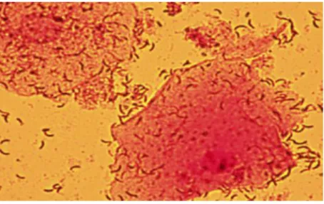
Figure 1.7. Gram staining of vaginal smear of clinical sample that we received shows curved rods,
morphotypes associated with Mobilluncus spp. (original magnification, x1000).
The presence of Mobiluncus spp., specially M. curtisii and M. mulieris is highly specific, although not sensitive, for the diagnosis of BV (Roberts et al., 1985). The
Chapter 1 | 13
presence and persistence of M. curtisii, detected by Polymerase Chain Reaction (PCR), was found to be strongly associated with recurrence of BV. Whether this organism is truly pathogenic or simply a marker for greater disturbances of vaginal flora remains unknown (Meltzer et al., 2008).
To better understand BV there is a need to isolate more BV-associated bacteria, such as the species detected by molecular methods (Table 1.4).
Table 1.4. Detailed composition of the vaginal microflora of 515 vaginal swab samples, as determined by
culture and tDNA intergenic spacer PCR (tDNA-PCR) based identification. Adapted from Verhelst et al., (2005).
Bacterial vaginosis-related anaerobe organisms
Actinomyces neuii Gemella morbilloriumb
Aerococcus christensenii Mobiluncus curtisii
Anaerococcus tetradiusb Mycoplasma hominis
Anaerococcus vaginalisb Peptoniphilus spicies.b
Atopobium vaginae Peptostreptococcus sp.
Bacteroides ureolyticus Prevotella bivia
Dialister species Prevotella ruminicola
Finegoldia magnab Prevotella species
Gardnerella vaginalis Varibaculum cambriense
b Formerly known as Peptostreptococcus
1.4. Vaginal microbial diversity
1.4.1. Isolation - Culture perspective
Culture studies provide critical results on the phenotypic characteristics of the microorganisms that are not easily obtained from molecular studies. Moreover, a microorganism culture allows manipulation of these organisms in experimental laboratory and testing hypotheses about their pathogenesis and virulence factors. Thus, studies using the culture of microorganisms remain an important area of research in vaginal microbiology, despite the limitations of this approach (Donachie et al., 2007).
Burton and Reid published a study which involved the isolation of the microorganisms in culture using selective and non-selective media and subsequent identification by phenotypic techniques. The use of a wide range of PCR primers helped to maximize the diversity of species detected and a large number of media and growth
14 | Chapter 1
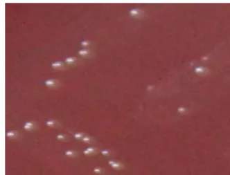
conditions may be required for optimal isolation of diverse bacterial species (Burton and Reid, 2002). Selective media such as MacConkey agar, mannitol and Tryptic Soy Agar (TSA) with 5% sheep blood may be useful to estimate the number of aerobic and anaerobic bacteria in vaginal samples. Some of the selective media/ semi-selective agar include Rugosa, De Man-Rugosa and Sharpe (MRS) agar and Human bilayer Tween (HBT) and Columbia Blood Agar (CBA) with 5% horse blood are used to help identify microorganisms presents in vaginal swabs samples (Figure 1.8). In some of the media, such as the CBA it is possible to observe the hemolysis arising from the growth of some microorganism (eg. bacterium G. vaginalis shows β-hemolysis in CBA) (Totten et al., 1982).
Figure 1.8. Growth of the bacterium G. vaginalis AMD in CBA selective medium.
Thus, using approaches based on the culture of organisms, it is possible to identify
G. vaginalis and anaerobic bacteria such as Neiserria gonorrhea, Streptococcus spp., Escherichia coli, Trichomonas vaginalis, Prevotella, Porphyromonas, Peptostreptococcus, Mobiluncus and Mycoplasma, which may be largely associated with
disturbances in the microbial flora in women with BV (Kalra et al., 2007).
1.4.2. Isolation - Molecular Perspective
Results of different research groups confirmed that the human vagina hosts numerous species of bacteria which are not yet cultured or are not easily identified by culture methods (Srinivasan and Fredericks, 2008).
With advances in technology and decreasing costs of sequencing, there is a better knowledge of the microflora of the human vagina. As the conditions of the vagina may be transient and dependent on many factors, molecular studies provide results able to demonstrate features of the vaginal microbiota under specific conditions (Hugenholtz et
Chapter 1 | 15
Burton and Reid, were also the first researchers to examine the microbiota of the vaginal niche using a wide range of molecular methods such as PCR and Denaturing Gradient Gel Electrophoresis (DGGE) and found that different women with BV had different profiles indicating heterogeneity in bacterial composition in subjects with BV (Burton and Reid, 2002). Many researchers such as Verhelst et al., (2004), Fredericks et
al., (2005), contributed to the current knowledge of BV, and there are many studies that
have been performed in order to intensively investigate the bacterial flora of the human vagina. These studies, using methods of cultivation and molecular approaches aimed to reveal which microorganisms are the most persistent in BV.
Although progress has occurred in defining the composition of vaginal flora using Deoxyribonucleic acid (DNA) amplification, the rapid accumulation of data resulted in no therapeutic advantage. Treatment options remain limited and often have unsatisfactory results, especially in relation to the frequent relapse of symptomatic disease, leading to great frustration among patients and professionals (Srinivasan and Fredericks, 2008).
1.4.3. Virulence factors of G. vaginalis
The normal flora of the human vagina is a diverse set of microorganisms that live in a dynamic relationship in the colonized epithelium. Although the colonization of the lower female genital tract is usually benign, it is noted that the vast majority of infections involving female pelvic structures arise from organisms that are members of the normal flora (Larsen, 1994) due to a shift in the microbial balance caused by reasons discussed previously.
Although there is much controversy surrounding the etiology of BV, G. vaginalis remains the most studied organism and potential primary agent of BV. The following factors its pathogenic potential.
1.4.3.1. Initial adhesion to epithelium
Microorganisms can improve their virulence factors by producing compounds which act against host defense or in response to them. This virulence attributes include: interacting with the host defense, adhesion to host tissue, adjustment of the characteristics of virulence and production of toxins (Larsen, 1994). The organism adhesion to the epithelial surface is a significant aspect in the life of microorganisms in the vagina. The bacterium G. vaginalis, which is present in asymptomatic women and in abundance in
16 | Chapter 1
individuals with BV, is known for its ability to adhere to epithelial cells (Patterson et al., 2010). Boustouller et al. attribute the responsibility of adhering to the epithelial cells to pilli, while Scott and Smyth believe that the fibrillar matrix is responsible for foreign citoadhesion, with pilli functioning as a hemagglutinin (Scott and Smyth, 1987; Boustouller et al., 1989). It should be considered that although cell adhesion is important in the disease process, the fixation to the tissue does not characterize an organism as pathogenic. In fact, vaginal lactobacilli have adhesins that probably serve to promote the stable colonization of the vaginal epithelium, possibly with beneficial effects for the host (Nagy et al., 1992). Thus, if a microbial species is a highly virulent or only modestly virulent it is due to the interaction with the host tissue through adhesives factors.
Clearly, organisms that are usually associated with the host, described as belonging to the normal commensal flora have had little attention as producers of toxins. Although, the production of toxins with subtle effects on the physiology of the host, or his immunity, may play a role in the virulence of this microorganism (Larsen, 1994).
1.4.3.2. Cytotoxicity of G. vaginalis
While G. vaginalis can be isolated in 95% of cases of BV, studies with healthy subjects indicate that pure cultures of bacteria do not always cause BV, perpetuating the exiting doubts about it is potential as a pathogen (Catlin, 1992; Marrazzo et al., 2008). However, the connection between the cytolysin secreted by G. vaginalis, called recently vaginolysin (VLY), and BV has been made (Gelber et al., 2008). The production of the cytolysin by G. vaginalis was first reported in 1990 but was only recently named VLY. VLY, a cholesterol-dependent cytolysin recognizes the complement of the molecule CD59 on the target cell surface, which accounts for the specificity for human erythrocytes epithelial cells (Madden et al., 2001), leading to cell death (Gelber et al., 2008). Immunoglobulin A (IgA) antibodies against VLY have been linked to the mucosal immune response during BV, increasing the emphasis on the role of VLY in the pathogenesis of BV (Cauci et al., 2002). Studies have examined the levels of cytotoxic activity of anaerobes associated with BV, and found that only the bacteria G. vaginalis was able to induce lysis of vaginal epithelial cells (Patterson et al., 2010).
Other virulence factors produced by G. vaginalis include sialidase and prolidase, which are two hydrolytic enzymes that may play a role in the degradation of many
Chapter 1 | 17
mucosa protective factors, such as mucins and contribute to the detachment and exfoliation of epithelial cells (Cauci et al., 2008).
1.4.3.3. Formation of biofilm
The adherence to the epithelium is the first step in the pathogenesis of a biofilm-forming pathogen as it must first adhere to host tissues in order to avoid the clearance by means of host defense mechanisms, such as urine flow and the flow of vaginal secretions, and be able to form a biofilm (Patterson et al., 2010). Biofilm formation is an important virulence factor because it confers tolerance to antibiotics (Figure 1.9) and increased resistance to host immune defense (Cantón et al., 2005).
Figure 1.9. Proposed mechanisms for antibiotic resistance of biofilms. Yellow region: is characterized by
low penetration, the antibiotic (yellow) may fail to penetrate the surface layers of the biofilm. Green region: some of the bacteria can differentiate themselves to protect the phenotypic state (green). Red region is in regions of lack of nutrients or accumulation of waste products (red), the action of the antibiotic can be antagonized. Adapted from Stewart and Costerton, (2001).
Microbial biofilms are communities of microorganisms that are closely associated with each other in a way that they are attached to a surface, in this case to vaginal epithelial cells, forming a porous structure where the microbial cells are involved in extracellular matrix polymers and are able to concentrate products of their own
Antibiotic (yellow) may fail to penetrate beyond the surface layers of the biofilm
Some of the bacteria may differentiate into a protect phenotypic state (green)
In zones of nutrient depletion or waste product accumulation (red), antibiotic action may be antagonized
18 | Chapter 1
metabolism, together with ions and nutrients that are captured from the environment that surrounds them (Costerton, 1995). This matrix consists of several components including proteins, extracellular polysaccharides (EPS), nucleic acids and other substances (Davey and O´toole, 2000) and represents approximately 85% of the biofilm (Donland and Costerton, 2002). The synthesis of polymer matrix appears to be regulated by a variety of factors in which the cell surface adhesion is very important. The exopolymers serve two main functions within the biofilm: first, the polysaccharides are produced in large amounts after initial fixation of the cell surface, being suggested to act as ―cement‖ with which bacteria can reinforce the primary mechanisms of adhesion (Figure 1.10). Then, due to the existence of an extracellular polymer matrix (Gilbert et al., 1997) bacteria become protected against biocides, antibiotics and surfactants, by limiting diffusion of the agents of the surrounding medium through a combination of ionic interactions and molecular events, and are also protected from bacteriophages and other phagocytic predators such as protozoa (Dunne, 2002). Biofilms can release antigens and stimulate production of antibodies, but the bacteria living in biofilms are generally resistant to these defense mechanisms, providing the basis for persistent infections (Costerton, 1995). The microenvironment generated allows a symbiotic relationship of the species that make up the biofilm, providing necessary nutrients and allowing in some cases, due to the close proximity between different species involved, the transmission of genetic elements (Roberts et al., 1999).
Biofilms can be composed of populations derived from a single species or a community of diverse species but, in both cases, the development of biofilm behavior requires the multistage approach described above (Davey and O´Toole, 2000). Biofilm formation and its structure are affected by many conditions such as surface properties, nutrient availability, hydrodynamics and microbial community (Davey and O´Toole, 2000). The biofilm cells have different metabolic activities, depending on their position within the biofilm and can change over time.
Many species have been shown to use the same steps in biofilm formation, which include 1) an initial adhesion to the surface, 2) formation of the microcolonies 3) ripening microcolonies in the developing matrix thereby allowing the maturation of biofilm and 4) dispersion of planktonic bacteria (Figure 1.10). It is believed that the biofilm formation bacteria start when certain environmental factors trigger the fluctuations in cell-population density (Miller and Bassler, 2001) through a mechanism called quorum
Chapter 1 | 19
sensing (O´Toole et al., 2000). Since these communities are dynamic, there is also a great exchange of information through the mechanism of quorum sensing, which allows them to better adapt their phenotype and gene expression to changes in the surrounding environment. Many bacteria use this mechanism to regulate a diverse array of physiological activities (Miller and Bassler, 2001).
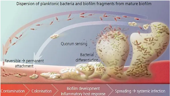
Figure 1.10. Schematic representation of the stages of microbial biofilm forming multiple bacteria
development. The progression of biofilm, initiated with bacterial contamination, followed by colonization, leading to critical colonization and systemic infection. Adapted from Phillips et al., (2008).
In fact, direct evidence of microscopic analysis of biopsies performed on women with BV revealed the presence of bacterial biofilm on the vaginal epithelium (Swidsinski
et al., 2005), and examining the composition and structural organization of the vaginal
biofilm, has revealed that G. vaginalis accounted for 60 - 95% of the biofilm mass, A.
vaginae accounted for the 1 - 40% of the biofilm mass whereas Lactobacillus maked up
only 5% of the biofilm (Polatti, 2012). Studies using therapeutic treatments, like the antimicrobial agent metronidazole, revealed that biofilm formation is a decisive factor in the virulence of BV as it could not act in biofilm leading to relapse (Polatti, 2012). Because microorganisms in biofilms react differently to antibiotic treatment when compared with their planktonic counter-parts (Fux et al., 2005), antibiotic resistance is postulated as one of the reasons for persistent and recurrent BV (Polatti, 2012).
20 | Chapter 1
In vitro experiments show that G. vaginalis has a propensity to form biofilm
(Figure 1.11). Patterson et al., (2010) reported to presence of a protein associated with biofilm homologous to other biofilm-associated proteins (BAP) and BV. In G. vaginalis isolates the gene identified by Patterson et al., (2010), is called by Bapl - Like (BapL). BAP are large proteins, which are anchored to the cell wall with the capacity to mediate adherence to host cells and intracellular adhesion, thus contributing to biofilm formation (Latasa et al., 2006).
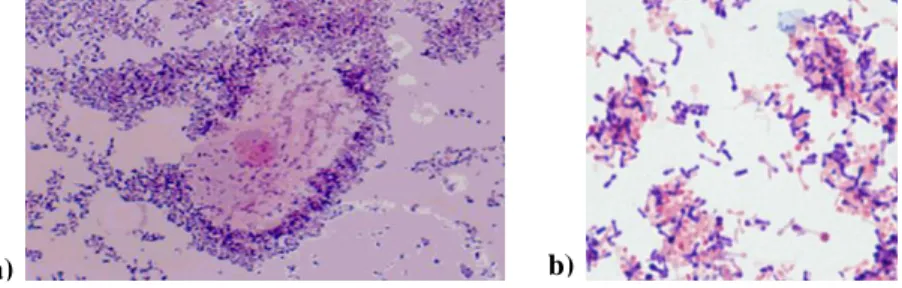
Figure 1.11. Biofilms formation of strains of G. vaginalis and anaerobic bacteria associated with BV. a)
Bacteria were grown anaerobically in Brain Heart Infusion Broth supplemented with glucose (sBHIG) at 37°C for 24 h. The adhered cells were stained with safranin; b) Quantitative assessment of the capability of formation of the biofilm formation was made by dissolving safranin stain in 33% acetic acid and measuring
optical density (OD) OD562. Adapted from Patterson et al., (2010).
Summarizing, there is strong evidence that G. vaginalis has an innate pathogenic potential compared to other BV-associated anaerobes. However there may still be many virulence factors not yet described, or maybe G. vaginalis has the potential of becoming more pathogenic in the presence of other species. It is also likely that the main agent of BV, if any, varies from case to case, however, several studies suggest that due to the many factors of virulence present in G. vaginalis, it is likely that the key agent in certain cases of BV is this bacterial species.
b) a)
Chapter 1 | 21
1.5. Outline and objectives of this thesis
In one study publish this year it was shown that BV is a prevalent gynaecological disorder in Portugal and that additional studies should be done to better characterize it in epidemiological and microbiological terms (Henriques et al., 2012). Thus, the characterization of vaginal microflora is a very important tool for the understanding of the vaginal flora associated with BV and can help investigate the significance of this condition in clinical pathology and to guide treatment(Srinivasan and Fredericks, 2008).
The main objective of this thesis is the study of clinical vaginal swab samples of Portuguese patients healthy or with BV. For this, this thesis is divided into 4 chapters. The first includes a general introduction to the work and includes the major implications and treatment of this condition. The methods, results and their discussion are separated into different sections, each one corresponding to a part of the experimental work. Chapter 2 concerns the studies of isolation the G. vaginalis and others microorganisms belonging to the vaginal flora, followed by characterization of the biofilm formation capacity and susceptibility of these strains by determining the MIC in chapter 3. The last chapter outlines the main conclusions and suggestions for future work.
Knowing more about how bacterial communities in the human vagina promoting health or facilitate disease is important, in order to optimize reproductive health.
CHAPTER 2
Isolation and identification of microorganisms from clinical
Chapter 2 | 25
2.1. Introduction
The identity and diversity of populations of vaginal microflora remains largely obscure and the complex interactions of the various members of the vaginal flora are still poorly understood. However, as described in chapter 1, in recent years several new fastidious bacteria have been identified that show a high specificity for BV (Fethers et
al., 2012). These microorganisms have been evaluated using cultivation-dependent
methods of vaginal fluid samples and it was observed that most species require special conditions of culture media and incubation. Traditional methods of biochemical characterization such as oxidase, catalase reactions and Gram staining allow the identification of various microorganisms but fail to identify many vaginal microorganisms (Ledger and Witkin, 2007). On the other hand, recently, molecular biology techniques (cultivation-independent) have provided new insights regarding bacterial diversity in vaginal flora, particularly in women with BV (Zhou et al., 2004). What is currently accepted is that healthy women are mainly colonized by lactobacilli, though a variety of other bacteria may be present in BV (Fethers et al., 2012).
2.2. Objectives
The main objectives of the work described in this chapter were the isolation and identification of microorganisms from clinical vaginal samples in women with or without BV. The isolation of these microorganisms was necessary to enable phenotypic characterization of selected isolates in the next chapter of this thesis.
2.3. Materials and Methods
2.3.1. Study population and sample collection
The collection of clinical vaginal samples from patients with suspected BV and healthy vaginal flora was performed with the collaboration of some Portuguese gynecologists who performed the collections at their private clinics. All volunteers were asked to complete a questionnaire form which data was analyzed by another member of our research group (Salgueiro, 2012) and clinically examined. The doctors proceeded to the collection of the samples using an unlubricated sterile swab with Amies transport medium provided with coal (VWR) (Al-Muk and Hasony, 2001). The swabs were transported, within 48 h of collection, to the laboratory. As soon as the clinical samples
26 | Chapter 2
arrived at the Laboratory of Applied Microbiology 1 (LAM 1), of the Department of Biological Engineering, University of Minho, they were processed (Kharsany et al., 1993).
2.3.2. Treatment of the clinical samples
Each sample was used to make a smear for Gram stain (as described in section 2.3.3.2) and the evaluation of vaginal microflora of each patient and BV diagnosis were based on the method described by Nugent et al., (1991). After this, each sample was placed in selective culture media (as described in 2.3.3.1) and the swab was inserted into a plastic tube containing 2 ml of sterile saline solution of 0.9% sodium chloride (NaCl) (ProLAbo) which was vortexed vigorously until the solution becomes cloudy, so as to recover all the bacterial cells still present in the swab. Then, 1 mL was transferred into 2 eppendorfs, and centrifuged at 10,000 rpm for 5 min. At the end, the supernatant was discarded leaving only the pellet in eppendorfs (BIOplastics); one of them was used to prepare suspensions to be used for PCR (for selection of positive samples for G.
vaginalis, as described in 2.3.3.3) by resuspending the pellet in ultra-pure water and the
remaining pellet was used to prepare a cryovial containing the pellet resuspended in 800 µL of Brain Heart Infusion (BHI) broth (Oxoid) with 200 µL of pure sterile glycerol (Panreac) (final concentration of glycerol 20%). All samples were then stored in cryovials (VWR) properly identified and stored at -80°C. The cryopreservation of samples allows the preservation of the genotypic and phenotypic characteristics of bacteria, ensuring that the characteristics remain intact for further studies, decreasing the risk of bacterial contamination or fungal.
2.3.3. Isolation and identification methods 2.3.3.1. Culture methods
Media and growth conditions
Selected samples were cultured on Columbia Blood Agar Base (CBA) (Liofilchem) supplemented with 5% of defibrinated horse´s blood (Probiologica) with
G. vaginalis selective supplement (Oxoid) or CBA without supplement; Man-Rogosa
and Sharpe Agar (MRS) (Liofilchem) that were prepared according to the manufacturer´s instructions and Bromocresol Purple Starch Agar (Purple) which is