ORIGINAL
Resumo
Abstract
A associação do treinamento de força (TF) e aeróbio (TA), conhecido com treinamento concorrente (TC), parece diminuir os ganhos de força e hipertrofi a muscular quando com-parado ao TF isolado. Dessa forma, esse estudo teve como objetivo comparar os efeitos de 16 semanas de TF e TC sobre os indicadores de hipertrofi a e a força muscular em mulheres de meia-idade na pós-menopausa. Participaram 24 mulheres, não ativas fi sicamente, sub-divididas em três grupos: Grupo TC (n=8), Grupo TF (n=8) e Grupo Controle (n=8). Os trei-namentos foram compostos de duas etapas (E1 e E2) com duração de oito semanas cada, e frequência de três sessões/semana (TF: 10 exercícios com 3 x 8-10 RM; TC: 6 exercícios com 3 x 8-10 RM, seguido de 30 min de caminhada ou corrida a 55-85% VO2pico). Foram avalia-das a área muscular de coxa (AMC), força máxima e consumo pico de oxigênio (VO2pico). Os resultados demonstraram aumento na força máxima nos exercícios leg press, supino reto e rosca direta para o TF e TC, sem diferença entre eles. Com relação aos indicadores hipertró-fi cos não houve aumento na AMC para o TF e TC. Houve aumento do VO2pico somente para o TC. Dessa forma, podemos concluir que o TC, realizado com as recomendações mínimas de TA preconizadas pelo American College of Sports Medicine (ACSM), não promoveu efeito de interferência na força máxima e hipertrofi a muscular de mulheres de meia-idade na pós-menopausa.
Palavras-chave: Pós-menopausa; Composição corporal; Força muscular; Treinamento de
força; Exercício.
The combination of strength (TF) and aerobic training (TA), known as concurrent training (TC), seems to diminish the muscle strength and hypertrophy gains when compared with isolated TF. This study aimed to compare the eff ects of 16 weeks of concurrent training (TC) and resistance training (TF) on hypertrophic indicators and muscle strength of middle-aged postmenopausal women. Participated 24 non-active women randomly assigned in three groups: TC (n=8), TP (n=8) and control group (GC, n=8). Both training protocols were divided in two phases lasting eight weeks with a three weeks sessions frequency (TF: 10 exercises, 3x8-10 RM; TC: 6 exercises, 3x8-10 RM followed by 30 min of walking or running at 55-85% VO2peak). It were assessed thigh muscle area (AMC), muscle strength and maximal oxygen uptake (VO2peak). Our data showed that both training protocols (i.e., TF and TC) signifi cantly increased maximal strength in leg press, bench press and arm curl without diff erences between groups. Regarding the hypertrophic indicators there was no diff erence in AMC for both training groups. VO2peak signifi cantly increased only for TC. Thus, our data showed that when TC is held closely to the minimum of American College of Sports Medicine (ACSM) recommendation for aerobic training, no interference eff ect is observed in muscle strength and hypertrophic indicators in middle-aged postmenopausal women.
Keywords: Postmenopausal; Body composition; Muscle strength; Resistance training;
Exercise.
Eff ect of concurrent training on muscle
hypertrophy and strength of postmenopausal
women
Efeito do treinamento concorrente sobre a força e hipertrofi a muscular de
mulheres na pós-menopausa
Manoel Emílio Lixandrão1
Valéria Bonganha1
Miguel Soares Conceição1
Cleiton Augusto Libardi1,2
Ricardo Paes de Barros Berton1
Claudia Regina Cavaglieri1
Mara Patrícia Traina Chacon-Mikahil1
Vera Aparecida Madruga1
1. Laboratory of Exercise Physiology (FISEX), School of Physical Education (FEF), State University of Campinas (UNICAMP). Campinas, SP, Brazil.
2. Research and Study Group of
Neuromuscular Adaptations (GEPAN), School of Physical Education and Sports (EEFE), University of São Paulo (USP). São Paulo, SP, Brazil.
Manoel Emílio Lixandrão
Laboratório de Fisiologia do Exercício – FISEX. Faculdade de Educação Física Universidade Estadual de Campinas – UNICAMP, Campinas, Brazil Av. Érico Veríssimo, 701 CEP 13083-851 – Caixa postal 6134 e-mail: [email protected]
ENDEREÇO PARA CORRESPONDÊNCIA
• Recebido: • Re-submissão: • Aceito: 21/12/2011 04/02/2012 20/03/2012 21/06/2012
INTRODUCTION
Menopause is characterized by the reduction in the pro-duction of female hormones1. During this stage of life, women
have an increase in total fat mass and redistribution of peri-pheral fat to the central region of the body, with a concomi-tant reduction in skeletal muscle mass2. This reduction has
been associated with a decrease in daily physical activity, the capacity to generate muscle strength and aerobic fitness in the elderly (i.e. maximal oxygen uptake, VO2max.)3. In their turn,
these reductions are associated with a higher prevalence of both degenerative chronic diseases such as insulin resistan-ce, type 2 diabetes4 and metabolic syndrome5, and accidental
deaths from falls6.
For this reason, physical activity practice has been widely recommended as a strategy to revert/minimize the harmful effects of aging on muscle mass and strength. Several directi-ves on physical exercise practice recommend that concurrent training (CT, i.e. strength training – ST and aerobic training – AT in the same training session or on alternate days) should be performed by men and women during the aging process7, 8. However, some studies have shown that CT lessens the
adaptive response to physical training, as it seems to reduce muscle strength and hypertrophy gains in exercised muscles, when compared to isolated ST. This phenomenon is known as “interference effect”9, 10.
Although many hypotheses about interference effect have been reported in the literature9, 11, 12, the mechanism
responsible for such phenomenon has not been fully un-derstood. One of these hypotheses suggests that, when the volume of CT performed is higher than that of isolated ST, the interference effect is more frequently observed. As an example, Karavirta et al. (2011) only observed an increase in the cross-sectional area of type 2 muscle fibers in the thigh of middle-aged men who performed ST (2x/week), whereas tho-se who performed CT (ST 2x/week and AT 2x/week) showed no changes. The interference effect on the lean mass of lower limbs has also been described by Sillampää et al. (2008)13,
using Dual-energy X-ray absorptiometry (DXA). However, in this same study, ultrasonography showed that the thickness of the vastus lateralis and intermedius muscles were not sig-nificantly different between the ST (11%) and CT (9%) groups. It should be emphasized that the above mentioned studies found that the volume of CT increased, when compared to that of ST, due to the inclusion of AT sessions lasting up to 90 minutes on a cycle ergometer. On the other hand, when the CT was performed with a weekly volume (1x/week of ST and 1x/week of AT) lower than that of isolated ST (2x/week), there was a similar increase in the cross-sectional area of the thigh when both training regimens were compared (ST and CT)14, 15.
Although the interference effect did not appear in the studies conducted by Izquierdo et al. (2004; 2005), the weekly volume of AT during the CT performed was lower than the minimum values recommended by the American College of Sports Me-dicine (ACSM – 2007; 2009)7, 8.
Recently, researchers of our group showed that CT ena-bled muscle strength gains similar to those of ST among mi-ddle-aged men, when AT was performed at values close to the ACSM recommmendations16. Consequently, this may also
take place among postmenopausal women, although it is not known whether the interference effect will occur with muscle hypertrophy as well.
In this sense, the present study aimed to verify whe-ther CT with a low volume of AT, despite meeting the mi-nimum ACSM recommendations, has an interference effect
on muscle strength and hypertrophy in postmenopausal women. The hypothesis is that CT does not have an interfe-rence effect on muscle strength and hypertrophy after 16 weeks of training.
METHODS
SampleA total of 24 middle-aged menopausal women partici-pated in this study (five of which were hysterectomized), all clinically healthy and not performing physical activities regu-larly (i.e. fewer than two times per week in a non-systematic way). These women were sub-divided into three groups: CT Group (n=8, age 53.0 ± 6.0), ST Group (n=8, age 54.0 ± 3.6) and Control Group (CG – n=8, age 51.0 ± 6.0). The following inclusion criteria were considered: to be postmenopausal (12 months without menstruations); not to have performed regu-lar ST programs for at least six months before the beginning of the study; not to be undergoing hormonal therapy (i.e. one year prior to the study); not to have any cardiovascular and/ or orthopedic diseases; not to use medications that could interfere with physiological responses (i.e. muscle strength, aerobic capacity and power, and body composition); and a level of adherence higher than 85% of all expected training sessions. Volunteers were instructed not to change the pat-tern of their eating habits during the experimental period, al-though no control over the diet was performed. After being informed about the study proposal and procedures to which they would be submitted, volunteers signed an informed con-sent form. The precon-sent study was approved by the university’s Research Ethics Committee (248/2004).
Anthropometric assessment and body composition
A platform scale (Filizola, São Paulo, Brazil) was used to measure body mass and a wooden stadiometer with a 0.1cm accuracy was used to measure height. The Body Mass Index (BMI) was obtained by dividing body mass by the square of height. The skinfold thickness of the right thigh was assessed with a calibrated adipometer (LANGE, Cambridge, Maryland, USA). In addition, right thigh circumference measurements were also taken. The femoral condyle diameter was measured using a blunt-pointed compass with a 0.1cm accuracy. All an-thropometric assessment and skinfold measurement proce-dures were performed according to the techniques described by Heyward17.
Thigh muscle area
Thigh muscle area (TMA) was calculated with Knapik’s equation18. TMA (cm²) = 0.649 x [TC/π – TS)² - (0.3 – FD)²],
whe-re TC = right thigh circumfewhe-rence; TS = thigh skinfold thick-ness; and FD = femoral diameter. The TMA equation’s level of error is approximately 6%18.
Muscle strength
Prior to the beginning of the muscle strength assessment, two familiarization sessions were performed with the following exercises: leg press, knee extension, knew flexion, bench press, pulldown, lateral raise, elbow flexion, elbow extension on the pulldown, and abdominal exercise (RIGUETTOequipment, São Paulo, SP). During these two sessions, volunteers were expec-ted to perform two series of ten sub-maximal repetitions, with 60 seconds of interval between series and exercises. Muscle strength was measured with the maximum repetition test
(1-RM) when they performed bench press, leg press and elbow flexion19. All exercises were preceded by a warm-up series of ten
repetitions with approximately 50% of the weight expected on the first attempt of each 1-RM test. Tests began three minutes after warm-up. Volunteers were subsequently instructed to per-form only one repetition with the weight expected for the 1-RM. If two repetitions were completed on the first attempt or even if one repetition could not be completed, a second attempt was made after an interval of three to five minutes with a weight (kg) higher (first attempt) or lower (second attempt) than that used on the previous attempt. A third and last attempt was made if the weight of a single maximum repetition could not be determined. All participants performed two sessions of tests with a 48-hour interval between them, aiming to familiarize these participants with the exercises and consequently reduce learning effects. The highest weight obtained from the analyses was taken into consideration. The intra-class correlation coeffi-cient (ICC) was used to analyze test and re-test reliability of the 1-RM when the bench press (0.94), leg press (0.97) and biceps curl (0.99) were performed.
Cardio-respiratory assessment
Volunteers performed a protocol test on a treadmill (Quin-ton TM55, Bothell, Washing(Quin-ton, USA), where respiratory gas ex-changes were continually monitored, breath by breath, using a metabolic gas analysis system (CPX, Medical Graphics, St. Paul, Minnesota, USA).
The protocol consisted in an initial warm-up speed of 4 km/h for two minutes, followed by increases of 0.3 km/h every 30 seconds, with a constant inclination of 1% until physical exhaustion. Next, there was a period of four minutes for reco-very, the first minute at 5 km/h and each subsequent minute, 1 km/h lower20.
The cardio-respiratory assessment was performed in three moments: before, after eight weeks, and after 16 weeks of the experimental period. The assessment after eight weeks was performed to readjust the intensity of aerobic training in the CT group.
Aerobic capacity
The aerobic capacity was determined using graphic visu-al anvisu-alysis21, performed by three previously trained observers
who were familiarized with the Medical Graphics’ CPX system. The anaerobic limit (AL) was characterized as the first inflec-tion point of ventilainflec-tion curves (VE), respiratory equivalent of O2 (VE/O2) and partial pressure of O2 (PETO2), without a con-comitant increase in the respiratory equivalent of CO2 (VE/ VCO2)21. In contrast, the respiratory compensation point (RCP)
was determined considering the second break in the linearity of VE, increase in the ventilatory equivalent of CO2 (VE/VCO2), and decrease in the partial pressure curve of CO2 (PETCO2)21, 22.
The majority of volunteers in this study did not show a plateau of oxygen uptake, considered to be a criterion to cha-racterize VO2max23
. Consequently, peak oxygen uptake (VO2peak)
was the term used. This was expressed according to the
(VO-2peak) considered to be the mean of values from the last 30
se-conds of the cardio-respiratory assessment24. At least two of
the following three criteria were adopted to guarantee that volunteers made a maximum effort: (1) a plateau of VO2, i.e. no or little variation in VO2 (< 2.1 mL.kg-1.m1) despite the in-crease in exercise intensity; (2) ratio of respiratory exchanges higher than 1.10; (3) heart rate (HR) higher than 90% of the maximum value expected for age23.
Strength training (ST)
The ST program was divided into two stages. In stage one (S1), participants performed ten exercises (leg press, knee extension, knee flexion, bench press, pulldown, shoulder la-teral raise, triceps pulley, biceps curl, abdominal exercise and calf raise). Exercises alternated as follows: first, an exercise was performed for the upper limbs, followed by another for the lo-wer limbs. The abdominal exercise was always the last one to be performed. The ST was prescribed per target region of maxi-mum repetitions, including three series of ten repetitions, with a pause of 60 minutes between series and exercises. In stage two (S2), the same exercises as S1 were performed. They were ordered by joint: first, the lower limb exercises, followed by upper limb exercises, including three series of eight maximum repetitions with a pause of 90 seconds between series and exercises. The total duration of each session was approxima-tely 60 minutes16.
Concurrent training (CT)
AT and ST were performed in the same CT session, divi-ded into two stages. In S1, participants initially performed the ST comprised of six exercises (leg press, knew extension, knew flexion, bench press, pulldown and biceps curl, including three series of ten repetitions, pause of 60 seconds, and session du-ration of approximately 30 minutes. The order of exercises was alternated per sections in this stage. Next, volunteers perfor-med 30 minutes of AT, including walking and/or running with a varying intensity, five minutes below the AL, ten minutes at the AL, ten minutes above the AL and below the RCP, and five minutes below the AL. These intensities were between 55-85% of the VO2peak or heart rate reserve (HRR) according to the ACSM, (1998)25. In S2 of the CT, the ST session was performed
with the same exercises and series as S1, although with three series of eight repetitions, pause of 90 seconds and duration of approximately 30 minutes per session. In this stage, exerci-ses were ordered by joint. In addition, there was an increase in the intensity of AT: five minutes below the AL, ten minutes above the AL and below the RCP, ten minutes at the RCP and five minutes below the AL, totaling 30 minutes. The total dura-tion of the CT session was approximately 60 minutes16.
Exercises with weights were performed at a rate of two seconds for the concentric phase and two seconds for the ec-centric phase. For these exercises, both the ST and CT showed a weekly increase in the previously used weight, as described by Libardi et al., (2011)16. For the AT, the intensity of training
referring to the AL and RCP was monitored using the speed of walking, running and heart rate obtained from the test perfor-med on the treadmill (before the training period and after ei-ght weeks) with an inclination of 1% to reproduce the training conditions of running tracks20.
Statistical analysis
Shapiro-Wilk test was used to verify sample normality. Levene’s test and Mauchlys’ sphericity test were used to ob-serve homogeneity and sphericity, respectively. The analysis of variance for repeated measures (two-way ANOVA) was used to compare inter- and intra-groups of maximum strength and TMA. One-way ANOVA was used to compare values of analysis of the baseline and percentage of change of VO2peak. Upon the occurrence of significant F values, Tukey’s test was performed for multiple comparisons. The results were described as mean values and standard-deviation. A P-value < 0.05 was conside-red to be significant.
Tabela 1 Anthropometric variables, muscle hypertrophy indicators and VO2peak, before and after 16 weeks of
strength training (ST), concurrent training (CT) and control group (CG).
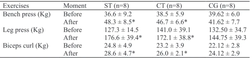
Tabela 2 Maximum strength before and after 16 weeks of strength training (ST), concurrent training (CT) and control group (CG).
Figura 1 Values of percentage of change in VO2peak among study stages.
RESULTS
Maximum strength
ST and CT showed significant increases in maximum strength for leg press exercises (F = 6.3; P = 0.0001; 37.6% and P = 0.0045; 26.3%, respectively), bench press (F =18.7; P = 0.0001; 34.4% and P = 0.0001; 21.9%, respectively) and biceps curl (F = 1.5; P = 0.0005; 16.0% and P = 0.01; 13.7%, respective-ly), without significant differences between groups (P > 0.05) (Table 2).
Muscle hypertrophy indicator (TMA)
TMA did not show significant differences in any of the in-tervention protocols (F= 4.9; P > 0.05) (Table 1).
Peak oxygen uptake (VO2peak)
There were no significant differences in pre-intervention VO2peak values between CT (26.1 ± 3.0 ml/kg/min.), ST (29.4 ± 2.3 ml/kg/min.) and CG (27.7 ± 2.5 ml/kg/min.) (Table 1). There
was only an increase in VO2peak values for CT (28.40 ± 2.3 ml/kg/ min.; 8.8%), which was significantly different from ST (26.60 ± 6.10 ml/kg/min.; -9.9%; P = 0.009) and CG (24.20 ± 2.80 ml/kg/ min.; -12.5%; P = 0.003) (F = 8.5) (Figure 1).
DISCUSSION
The present study aimed to compare the effects of CT and ST on the maximum muscle strength and hypertrophy among postmenopausal women. Its main results confirmed the hypothesis that CT performed at levels close to the mini-mum ACSM recommendations does not have an interference effect either on strength or muscle hypertrophy in postmeno-pausal women. There was a similar increase in muscle streng-th between ST and CT, wistreng-thout changes in streng-the TMA after 16 weeks of training.
Few studies have been exclusively conducted in middle--aged postmenopausal women26. Additionally, comparisons
CT are hindered by discrepancies in the variables found, such as volume, intensity and weekly frequency9, 11. As an example,
Sillampää et al. (2008, 2009) observed a similar increase in muscle strength between ST (2x/week) and CT (ST 2x/week and AT 2x/week) among middle-aged men and women. On the other hand, the interference effect was observed when ST and AT are performed in the same session27. Cadore et al.
(2010) pointed out that the order that training programs are performed (AT before ST) can be influenced by the results, indicating that the interference effect can be probably attri-buted to muscle fatigue induced by the first activity, which reduces the effectiveness of the physiological adaptations of the activity subsequently performed. This hypothesis can be confirmed with the findings from Lemos et al., (2009), who observed that the performance of high-intensity aerobic exer-cises (80% HRmax) prior to strength exercises decreases the vo-lume of the subsequent ST series28.
However, in the present study, the performance of AT after ST did not have an interference effect on the strength gains of lower limbs in the CT (Table 2). Recently, this group of researchers did not observe the interference effect on mi-ddle-aged men in a CT protocol similar to that of such study either16. In fact, the order in which ST and AT are performed
can somehow influence the absence or not of the interference effect. It could be speculated that although ST and AT were performed in the same session, the low volume of AT could have contributed to the prevention of the interference effect on maximum strength gains and also to the increase in VO2peak.
Although not finding an interference effect on the mus-cle strength of lower limbs after CT was performed13, 29, some
studies found this effect on muscle hypertrophy. In addition, they have indicated that the interference effect could be as-sociated with a volume of CT (ST 2x/week and AT 2x/week) higher than that of ST (ST 2x/week). On the other hand, when CT was performed with a weekly volume (1x/week ST and 1x/week AT) lower than that of isolated ST (2x/week), there was a similar increase in the cross-sectional area of the thigh, comparing both training regimens, i.e. ST and CT.14, 15. In the
present study, even with a low volume of AT, there was not an increase in the muscle area of lower limbs in any of the groups studied. It should be emphasized that the thigh muscle area was measured with an indirect method. However, this method is highly correlated with measures such as MRIs (r = 0.96)30 and
CT scans (r = 0.97).18
Although there are certain variables that seek to explain the different responses to CT programs (intensity, volume, fre-quency, duration)11, the literature has not reached a consensus
on if and how these variables regulate the interference effect on muscle strength and hypertrophy. Thus, based on the re-sults of the present study, it could be concluded that the CT performed with a volume of AT close to the minimum ACSM values recommended did not have an interference effect on the muscle strength of lower limbs, after 16 weeks of interven-tion in middle-aged postmenopausal women.
REFERENCES
1. WHO. Research on the menopause in the 1990s. Report of a WHO Scientific Group. World Health Organ Tech Rep Ser. 1996;866:1-107.
2. Roghani T, Torkaman G, Movasseghe S, et al. Effects of short-term aerobic ex-ercise with and without external loading on bone metabolism and balance in postmenopausal women with osteoporosis. Rheumatol Int. 2012.
3. Hunter GR, McCarthy JP, Bamman MM. Effects of resistance training on older adults. Sports Med. 2004;34(5):329-48.
4. Guillet C, Boirie Y. Insulin resistance: a contributing factor to age-related mus-cle mass loss? Diabetes Metab. 2005;31 Spec No 2:5S20-5S26.
5. Kuzuya M, Ando F, Iguchi A, et al. Age-specific change of prevalence of meta-bolic syndrome: longitudinal observation of large Japanese cohort. Athero-sclerosis. 2007;191(2):305-12.
6. Lipsitz LA, Nakajima I, Gagnon M, et al. Muscle strength and fall rates among residents of Japanese and American nursing homes: an International Cross-Cultural Study. J Am Geriatr Soc. 1994;42(9):953-9.
7. Chodzko-Zajko WJ, Proctor DN, Fiatarone Singh MA, et al. American College of Sports Medicine position stand. Exercise and physical activity for older adults. Med Sci Sports Exerc. 2009;41(7):1510-30.
8. Nelson ME, Rejeski WJ, Blair SN, et al. Physical activity and public health in older adults: recommendation from the American College of Sports Medicine and the American Heart Association. Circulation. 2007;116(9):1094-105. 9. Docherty D, Sporer B. A proposed model for examining the interference
phe-nomenon between concurrent aerobic and strength training. Sports Med. 2000;30(6):385-94.
10. Cadore EL, Pinto RS, Lhullier FL, et al. Physiological effects of concurrent train-ing in elderly men. Int J Sports Med. 2010;31(10):689-97.
11. Leveritt M, Abernethy PJ, Barry BK, et al. Concurrent strength and endurance training. A review. Sports Med. 1999;28(6):413-27.
12. Nader GA. Concurrent strength and endurance training: from molecules to man. Med Sci Sports Exerc. 2006;38(11):1965-70.
13. Sillanpaa E, Hakkinen A, Nyman K, et al. Body composition and fitness dur-ing strength and/or endurance traindur-ing in older men. Med Sci Sports Exerc. 2008;40(5):950-8.
14. Izquierdo M, Hakkinen K, Ibanez J, et al. Effects of combined resistance and cardiovascular training on strength, power, muscle cross-sectional area, and endurance markers in middle-aged men. Eur J Appl Physiol. 2005;94(1-2):70-5. 15. Izquierdo M, Ibanez J, K HA, et al. Once weekly combined resistance and cardio-vascular training in healthy older men. Med Sci Sports Exerc. 2004;36(3):435-43.
16. Libardi CA, Souza GV, Gaspari AF, et al. Effects of concurrent training on inter-leukin-6, tumour necrosis factor-alpha and C-reactive protein in middle-aged men. J Sports Sci. 2011;29(14):1573-81.
17. Heyward V. ASEP methods recommendation: body composition assessment. Journal of Exercise Physiology - online. 2001;4:1-12.
18. Knapik JJ, Staab JS, Harman EA. Validity of an anthropometric estimate of thigh muscle cross-sectional area. Med Sci Sports Exerc. 1996;28(12):1523-30. 19. Brown L, Weir J. Procedures recommendation I: Accurate assessment of mus-cular strength and power. Journal of Exercise Physiology - online. 2001;4:1-21. 20. Jones AM, Doust JH. A 1% treadmill grade most accurately reflects the
ener-getic cost of outdoor running. J Sports Sci. 1996;14(4):321-7.
21. Wasserman K, Whipp BJ, Koyl SN, et al. Anaerobic threshold and respiratory gas exchange during exercise. J Appl Physiol. 1973;35(2):236-43.
22. McLellan TM. Ventilatory and plasma lactate response with different exercise protocols: a comparison of methods. Int J Sports Med. 1985;6(1):30-5. 23. Howley ET, Bassett DR, Jr., Welch HG. Criteria for maximal oxygen uptake:
re-view and commentary. Med Sci Sports Exerc. 1995;27(9):1292-301. 24. Heubert RA, Billat VL, Chassaing P, et al. Effect of a previous sprint on the
pa-rameters of the work-time to exhaustion relationship in high intensity cycling. Int J Sports Med. 2005;26(7):583-92.
25. ACSM. American College of Sports Medicine Position Stand. The recommend-ed quantity and quality of exercise for developing and maintaining cardiore-spiratory and muscular fitness, and flexibility in healthy adults. Med Sci Sports Exerc. 1998;30(6):975-91.
26. Sillanpaa E, Laaksonen DE, Hakkinen A, et al. Body composition, fitness, and metabolic health during strength and endurance training and their combina-tion in middle-aged and older women. Eur J Appl Physiol. 2009;106(2):285-96. 27. Cadore EL, Izquierdo M, Goncalves Dos Santos M, et al. Hormonal Responses to Concurrent Strength and Endurance Training with Different Exercise Orders. J Strength Cond Res. 2012.
28. Lemos A, Simao R, Polito M, et al. The acute influence of two intensities of aero-bic exercise on strength training performance in elderly women. J Strength Cond Res. 2009;23(4):1252-7.
29. Karavirta L, Hakkinen A, Sillanpaa E, et al. Effects of combined endurance and strength training on muscle strength, power and hypertrophy in 40-67-year-old men. Scand J Med Sci Sports. 2011;21(3):402-11.
30. Heymsfield SB, McManus C, Smith J, et al. Anthropometric measurement of muscle mass: revised equations for calculating bone-free arm muscle area. Am J Clin Nutr. 1982;36(4):680-90.