https://doi.org/10.1177/1756284819869141 https://doi.org/10.1177/1756284819869141 Ther Adv Gastroenterol
2019, Vol. 12: 1–13 DOI: 10.1177/ 1756284819869141 © The Author(s), 2019. Article reuse guidelines: sagepub.com/journals-permissions
Therapeutic Advances in Gastroenterology
journals.sagepub.com/home/tag 1
Soluble human Suppression of
Tumorigenicity 2 is associated with
endoscopic activity in patients with
moderate-to-severe ulcerative colitis
treated with golimumab
Fernando Magro , Susana Lopes, Marco Silva, Rosa Coelho, Francisco Portela, Diogo Branquinho, Luís Correia, Samuel Fernandes, Marília Cravo, Paulo Caldeira, Helena Tavares de Sousa, Marta Patita, Paula Lago, Jaime Ramos, Joana Afonso, Isabel Redondo, Patrícia Machado, George Philip, Joanne Lopes and
Fátima Carneiro on behalf of GEDII Abstract
Background: Suppressor of Tumorigenicity 2 (ST2) is an IL33 receptor detected in the mucosa and serum of ulcerative colitis (UC) patients. We evaluated soluble ST2 (sST2) as a surrogate biomarker of disease outcome and therapeutic response, in moderate-to-severe UC patients treated with golimumab.
Methods: We conducted an open-label single-arm multicentre prospective study. At screening/baseline, week 6 (W6) and week 16 (W16), clinical and endoscopic activity (total Mayo score), histologic activity (Geboes index) and biomarkers were evaluated.
Results: From 38 patients, 34 (89.5%) completed W6 and 29 (76.3%) completed W16. Mean age (±SD) was 34.6 ± 12.6 years; 55.9% were female. At W16, 62.1% achieved clinical response. Patients with endoscopic activity at W6 (n = 20) had higher baseline sST2 (median, 24.5 versus 18.7 ng/ml, p = 0.026) and no decrease from baseline (median change, 0.8 versus −2.7, p = 0.029). At W6, sST2 levels correlated with endoscopic activity (rs = 0.45, p = 0.007) but not with histological activity (rs = 0.25, p = 0.151). The best cut-offs for endoscopic activity were sST2 = 16.9 ng/ml (sensitivity = 85%; specificity = 71%) and faecal calprotectin (FC) = 353 μg/g (sensitivity = 90%, specificity = 67%). Patients with histological activity at W6 (n = 27) had higher baseline ST2 levels (median, 23.0 versus 13.7 ng/ml, p = 0.035). sST2 did not correlate with FC or serum C-reactive protein. FC levels correlated with histological activity and baseline FC were higher when Geboes ⩾3.1 at W6.
Conclusions: sST2 may be a surrogate biomarker of UC activity and therapeutic response as it correlates with endoscopic and clinical activity at W6 of golimumab treatment, and subjects with endoscopic and histological activity at W6 had higher baseline ST2 levels.
Keywords: endoscopic activity, golimumab, histological activity, serum soluble ST2, ulcerative colitis
Received: 24 April 2019; revised manuscript accepted: 17 July 2019.
Correspondence to:
Fernando Magro Institute of Pharmacology and Therapeutics, Faculty of Medicine, University of Porto, Alameda Prof. Hernâni Monteiro, 4200-319 Porto, Portugal [email protected] Susana Lopes Marco Silva Rosa Coelho Joana Afonso Centro Hospitalar São João, Porto, Portugal Francisco Portela Diogo Branquinho Centro Hospitalar Universitário de Coimbra, Portugal Luís Correia Samuel Fernandes Centro Hospitalar Lisboa Norte, Portugal Marília Cravo Hospital Beatriz Ângelo, Loures, Portugal Paulo Caldeira Helena Tavares de Sousa Centro Hospitalar Universitário do Algarve, Faro, Portugal Departamento de Ciências Biomédicas e Medicina, Universidade do Algarve, Faro, Portugal ABC–Algarve Biomedical Centre, Universidade do Algarve, Faro, Portugal Marta Patita Hospital Garcia de Orta, Almada, Portugal Paula Lago Centro Hospitalar do Porto, Porto, Portugal Jaime Ramos Centro Hospitalar Lisboa Central, Lisboa, Portugal Isabel Redondo Patrícia Machado MSD Portugal, Medical Affairs, Paço de Arcos, Portugal
Introduction
Ulcerative colitis (UC) is an inflammatory bowel disease (IBD) causing continuous mucosal inflammation of the colon without granulomas, and with a relapsing and remitting course.1
Current IBD biomarkers include serological lev-els of specific antibodies (e.g. ASCA, ANCA, anti-OmpC, anti-Cbir, and antiglycans), serum C-reactive protein (CRP), cytokines and faecal proteins (calprotectin and lactoferrin).2
Never-theless, most of these markers show low sensitiv-ity and specificsensitiv-ity and do not adequately reflect intestinal damage.2,3
Mucosal healing has become a major objective of IBD treatment but endoscopy is still the ‘gold standard’ for assessing inflammation of intestinal mucosa.4 Faecal calprotectin (FC) levels
signifi-cantly correlate with endoscopic activity in IBD and are currently one of the best surrogate mark-ers of intestinal inflammation.5,6 A meta-analysis
reported FC pooled sensitivity and specificity esti-mates of 0.88 and 0.79, respectively, regarding UC endoscopic activity.7 CRP is an acute-phase
marker produced in the liver and in the mesenteric adipocytes, and is a potential IBD biomarker.8 FC
seems to be more sensitive in UC than in Crohn’s disease, while CRP sensitivity in UC (50–60%) seems to be lower than in Crohn’s disease (70– 100%).7,8 Other simple, rapid, sensitive, specific,
inexpensive and noninvasive markers are still needed to detect and monitor intestinal inflamma-tion, especially during early treatment.9,10
IL-33 is a member of the IL-1 family that binds to its receptor, ST2 (human Suppression of Tumorigenicity 2), with the complex IL-33/ST2 leading to cytokine inactivation.11 However, the
IL-33/ST2 axis seems to have a dual and dichoto-mous role in the pathogenesis of IBD. On the one hand, proinflammatory cytokine stimuli (e.g. TNF) result in an increase of IL-33 in epithelial cells. On the other hand, IL-33 may be released by injured epithelial cells to induce production of proinflammatory cytokines through activation of ST2 in mast cells, macrophages, eosinophils and neutrophils.12 In mucosal tissue of mouse models,
IL-33 levels correlate positively with the severity of gut inflammation.12 Similarly, the expression
of ST2 seems to be increased in both colonic wall and serum of UC patients, and levels of serum soluble ST2 (sST2) also correlate positively with the severity of colonic mucosal disease and inflammatory cytokines.3,11,12
Treatment with TNFα inhibitors modulates IL-33 and sST2 levels. In a small cohort of IBD patients, Pastorelli and colleagues evaluated inf-liximab’s effects in a group of 9 UC patients and 11 subjects with Crohn’s disease, observing a decrease of IL-33 and of the IL-33/sST2 ratio, especially in UC patients.12 However, few studies
have correlated the levels of sST2 with endo-scopic and histological activity in UC patients receiving anti-TNFα therapy.
Golimumab is a fully human anti-TNFα mono-clonal antibody that binds with high affinity to human TNFα. It has been shown to induce a clinical response in 51% of patients with moder-ate-to-severe UC at week 6, with remission and mucosal healing being achieved by 18% and 42% of patients, respectively.13,14
The present work was an exploratory study aim-ing to evaluate sST2 as a surrogate biomarker of disease outcomes and therapeutic response in subjects with moderately-to-severely active UC who had started treatment with golimumab. The correlation of sST2 levels with clinical activity (total Mayo score), endoscopic activity (Mayo endoscopic subscore) and histological activity (Geboes index) were evaluated in this population. In addition, the association between sST2, FC and CRP levels was studied, as well as the perfor-mance of these biomarkers in predicting endo-scopic and histological activity in UC patients treated with golimumab.
Methods
This was an exploratory, multicentre, open-label, prospective, interventional, single-arm study, conducted in nine reference sites in Portugal (Clinical Trial Identification: EudraCT 2014– 003262–25). All procedures complied with the ethical principles of the Declaration of Helsinki and Good Clinical Practice requirements. The protocol and informed consent procedures were approved by the National Ethics Committee for Clinical Research and the Data Protection Authority.
Eligible subjects were aged ⩾18 and ⩽65 years old, with a previous diagnosis of moderate-to-severely active UC, who have had an inadequate response to conventional therapy (including corticosteroids and 6-mercaptopurine or azathioprine) or were intolerant to, or had medical contraindications for,
George Philip Merck & Co., Inc., Kenilworth, NJ, USA Joanne Lopes Faculdade de Medicina, Universidade do Porto, Porto, Portugal Fátima Carneiro Faculdade de Medicina, Universidade do Porto, Porto, Portugal Instituto de Patologia e Imunologia Molecular da Universidade do Porto (Ipatimup), i3S-Instituto de Investigação e Inovação em Saúde, Universidade do Porto, Porto, Portugal
such therapies. Subjects were naïve to TNFα inhibitors, and eligible to start golimumab accord-ing to its approved indication and dosaccord-ing regimen. Subjects who had extensive severe colitis, sympto-matic colonic or small bowel obstruction, history of colonic mucosal dysplasia, presence of adeno-matous polyps, who had used apheresis or had rectal corticosteroids or 5-ASA compounds within 2 weeks prior to the study were excluded. In addi-tion, subjects who received cyclosporine, tacroli-mus, sirolitacroli-mus, or mycophenolate mofetil within 8 weeks prior to the study, who received natali-zumab or any agent that depletes B or T cells within 12 months prior to the study, with history of, or ongoing, chronic infectious disease, immune deficiency, malignancy, chronic heart failure or other severe, progressive or uncontrolled disease, were also excluded.
Study treatment
Golimumab treatment followed the summary of product characteristics. The first dose of subcuta-neous golimumab (200 mg) was administered at the trial site at baseline. Subsequent dosing was done by the subject (i.e. unsupervised at their home) at week 2 (100 mg) and every 4 weeks there-after (50 mg if weight <80 kg or 100 mg if ⩾80 kg).
Procedures and definitions
After signing informed consent, subjects entered a screening period of up to 42 days, for confirmation of eligibility criteria. Eligible subjects had initiated treatment with golimumab at the baseline visit. Subjects were followed for 16 weeks for clinical assessments and data collection, including stool frequency, amount of blood in stool, the physi-cian’s global assessment and clinical classifications. Blood and stool samples were collected at baseline and at subsequent visits at week 6 (end of induc-tion) and week 16 (short-term maintenance). Soluble ST2 levels (expressed in ng/ml) were measured using an ELISA kit for human ST2 (DuoSet, R&D Systems, Minneapolis, MN, USA) according to the manufacturer’s instruc-tions. Blood samples were centrifuged, and serum was stored at −80°C. FC was measured using the Quantum Blue® Calprotectin test
(Bühlmann, Schönenbuch, Switzerland). Stool samples were kept at 4°C for a maximum of 48 h before being sent to the Central Laboratory (Department of Biomedicine, Unity of
Pharmacology and Therapeutics, Faculty of Medicine of University of Porto, Portugal). FC was extracted from stools within 7 days after collection using a specific preparation kit (Roche Diagnostics, Mannheim, Germany), and stored at −80°C until quantification.
At screening, week 6 (W6) and week 16 (W16), all subjects were evaluated for endoscopic activity (through fibrosigmoidoscopy) and rectum and sigmoid biopsies (two samples of each) were taken to be evaluated by two independent pathol-ogists. The samples were formalin fixed and sent to the Central Laboratory. Histological activity was graded using the Geboes index, and blinded for patient’s disease status and endoscopic score.15
The biopsy with most severe inflammatory activ-ity (according to the Geboes index) was selected for analysis of histological score. The Geboes index is a validated score for evaluating histologic disease activity in UC. Active histological disease was defined as a Geboes score >3.0 (presence of epithelial neutrophils with or without crypt destruction or erosions).15 The Geboes score was
also converted into the Robarts histopathology index (RHI).16 First, we treated the Geboes index
as a continuous scale, as previous established in the literature.17,18 Then we extrapolated the RHI
values by applying a simple rule of three to the continuous version of the Geboes index. Active histological disease was defined as RHI > 6.16
The total Mayo score is a scale for assessing UC activity that results from the sum of four sub-scores and ranges from 0 to 12 points.19,20 Clinical
response was defined as a reduction in the Mayo score of at least 3 points and a decrease of at least 30% from the baseline score, in addition to a decrease of at least 1 point in the rectal bleeding scale or a rectal bleeding score of 0 or 1.21 Clinical
remission was defined as Mayo score ⩽2, with no individual subscore >1.19 The partial Mayo score
results from the exclusion of the endoscopy sub-score of the Mayo sub-score and its values range from 0 to 9 points.22 Endoscopic response (i.e. mucosal
healing) at W6 and W16 was defined as Mayo endoscopic subscore of 0 or 1. Endoscopic active disease was defined as endoscopic subscore >1.23
Statistical analysis
This was an exploratory study, with no formal hypothesis testing. It was planned to include 37 subjects based on the recruitment capacity of the
centres. Statistical analyses were based on the full analysis set (FAS), defined as all subjects who received study medication and had at least one valid postbaseline assessment for the pri-mary endpoint, that is, the correlation between sST2 and endoscopic/histological activity at W6 of golimumab treatment. The statistical signifi-cance of the correlation between sST2 levels and other markers of UC activity at W6 and W16 was assessed through Spearman’s correlation coefficient (rs) with the correspondent 95% two-sided confidence intervals (CI). Receiver operat-ing characteristic (ROC) curve analysis was performed, and sensitivity, specificity, and posi-tive and negaposi-tive predicposi-tive values were esti-mated. The comparison of sST2, FC and CRP levels between groups of UC activity at W6 and W16 were performed by t-test or the Mann– Whitney nonparametric U test. Within-group changes were compared by paired t-test or equivalent nonparametric tests (signed rank and Wilcoxon signed-rank test). Statistical analysis assumed a significance level of 0.05, and was performed with SAS® (version 9.4, SAS Institute
Inc., Cary, NC, USA). Results
Patient characteristics
A total of 45 participants were screened and 38 were included. All enrolled participants received at least one dose of golimumab, and 29 (76.3%) sub-jects completed follow up until W16 (Figure S1); 34 subjects had data at W6 (FAS population). Subjects had a mean age (± SD) of 34.6 ± 12.6 years and 55.9% were female. The median time since UC diagnosis was 5.0 years. At baseline, 52.9% had extensive colitis and 44.1% presented left-sided colitis; 79.4% were receiving immunosup-pressants. Additional information about included patients was described previously.24
Clinical, endoscopic, histological and biomarker evolution of UC
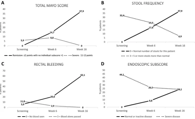
At W16, the clinical response rate was 62.1% (95% CI: 42.3–79.3%) and clinical remission 37.9% (95% CI: 20.7–57.7%). Total Mayo score decreased significantly over time, from a median score of 8 points before golimumab administra-tion to 4 points at W16. The dimensions of Mayo score also improved during the study, as well as the rate of endoscopic healing (Figure 1, Table
S1). Regarding histological activity, 97.0%, 79.4% and 75.9% subjects had histologically active UC (Geboes index >3.0) at screening, W6 and W16, respectively (Table S2). Changes in biomarker (sST2, FC and CRP) levels during the study were not statistically significant (Figure 2).
Baseline levels of biomarkers by clinical response, endoscopic and histological activity at week 6 and week 16
Subjects with endoscopic activity at W6 had higher baseline levels of sST2 (median, 24.5 ver-sus 18.5 ng/ml, p = 0.026) (Figure 3), and a differ-ent change of sST2 levels from baseline [median change (interquartile range), 0.8 (−3.2–7.8) ver-sus −2.7 (−9.2 to −0.3) ng/ml, p = 0.029]. In addition, baseline sST2 levels were higher among subjects with endoscopic activity at W16 (median, 11.3 versus 6.0 ng/ml, p = 0.025) (Table S3). With regards to histological results, base-line sST2 levels were higher among subjects with histological activity (assessed by Geboes index) at W6 (median, 23.0 versus 13.7 ng/ml, p = 0.035) (Figure 3) and W16 (median, 21.5 ver-sus 11.7 ng/ml, p = 0.016) (Table S3). Baseline sST2 levels were also higher among subjects with histological activity at week 16 as assessed by RHI: median, 20.8 versus 12.7 ng/ml, p = 0.038). No statistically significant differences were observed regarding baseline sST2 levels by clini-cal response at W6 and W16.
Subjects with histological activity (as assessed by Geboes index) at W6 had higher FC baseline lev-els (median, 884 versus 414 µg/g, p = 0.010) but no statistically significant different CRP baseline levels (Figure 3). Similar results were observed when classifying histological activity at W6 through the RHI (Table S3). Subjects with histo-logical activity (as assessed by Geboes index) at W16 had higher FC baseline levels (median, 831 versus 300 µg/g, p = 0.006) and higher CRP base-line levels (median, 4.2 versus 0.8 ng/ml, p = 0.033) (Table S3). When considering the RHI classifica-tion, FC baseline levels were higher among sub-jects with histological activity at W16 (median, 3.1 versus 0.3 ng/ml, p = 0.003) but no statistically significant differences were observed on CRP baseline levels (Table S3). Baseline levels of FC and CRP were not statistically significant differ-ent when comparing subjects by clinical response or endoscopic activity, at W6 (Figure 3) and W16 (Table S3).
Figure 1. Proportion (%) of patients by total Mayo score (A), stool frequency (B), rectal bleeding (C) and endoscopic Mayo subscore (D) during the study.
Figure 2. Evolution of CRP (A), FC (B) and sST2 (C) levels during the study: median and interquartile range
(grey area).
Comparison of biomarkers’ levels at week 6 and week 16, by endoscopic and histological activity and clinical response
At W6, subjects with endoscopic findings had sig-nificantly higher levels of sST2 than subjects without endoscopic activity (median, 24.1 versus
11.9 ng/ml, p = 0.004) (Figure 4, Table S3). The optimal sST2 cut-off to discriminate endoscopic activity at W6 was 16.9 ng/ml (AUC = 0.80, p < 0.001), with a sensitivity of 85%, specificity of 71% and a positive predictive value (PPV) of 81% (Figure S2 and Table 1). At W16, no statistically
Figure 3. Levels of sST2, FC and CRP at baseline, by endoscopic and histological activity and clinical response
at week 6.
significant differences were observed regarding sST2 levels by endoscopic activity and, at W6 and W16, no statistically significant differences were observed by clinical response or histological activ-ity (Table S3).
No statistically significant differences were observed regarding FC levels at W6, for subjects
with clinical response at the same moment (Figure 4, Table S3). Subjects with endoscopic activity at W6 had higher FC levels (median, 983 versus 180 µg/g, p = 0.037) (Figure 4, Table S3). The optimal cut-off for endoscopic activity was 353 µg/g (AUC = 0.73, p = 0.049), with a sensitivity of 90%, specificity of 67% and a PPV of 81% (Figure S2 and Table 1). Subjects with histological activity at
Figure 4. Levels of sST2, FC and CRP at week 6, by endoscopic and histological activity and clinical response.
W6 had higher FC levels than subjects without active UC (median, 983 versus 132 µg/g, p = 0.002) (Figure 4, Table S3). The optimal FC cut-off for histological activity was 353 µg/g (AUC = 0.92, p < 0.001), with a sensitivity of 84%, specificity of 100% and a PPV of 72% (Figure S2 and Table 1). At W16, subjects with histological activity also had higher FC levels at the same timepoint (median, 771 versus 46 µg/g, p = 0.001); no statistically sig-nificant differences were observed by clinical response or endoscopic activity (Table S3).
CRP levels at W6 were lower among subjects with clinical response (0.9 versus 4.8 mg/l, p = 0.020), no endoscopic activity (0.8 versus 4.0 mg/l, p = 0.026) and no histological activity (0.5 versus 4.2 mg/l, p = 0.011) (Figure 4). The optimal cut-off for endoscopic activity was 0.7 mg/l (AUC = 0.73, p = 0.016), with a sensitivity of 95%, specificity of 50% and a PPV of 72% (Figure S2 and Table 1). Regarding discrimination of histological activity, the optimal cut-off was 4.4 mg/l (AUC = 0.82, p < 0.001), with a sensitivity of 50%, specificity of 100% and a PPV of 100% (Figure S2 and Table 1). At W16, subjects with histological activity had higher CRP levels (median, 3.5 versus 0.4 mg/l,
p < 0.001); no statistically significant differences were observed by clinical response or endoscopic activity at the same moment (Table S3).
Correlation of biomarkers levels and clinical, endoscopic and histological activity
At W6, the correlations between sST2, FC and CRP levels were poor and not statistically signifi-cant (Table 2). At W16, the correlation between FC and CRP levels was positive (rs = 0.60, p < 0.001). No statistically significant correlations were observed between sST2 and FC or CRP levels at W16.
At W6, a positive correlation between sST2 levels and total Mayo score (rs = 0.40, p = 0.018) and endoscopic activity (rs = 0.45, p = 0.007) was observed (Table 2). The correlation of sST2 with histological activity (both Geboes index and RHI classifications), partial Mayo score and rectal bleeding subscore were poor and not statistically significant. FC levels at W6 correlated with statis-tical significance with histological activity assessed by Geboes index only (rs = 0.47, p = 0.008), but not with histological activity assessed by RHI,
Table 1. Accuracy of sST2, FC and CRP measurement in predicting endoscopic and histological activity at
week 6.
AUC 95% CI p value SEN SPE PPV NPV
Serum soluble ST2 endoscopic activity
(cut-off value ⩾16.9 ng/ml) 0.80 0.65–0.95 <0.001 85% 71% 81% 77%
histological activity
(cut-off value ⩾15.5 ng/ml) 0.67 0.46–0.88 0.111 74% 71% 91% 42%
Faecal calprotectin endoscopic activity (cut-
off value ⩾353 μg/g) 0.73 0.50–0.96 0.049 90% 67% 81% 80% histological activity
(cut-off value ⩾353 μg/g) 0.92 0.82–1.00 <0.001 84% 100% 100% 60% C-reactive protein
endoscopic activity
(cut-off value ⩾0.7 mg/l) 0.73 0.54–0.92 0.016 95% 50% 72% 88%
histological activity
(cut-off value ⩾4.4 mg/l) 0.82 0.65–0.99 <0.001 50% 100% 100% 35%
95% CI, 95% (two-sided) Confidence Interval; AUC, area under the curve; NVP, negative predictive value; PPV, positive predictive value; SEN, sensibility; SPE, specificity; ST2, Suppression of Tumorigenicity 2.
endoscopic activity, total/partial Mayo score or rectal bleeding. CRP levels correlated with total Mayo score (rs = 0.55, p = 0.001) and with endo-scopic (rs = 0.40, p = 0.021) and histological (Geboes: rs = 0.38, p = 0.031; RHI: rs = 0.48, p = 0.005) activity, as well as with partial Mayo
score (rs = 0.52, p = 0.002) and rectal bleeding Mayo subscore (rs = 0.39, p = 0.026).
At W16, FC and CRP levels were significantly correlated with histological activity assessed by Geboes index (rs = 0.65 and rs = 0.64, p < 0.001,
Table 2. Correlation between biomarker levels and clinical, endoscopic and histological activity, at week 6 and
week 16.
Week 6 p value Week 16 p value
rs (95% CI) rs (95% CI)
Serum soluble ST2 levels versus
Total Mayo score 0.40 (0.08; 0.65) 0.018 0.10 (−0.28; 0.45) 0.615
Endoscopic activity 0.45 (0.13; 0.69) 0.007 0.27 (−0.11; 0.58) 0.159 Histological activity (Geboes) 0.25 (−0.09; 0.54) 0.151 0.18 (−0.20; 0.51) 0.358 Histological activity (RHI) 0.33 (−0.01; 0.60) 0.059 0.29 (−0.09; 0.59) 0.128 Partial Mayo score 0.33 (−0.01; 0.60) 0.058 −0.02 (−0.38; 0.35) 0.922 Rectal bleeding Mayo subscore 0.23 (−0.12; 0.53) 0.187 −0.17 (−0.50; 0.21) 0.392 Faecal calprotectin −0.02 (−0.37; 0.34) 0.906 −0.14 (−0.49; 0.25) 0.478 C-reactive protein 0.20 (−0.15; 0.51) 0.264 0.02 (−0.35; 0.38) 0.936 Faecal calprotectin levels versus
Total Mayo score 0.20 (−0.16; 0.52) 0.275 0.27 (−0.11; 0.59) 0.163
Endoscopic activity 0.32 (−0.04; 0.61) 0.077 0.31 (−0.08; 0.61) 0.112 Histological activity (Geboes) 0.47 (0.14; 0.71) 0.008 0.65 (0.36; 0.82) <0.001
Histological activity (RHI) 0.28 (−0.08; 0.58) 0.126 0.47 (0.11; 0.72) 0.012
Partial Mayo score 0.13 (−0.24; 0.46) 0.501 0.19 (−0.20; 0.53) 0.330 Rectal bleeding Mayo subscore 0.02 (−0.34; 0.37) 0.916 0.27 (−0.12; 0.58) 0.172 C-reactive protein 0.26 (−0.12; 0.56) 0.174 0.60 (0.29; 0.80) <0.001
C-reactive protein results versus
Total Mayo score 0.55 (0.25; 0.75) 0.001 0.37 (0.00; 0.65) 0.052
Endoscopic activity 0.40 (0.07; 0.65) 0.021 0.34 (−0.03; 0.63) 0.070 Histological activity (Geboes) 0.38 (0.04; 0.64) 0.031 0.64 (0.35; 0.81) <0.001
Histological activity (RHI) 0.48 (0.17; 0.71) 0.005 0.59 (0.28; 0.78) 0.001
Partial Mayo score 0.52 (0.22; 0.73) 0.002 0.31 (−0.07; 0.61) 0.103 Rectal bleeding Mayo subscore 0.39 (0.05; 0.65) 0.026 0.14 (−0.24; 0.49) 0.456
95% CI, 95% confidence interval; RHI, Robarts Histopathology Index; rs, Spearman’s correlation coefficient. Note: correlations with statistical significance are shown in bold.
respectively) and by RHI (rs = 0.47, p = 0.012 and rs = 0.59, p = 0.001, respectively) (Table 2). At the
same time point, the correlations between these biomarkers versus total Mayo score and endo-scopic activity, as well as the correlations between sST2 and disease activity outcomes, were poor and without statistical significance.
Discussion
Previous studies have shown that sST2 levels cor-relate positively with the severity of colonic mucosal disease and inflammatory cytokines,3,11,12
but few have evaluated the potential of biomarkers in predicting response to biological therapy.25 We
observed that sST2 correlates moderately, although with statistical significance, with clinical and endoscopic activity at W6 of golimumab treatment, and that higher baseline levels of sST2 were associated with endoscopic and histological activity at W6 and W16. Furthermore, subjects without endoscopic activity at W6 had a decrease in sST2 levels from baseline, while subjects who maintained endoscopic findings at W6 showed almost no change. Together, these findings sug-gest that sST2 levels at baseline can predict endo-scopic response and histological remission after induction and at an early phase of maintenance treatment with golimumab.
After 6 weeks of treatment with golimumab, all biomarkers were significantly higher for subjects with endoscopic activity, but no statistically sig-nificant differences were observed in sST2 levels by clinical response or histological activity. In addition, FC levels were correlated only with his-tological activity assessed by Geboes index, in contrast with other studies.3,5,26,27 CRP levels
were significantly correlated with all UC activity outcomes at W6, as described by others.28,29
At W6, sST2 levels showed a good performance for discriminating endoscopic activity with a cut-off value of 16.9 ng/ml (sensitivity = 85%; speci-ficity = 71%; PPV = 81%), with a higher discriminating capacity (AUC = 0.80) than that observed for FC (AUC = 0.73) or CRP (AUC = 0.73). Díaz-Jiménez and colleagues esti-mated a sST2 cut-off of 74.87 pg/ml (sensitiv-ity = 83%, specific(sensitiv-ity = 83%) to discriminate endoscopic activity in UC patients.3 This lower
sST2 cut-off could be due to the inclusion of UC patients irrespective of disease activity or treat-ment.3 The FC cut-off (353 μg/g) resulted in a
PPV of 81% for endoscopic activity and, for his-tological activity (Geboes index), a specificity and PPV of 100%. This high specificity for histologi-cal activity was reported previously for a lower cut-off of 100 µg/g.30 However, few studies have
evaluated the correlation between FC and endos-copy at 6 weeks, that is, during induction, and the higher cut-off of 353 μg/g is probably more ade-quate for this treatment phase since the 100 μg/g was determined for asymptomatic patients in remission. Magro and colleagues reported median FC levels of 230 (40–425) μg/g at 8 weeks after induction with infliximab in UC patients.30
Hence, we hypothesize that different cut-offs should be used according to induction and main-tenance treatment phases.
CRP showed a high specificity and PPV for histo-logical outcome (Geboes index), suggesting that all subjects with CRP ⩾ 4.4 mg/l present histo-logical activity. However, this cut-off had low sensitivity (50%) and poor NPV (35%) for histo-logical activity and the cut-off of 0.7 mg/l had inadequate specificity (50%) for endoscopic activity. Consequently, CRP levels seem to have poor performance and utility for predicting endo-scopic and histological activity at W6.
Subjects with histological activity at W6 had higher baseline levels of sST2 and FC, and those with histological activity at W16 had higher base-line levels of sST2 (when classified by both Geboes and RHI) and CRP (Geboes only). These results suggest that sST2, FC and CRP are bio-markers of different manifestations of UC inflam-matory process during early treatment with golimumab. Although the mechanism is still to be clarified, sST2 expression seems to be upregu-lated in IBD patients and might reduce the pro-tective effect of IL-33 when combined to it, by reducing macrophage modulation, and conse-quently wound healing, in UC patients.31–33 For
that reason and based on our results, we hypoth-esize that sST2 may be useful as a surrogate bio-marker, both in terms of assessing endoscopic activity and when predicting early treatment response to golimumab treatment.
Of note, we did not observe any statistical correla-tion between FC and sST2 levels. FC is recog-nized as a useful marker of mucosal damage as its levels seem to be increased when neutrophils are present in the epithelium, the main marker of his-tological activity.6 Infiltration of neutrophils
through the inflamed mucosa occurs after local release of cytokines and compromises mucosal architecture, epithelial barrier and production of inflammatory mediators.34 Histological healing is
frequently incomplete and, compared with clinical and endoscopic response, takes a longer time to be observed after treatment initiation.35–37 In our
study, FC levels showed a stronger correlation with histological activity, which some consider the ultimate goal of UC treatment, as subclinical inflammation is predictor of UC relapses.38,39
With regards to CRP, its short half-life ensures that serum concentrations quickly decrease once the acute-phase stimulus is removed.40 CRP
lev-els correlated with all disease activity markers at W6 and were statistically higher among subjects with histological activity at W16. Therefore, CRP can provide additional support when investigat-ing subclinical inflammation, namely durinvestigat-ing maintenance treatment with golimumab, even though it is less specific to the intestinal inflam-matory process and inadequate for predicting endoscopic and histological findings.8,29
Even though clinical improvement (based on total Mayo score and its dimensions) was notice-able at W6, the follow-up period was probably insufficient to clearly observe the cellular inflam-matory response.6 In fact, more than two thirds of
the patients had histological active UC (Geboes >3.0, i.e. with presence of neutrophils) during the study period.
The study presents other limitations. Due to the exploratory nature of the study, no formal sample size was determined. Hence, the small sample size may have also limited the comparison between subgroups of disease activity. We cannot exclude that, due to the number of comparisons made, some results could have resulted from sta-tistical chance. Larger studies should include sST2 assessment to clarify its role as a predictor of endoscopic activity. Finally, we should be cau-tious with generalisation of results as the eligibil-ity criteria, aiming at a more homogeneous sample and the safety of the participants, may have com-promised the external validity of the study. The use of well-defined indexes of endoscopic, clinical and histological activity, which are com-monly used in the clinical practice, and the cor-relation between several biomarkers, are major strengths of this study. This is particularly
relevant with regards to the use of the Geboes index and RHI to assess histological activity, both of which have shown good reproducibility and are the most commonly used indices in UC.6,15,16
Recent research has identified histological healing as an important predictor of long-term bene-fit.36–38,41 Still, the STRIDE consensus does not
consider histological healing as a treatment tar-get; probably due to the cost and workload of adding pathological assessment.42 Therefore, a
biomarker that would be correlated with histo-logical activity would be useful in clinical prac-tice. Further research should be conducted with a longer follow-up period, control groups and larger samples to confirm the role of sST2 levels among other biomarkers, when treating moderately-to-severely active UC with golimumab.
Acknowledgements
The authors would like to thank to Eurotrials, now part of CTI, Clinical Trials and Consulting Services, for medical writing assistance (Luís Veloso and Milene Fernandes), study monitoring and statistical analysis (Mariana Aparício and Vera Vicente). Consultant were funded by Merck Sharp and Dohme, Lda, Portugal. Project man-agement was provided by Carolina Moura (PhD) of C3B–Clinical Research Lda, Portugal, to whom we would like to express our gratitude for her deep commitment to this study.
Author contributions
Fernando Magro was involved in the conception and design of the study, interpretation of data, and drafting and revising the manuscript. All other authors were responsible for data acquisi-tion. All authors read and approved the final manuscript.
Funding
This work was supported by Merck Sharp and Dohme, Lda, Portugal, which provided drug and financial support for the interventional study (Protocol Nr MK8259-22).
Conflict of interest statement
Fernando Magro received a fee for presenting from: AbbVie, Ferring, Falk, Hospira, PharmaKern, MSD, Schering, Lab. Vitoria, Vifor, OmPharma. Helena Tavares de Sousa reports expert fees and nonfinancial support from MSD, Abbvie, Ferring, Dr Falk Pharma, PharmaKern, Janssen and Takeda, outside the
submitted work. Patrícia Machado is employee of MSD Portugal; Philip G is employee of Merck and Co., USA; and Isabel Redondo was an employee of MSD Portugal at the time the study was conducted. All other authors: none to declare. ORCID iD
Fernando Magro https://orcid.org/0000- 0003-2634-9668
Supplemental material
Supplemental material for this article is available online.
References
1. Silverberg MS, Satsangi J, Ahmad T, et al. Toward an integrated clinical, molecular and serological classification of inflammatory bowel disease: report of a working party of the 2005 montreal world congress of gastroenterology. Can J Gastroenterol 2005; 19(Suppl. A): 5A–36A. 2. Lichtenstein GR and McGovern DPB.
Using markers in IBD to predict disease and treatment outcomes: rationale and a review of current status. Am J Gastroenterol Suppl 2016; 3: 17–26.
3. Díaz-Jiménez D, Núñez LE, Beltrán CJ, et al. Soluble ST2: a new and promising activity marker in ulcerative colitis. World J Gastroenterol 2011; 17: 2181–2190.
4. Harbord M, Eliakim R, Bettenworth D, et al. Third European evidence-based consensus on diagnosis and management of ulcerative colitis. Part 2: current management. J Crohn’s Colitis 2017; 11: 769–784.
5. Daperno M, Castiglione F, de Ridder L, et al. Results of the 2nd part scientific workshop of the ECCO (II): measures and markers of prediction to achieve, detect, and monitor intestinal healing in inflammatory bowel disease. J Crohn’s Colitis 2011; 5: 484–498.
6. Magro F, Lopes J, Borralho P, et al. Comparison of different histological indexes in the assessment of UC activity and their accuracy regarding endoscopic outcomes and faecal calprotectin levels. Gut 2019; 68: 594–603.
7. Mosli MH, Zou G, Garg SK, et al. C-reactive protein, fecal calprotectin, and stool lactoferrin for detection of endoscopic activity in
symptomatic inflammatory bowel disease patients: a systematic review and meta-analysis. Am J Gastroenterol 2015; 110: 802–819.
8. Magro F, Sousa P and Ministro P. C-reactive protein in Crohn’s disease: how informative is it? Expert Rev Gastroenterol Hepatol 2014; 8: 393–408. 9. Tibble J and Bjarnason I. Non-invasive
investigation of inflammatory bowel disease. World J Gastroenterol 2001; 7: 460–465.
10. Gisbert JP, González-Lama Y and Maté J. Role of biological markers in inflammatory bowel disease. Gastroenterol Hepatol 2007; 30: 117–129. 11. Beltrán CJ, Núñez LE, Díaz-Jiménez D, et al.
Characterization of the novel ST2/IL-33 system in patients with inflammatory bowel disease. Inflamm Bowel Dis 2010; 16: 1097–1107. 12. Pastorelli L, Garg RR, Hoang SB, et al.
Epithelial-derived IL-33 and its receptor ST2 are dysregulated in ulcerative colitis and in experimental Th1/Th2 driven enteritis. Proc Natl Acad Sci USA 2010; 107: 8017–8022.
13. Sandborn WJ, Feagan BG, Marano C, et al. Subcutaneous golimumab induces clinical response and remission in patients with moderate-to-severe ulcerative colitis. Gastroenterology 2014; 146: 85–95.
14. Sandborn WJ, Feagan BG, Marano C, et al. Subcutaneous golimumab maintains clinical response in patients with moderate-to-severe ulcerative colitis. Gastroenterology 2014; 146: 96–109.e1.
15. Geboes K, Riddell R, Ost A, et al. A reproducible grading scale for histological assessment of inflammation in ulcerative colitis. Gut 2000; 47: 404–409.
16. Mosli MH, Feagan BG, Zou G, et al. Development and validation of a histological index for UC. Gut 2017; 66: 50–58.
17. Shi HY, Chan FK, Chan AWH, et al. Fecal immunochemical test predicts histological healing in ulcerative colitis: a prospective study using the geboes score and nancy index. Gastroenterology 2017; 152: S209.
18. Zenlea T, Yee EU, Rosenberg L, et al. Histology grade is independently associated with relapse risk in patients with ulcerative colitis in clinical remission: a prospective study. Am J Gastroenterol 2016; 111: 685–690.
19. Rutgeerts P, Sandborn WJ, Feagan BG, et al. Infliximab for induction and maintenance therapy for ulcerative colitis. N Engl J Med 2005; 353: 2462–2476.
20. Magro F, Gionchetti P, Eliakim R, et al. Third European evidence-based consensus on diagnosis and management of ulcerative colitis.
Part 1: definitions, diagnosis, extra-intestinal manifestations, pregnancy, cancer surveillance, surgery, and ileo-anal pouch disorders. J Crohn’s Colitis 2017; 11: 649–670.
21. D’Haens G, Sandborn WJ, Feagan BG, et al. A review of activity indices and efficacy end points for clinical trials of medical therapy in adults with ulcerative colitis. Gastroenterology 2007; 132: 763–786.
22. Lewis JD, Chuai S, Nessel L, et al. Use of the noninvasive components of the Mayo score to assess clinical response in ulcerative colitis. Inflamm Bowel Dis 2008; 14: 1660–1666. 23. Schroeder KW, Tremaine WJ and Ilstrup DM.
Coated oral 5-aminosalicylic acid therapy for mildly to moderately active ulcerative colitis. N Engl J Med 1987; 317: 1625–1629.
24. Magro F, Lopes S, Silva M, et al. Low golimumab trough levels at week 6 are associated with poor clinical, endoscopic and histological outcomes in ulcerative colitis patients: pharmacokinetic and pharmacodynamic sub-analysis of the evolution study. J Crohn’s Colitis. Epub ahead of print 16 April 2019. DOI: 10.1093/ecco-jcc/jjz071. 25. Díaz-Jiménez D, De la Fuente M,
Dubois-Camacho K, et al. Soluble ST2 is a sensitive clinical marker of ulcerative colitis evolution. BMC Gastroenterol 2016; 16: 103.
26. Røseth AG, Aadland E, Jahnsen J, et al.
Assessment of disease activity in ulcerative colitis by faecal calprotectin, a novel granulocyte marker protein. Digestion 1997; 58: 176–180.
27. Sipponen T, Savilahti E, Kärkkäinen P, et al. Fecal calprotectin, lactoferrin, and endoscopic disease activity in monitoring anti-TNF-alpha therapy for Crohn’s disease. Inflamm Bowel Dis 2008; 14: 1392–1398.
28. Kim DB, Lee KM, Lee JM, et al. Correlation between histological activity and endoscopic, clinical, and serologic activities in patients with ulcerative colitis. Gastroenterol Res Pract 2016; 2016: 1–7.
29. Karoui S, Laz S, Serghini M, et al. Correlation of C-reactive protein with clinical and endoscopic activity in patients with ulcerative colitis. Dig Dis Sci 2011; 56: 1801–1815.
30. Magro F, Lopes SI, Lopes J, et al. Histological outcomes and predictive value of faecal markers in moderately to severely active ulcerative colitis patients receiving infliximab. J Crohn’s Colitis 2016; 10: 1407–1416.
31. Boga S, Alkim H, Koksal AR, et al. Serum ST2 in inflammatory bowel disease: a potential
biomarker for disease activity. J Investig Med 2016; 64: 1016–1024.
32. Seo DH, Che X, Kwak MS, et al. Interleukin-33 regulates intestinal inflammation by modulating macrophages in inflammatory bowel disease. Sci Rep 2017; 7: 851.
33. Pastorelli L, De Salvo C, Cominelli MA, et al Novel cytokine signaling pathways in inflammatory bowel disease: insight into the dichotomous functions of IL-33 during chronic intestinal inflammation. Therap Adv Gastroenterol 2011; 4: 311–323.
34. Zhou GX and Liu ZJ. Potential roles of neutrophils in regulating intestinal mucosal inflammation of inflammatory bowel disease. J Dig Dis 2017; 18: 495–503.
35. Magro F, Lopes S, Coelho R, et al. Accuracy of faecal calprotectin and neutrophil gelatinase B-associated lipocalin in evaluating subclinical inflammation in ulcerative colitis-the
ACERTIVE study. J Crohn’s Colitis 2017; 11: 435–444.
36. Villanacci V, Antonelli E, Geboes K, et al. Histological healing in inflammatory bowel disease: a still unfulfilled promise. World J Gastroenterol 2013; 19: 968–978.
37. Bryant RV, Winer S, Travis SP, et al. Systematic review: histological remission in inflammatory bowel disease. Is ‘complete’ remission the new treatment paradigm? An IOIBD initiative. J Crohn’s Colitis 2014; 8: 1582–1597.
38. Papi C, Fascì-Spurio F, Rogai F, et al. Mucosal healing in inflammatory bowel disease: treatment efficacy and predictive factors. Dig Liver Dis 2013; 45: 978–985.
39. Chew TS and Mansfield JC. Can faecal
calprotectin predict relapse in inflammatory bowel disease: a mini review. Frontline Gastroenterol 2018; 9: 23–28.
40. Vermeire S, Van Assche G and Rutgeerts P. C-reactive protein as a marker for inflammatory bowel disease. Inflamm Bowel Dis 2004; 10: 661–665.
41. Lobatón T, Bessissow T, Ruiz-Cerulla A, et al. Prognostic value of histological activity in patients with ulcerative colitis in deep remission: a prospective multicenter study. United Eur Gastroenterol J 2018; 6: 765–772.
42. Peyrin-Biroulet L, Sandborn W, Sands BE, et al. Selecting therapeutic targets in inflammatory bowel disease (STRIDE): determining therapeutic goals for treat-to-target. Am J Gastroenterol 2015; 110: 1324–1338.
Visit SAGE journals online journals.sagepub.com/ home/tag