Ana Catarina Oliveira Ferreira
Junho 2012
Searching for the role of lipocalin-2 in the
central nervous system
O papel da lipocalina-2 no sistema nervoso
central
UMinho|20 12 Ana Cat arina Oliv eir a F err eir a Searching for t he role of lipocalin-2 in t he central ner vous system O papel da lipocalina-2 no sis
tema ner
voso central
Universidade do Minho
Escola de Ciências da Saúde
Trabalho realizado sob a orientação da
Professora Doutora Fernanda Marques
e da
Professora Doutora Joana Palha
Ana Catarina Oliveira Ferreira
Dissertação de Mestrado
Mestrado em Ciências da Saúde
Searching for the role of lipocalin-2 in the
central nervous system
O papel da lipocalina-2 no sistema nervoso
central
Universidade do Minho
Escola de Ciências da Saúde
ii
DECLARAÇÃO
Nome: Ana Catarina Oliveira Ferreira
Endereço eletrónico: [email protected] Telefone: +351 912884159
Número do Bilhete de Identidade: 13172955
Título dissertação:
Searching for the role of lipocalin-2 in the central nervous system O papel da lipocalina-2 no sistema nervoso central
Orientadores:
Professora Doutora Fernanda Marques Professora Doutora Joana Palha
Ano de conclusão: 2012
Ramo de Conhecimento do Mestrado: Ciências da Saúde
É AUTORIZADA A REPRODUÇÃO INTEGRAL DESTA DISSERTAÇÃO APENAS PARA EFEITOS DE INVESTIGAÇÃO, MEDIANTE DECLARAÇÃO ESCRITA DO INTERESSADO, QUE A TAL SE COMPROMETE.
Universidade do Minho, Junho de 2012
Agradecimentos
A presente dissertação é resultado da minha investigação científica no ICVS, contudo tal não poderia ter acontecido sem ajuda de todos aqueles a quem, abaixo, não posso deixar de expressar a minha maior gratidão.
À Fernanda, por ser sido mais que uma orientadora, uma verdadeira companheira e amiga. Obrigada pelo apoio e presença incondicionais e por acreditar sempre que eu era capaz de chegar até aqui.
À Joana e ao Nuno, pelas diversas discussões científicas e importantes sugestões, pelos desafios propostos e pelo entusiasmo.
Ao João, por ter sido um verdadeiro mentor aquando da minha chegada ao ICVS e por sempre acreditar em mim. Um Muito Obrigada por tudo!
À Falcão, por me saber dar na cabeça no momento certo, por todas as ‘discussões’, fossem elas de que conteúdo, pela imensa ajuda e por ser uma ‘mini-chefe’ cheia de energia e boa disposição.
À Ashley e ao Sandro, por partilharem comigo mais do que a inexplicável sensação de cada nova descoberta, a vossa amizade e companheirismo. Obrigada por tudo!
À Susana Roque, Margarida e Nadine, pela partilha de ideias e importantes sugestões. Aos meus eternos companheiros de carteira, o ‘gang’ JPêgo: Paula, Ana Rita e António Melo. Obrigada pelas infinitas horas de convívio e descontração, pelas risadas e momentos que só nós sabemos!
Ao Zé Miguel, um obrigado pelas lições de cultura e sabedoria e por me dar a conhecer o Sr. BNST.
À Cristina (Tininha!), à Sofia, à Diana, ao Miguel e à Filipa, um muito obrigada pela partilha desta etapa comigo e pelos imensos momentos de diversão, companheirismo e discussões científicas.
A todos os NeRDs, pelo apoio e sugestões partilhadas e auxílio prestado. À Pipoca, pela amizade e por me acompanhar nesta e outras etapas.
Aos meus amigos, por nem sempre compreenderem as minhas ausências mas por me apoiarem mesmo assim.
Ao Dinis, pelo apoio incondicional, pela paciência e compreensão, e por todo o carinho e força. Sem ti tudo teria sido bem mais difícil! Obrigada por tudo.
Aos meus pais e avó, por suportarem as minhas muitas ausências sem questionarem, por toda a preocupação e apoio incondicional, pelo amor e exemplo de força. Muito Obrigada!
Searching for the role of lipocalin-2 in the central nervous system
Abstract
Lipocalin-2 (LCN2), a well described secreted protein, has a denotable function as part of the innate immune response. With a remarkable broad expression and rapid induction upon stimulus and in tissues more prone to infection, LCN2 is able to limit bacterial growth through iron-depletion strategies, since it binds iron-loaded siderophores secreted by bacteria during infection. On the other hand, the evidence for the existence of an endogenous mammalian siderophore, along with the capacity for LCN2 to interact with specific cell surface receptors, has brought into light its significance in mediating cellular processes through iron mechanisms, even in basal conditions. In fact, an iron delivery pathway mediated by LCN2 has been proposed with ultimate roles in cell proliferation and survival, tissue development and protection and cellular apoptosis. However, studies addressing LCN2 involvement in such processes are limited to the periphery, being its role in the central nervous system (CNS) context less explored. In fact, most of the reports show LCN2 importance in an inflammatory context, as an autocrine mediator of reactive astrocytosis or even in the apoptosis and deramification of activated microglia. Additionally, its behavior as an acute-phase protein at the brain barriers adds the involvement of LCN2 in the mechanism of immune defense in brain contexts. However, concerning the role of LCN2 in the physiological brain, there is still a gap that remains unfilled. Therefore, and considering the extensively described functions in the periphery of LCN2 as an iron-traffic protein, we sought to investigate the role of LCN2 in normal brain development and function. Taking advantage of a mouse strain with a target deletion of the Lcn2 gene (LCN2-null mice), we analyzed the impact of LCN2 in the CNS through behavioral, morphological and cellular approaches. Our findings propose for a putative participation of LCN2 in the development and maturation of specific brain regions related to spatial perception and coordination, whereas in adulthood, LCN2 was observed to trigger altered emotional states, as LCN2-null mice displayed both anxious and depressive-like behaviors. Concomitantly, morphological and cellular approaches evidenced the contribution of LCN2 in the regulation of emotional-related structures, namely the bed nucleus of the stria terminalis (BNST) and in the subgranular zone (SGZ) of the hippocampus. Respectively, LCN2-null mice presented an increased spine density in the BNST and a decreased cell proliferation at the SGZ. Therefore, LCN2 can be assumed to be involved in the modulation of both morphological and cellular mechanisms that, in turn, can culminate with the altered behaviors, namely anxious and depressive-like ones.
O papel da lipocalina-2 no sistema nervoso central
Resumo
A lipocalina-2 (LCN2), descrita como uma proteína secretada, tem uma importante função na resposta imunitária inata. Com uma notável e rápida indução aquando de um estímulo e em tecidos expostos a infeção, a LCN2 é capaz de limitar o crescimento bacteriano através de estratégias de depleção de ferro, uma vez que se liga a sideróforos bacterianos carregados com ferro durante uma infeção. Por outro lado, a existência de sideróforos mamíferos endógenos, juntamente com a capacidade da LCN2 de interagir com recetores na superfície celular, tem revelado a sua importância em mediar processos celulares através do transporte de ferro, mesmo em condições fisiológicas. De facto, uma via do metabolismo do ferro mediada pela LCN2 foi proposta, e consequentemente funções na proliferação celular, no desenvolvimento e proteção de tecidos, assim como na apoptose têm sido mostradas.
Contudo, a maioria dos estudos que demonstram o envolvimento da LCN2 na mediação de tais processos celulares são limitados à periferia, sendo que o seu papel no contexto do sistema nervoso central (SNC) é menos explorado. Aqui, a função da LCN2 tem sido mais demonstrada no contexto da inflamação, como um mediador autócrino da proliferação de astrócitos, e ainda na apoptose e desramificação da microglia ativada. Adicionalmente, o seu comportamento como uma proteína aguda nas barreiras do cérebro acrescenta a sua relevância em mecanismos de defesa do sistema imunitário também no SNC. No entanto, e no que diz respeito à função da LCN2 no cérebro em condições fisiológicas, muito ainda está por descobrir.
Tendo em conta a extensa caracterização na periferia da LCN2 como uma proteína transportadora de ferro, com o presente estudo procurou-se investigar qual o seu papel no normal desenvolvimento e funcionamento do cérebro. Usando como modelo animal um ratinho no qual o gene que codifica para a LCN2 foi removido, analisamos a importância da LCN2 no SNC através de uma análise comportamental, morfológica e celular. Os resultados demonstram o envolvimento da LCN2 na maturação de regiões específicas do cérebro envolvidas na aquisição da perceção espacial e na coordenação, enquanto numa fase adulta, a LCN2 está envolvida no desencadeamento de comportamentos emotivos, nomeadamente ansiosos e depressivos. Simultaneamente, a análise morfológica e celular evidenciam a contribuição da LCN2 na modulação destes processos em estruturas relacionadas com processos emotivos, nomeadamente no núcleo da estria terminal e na zona subgranular do hipocampo. Nestas regiões foi verificado que os animais nos quais o gene Lcn2 foi removido apresentam um aumento significativo de densidade de espinhas nos neurónios do núcleo da estria terminal e uma diminuição no total da proliferação celular no hipocampo. Consequentemente, podemos assumir que a LCN2 está envolvida na modulação de ambos processos morfológicos e celulares, os quais, por sua vez, culminam nas alterações comportamentais verificadas, nomeadamente na ansiedade e depressão.
TABLE OF CONTENTS
1. INTRODUCTION 1
1.1 Iron homeostasis 3
1.2 The lipocalin protein family 4
1.3 LCN2 – where and when? 5
1.4 LCN2 ligands 7
1.5 LCN2 major functions 7
1.5.1 The role of LCN2 in the innate immune response 8 1.5.2 LCN2 mechanism of action in iron delivery 10
1.6 LCN2 at the central nervous system 13
1.7 Research objectives 17
2. MATERIAL AND METHODS 19
2.1 Animals 21
2.2 Genotyping 22
2.3 Behavioral assessment 24
2.3.1 Developmental milestones 24
2.3.2 Adult behavior 30
i) Elevated Plus Maze 31
ii) Open Field 32
iii) Forced Swim Test 32
iv) Acoustic Startle 33
v) Cognitive Function Assessment 33
2.4 Serum corticosterone measurements 35
2.5 BrdU labeling in vivo 36
2.5.1 BrdU immunohistochemistry 36
2.5.2 Stereology 37
2.6 Golgi staining 37
2.7 Dendritic tree analysis 38
3. RESULTS 41
3.1 Impact of LCN2 in the normal brain development and function 43 3.1.1 Developmental milestones assessment 43
3.1.2 Adult behavior performances 46
3.2 Corticosteroids measurements 51
3.3 Morphological analysis of brain cytoarquitecture in LCN2-null mice 51 3.4 Assessment of LCN2 impact on cell proliferation 56
4. DISCUSSION 61
4.1 LCN2 during brain development 63
4.2 The adult brain function and the contribution of LCN2 65 4.3 The hypothalamic-pituitary-adrenal (HPA) axis in LCN2-null mice 67 4.4 Uncovering the role of LCN2 in neuronal morphology
and its implications in emotional behaviors
68
4.5 LCN2 modulation of cell proliferation at the neurogenic niches 70
5. CONCLUDING REMARKS 73
Abbreviations
ANGD Anogenital Distance
AS Acoustic Startle
BBB Blood Brain Barrier
BCSFB Blood-Cerebrospinal Fluid Barrier
BLA Basolateral Amygdala
BNST Bed Nucleus of the Stria Terminalis BrdU Bromodeoxyuridine
BSA Bovine Serum Albumin
CNS Central Nervous System
CP Choroid Plexus
CSF Cerebrospinal Fluid
DAB 3,3'-Diaminobenzidine Substrate
DG Dentate Gyrus
DNA Deoxyribonucleic acid
EPM Elevated Plus Maze
FST Forced Swim Test
HCl Hydrochloric Acid
Hetero Heterozygous
HPA Hypothalamic-Pituitary Adrenal
H2O2 Hydrogen Peroxide
LCN2 Lipocalin-2
LPS Lipopolysaccharide
MWM Morris Water Maze
NaCl Sodium Chloride
Neo Neomicin
NGAL Neutrophil Gelatinase Associated Lipocalin
OF Open Field
PCR Polymerase Chain Reaction
PFA Paraformaldehyde
PFC Prefrontal Cortex
PND Postnatal Day
RAM Radial Arm Maze
RMS Rostral Migratory Stream SEM Standard Error of the Mean
SEZ Subependymal Zone
SGZ Subgranular Zone
TBS-T Tris Buffer Saline-Tween
Wt Wild-Type
Figures Index
Figure 1 Schematic representation addressing how bacteria acquire iron
from the host
Figure 2 Possible effects of holo and apo-LCN2 on iron metabolism and
apoptosis, mediated by 24p3R
Figure 3 Generation of LCN2-null mice
Figure 4 Schematic representation of the genotype strategy for
LCN2-null mice
Figure 5 Schematic representation of the timeline used for adult
behavior
Figure 6 Impact of LCN2 deletion in brain development and maturity Figure 7 Anxious-like behavior analysis in the elevated plus maze
(EPM) test, as the percentage of time spent in each arm
Figure 8 Forced swim test (FST) for learned helplessness evaluation of
LCN2-null mice and respective littermates
Figure 9 Evaluation of spatial reference and working memory in the
radial arm maze (RAM)
Figure 10 Spatial reference memory in the Morris water maze (MWM)
task
Figure 11 Spine density assessment at the BNST neurons
Figure 12 Dendritic spines proportion of each type of spines analyzed at
the BLA and the BNST brain regions
Figure 13 Sholl analysis evaluation of the 3D reconstructed neurons, on
both BLA and BNST brain regions
Figure 14 3D reconstructions of BNST bipolar neurons of Wt and
LCN2-null mice in the 3D spatial dimensions
Figure 15 Representative total proliferation at the intermediate level of
the SEZ
Figure 16 Representative total proliferation at the SGZ of the DG
Tables Index
Table 1 Primers sequence for mice genotyping
Table 2 PCR Mix composition and reaction conditions Table 3 Data sheet for developmental milestones
Table 4 Conversion of time intervals registered for each test into
dichotomic scores
Table 5 Summary of each test analyzed and the respective dimension
that it evaluates
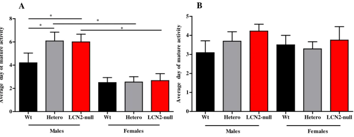
Table 6 Summary of the average days of mature response obtained in
each genotype, both in males and females, during the developmental milestones assessment
Table 7 Acoustic startle data represented as the startle amplitude
response of the animals in the presence of increased acoustic stimulus (dB)
Table 8 Influence of LCN2 in the morphometric analysis of dendrites
of neurons of the BLA and BNST
Table 9 Dendritic spines proportion assessment at the BLA and the
BNST brain regions
Table 10 Summary of all the results obtained for the morphological
assessments in all the animals
Table 11 Rates of proliferation in Wt, Hetero and LCN2-null mice,
along the dorso-ventral axis at the intermediate level of the SEZ (considering the anterior-posterior axis)
Table 12 Summary of the data obtained during this project regarding the
role of LCN2 in the CNS
1. INTRODUCTION
1.1 Iron homeostasis
Iron is a required and an essential element for fundamental biological processes. As part of enzymes, cytochromes and protein prosthetic groups, iron ensures cellular homeostasis, participating in processes that range from cell proliferation and survival to oxygen transport and energy metabolism (Hentze et al., 2004; Mesquita et al., 2012). Although essential, systemic iron imbalance, whether due to its deficiency or excess, can lead to the development of several pathological conditions, such as anemia, or iron deposition as in hemacromatosis and in some neurodegenerative disorders (i.e. Alzheimer’s and Parkinson’s diseases) (Mesquita et al., 2012).
Of interest, there is no biological mechanism to excrete iron from the body, apart from physiological bleeding in women and enterocyte renewal. Therefore, iron levels are tightly controlled at the absorption level. The liver assumes the control of this regulation through the production of a small peptide, hepcidin, (Krause et al., 2000)that in turn binds to ferroportin on the surface of the enterocytes, causing its internalization and degradation; this ultimately leads to a decrease in iron absorption to the bloodstream (Anderson and Frazer, 2005). In the blood, iron is mostly bound to plasma proteins, particularly to transferrin, responsible for both the transport and delivery of iron into the majority of cells (Gomme et al., 2005). After binding to receptors (mainly transferrin receptor 1), transferrin enters an endocytic pathway and, within endosomes, iron dissociates from the receptor and is transported into the cell cytoplasm where it can be stored bound to ferritin (Harrison and Arosio, 1996).
In the central nervous system (CNS), the mechanism through which iron homeostasis is regulated is less known, but must be specific and precise since when iron accumulates in peripheral organs, such as in the case of hemacromatosis, the brain seems spared. The brain parenchyma is protected from the blood content by two main barriers, the brain-blood barrier (BBB) and the blood-cerebrospinal fluid barrier (BCSFB). Both have been showed to mediate iron uptake into the brain through transferrin-mediated endocytosis (Moos et al., 2006). Once inside the endothelial cell of the BBB or the choroid plexus (CP) epithelial cell of the BCSFB, iron is released through ferroportin into the interstitial fluid of the brain parenchyma, or to the cerebrospinal fluid (CSF), respectively, where it binds to transferrin and is taken up by neurons and glia cells
(Moos et al., 2006). Interestingly, the CP has been recently suggested to play a specific role in regulating brain iron homeostasis, at least in particular circumstances such as in response to peripheral inflammation, where the CP is able to produce and secrete hepcidin (Marques et al., 2009).
Of notice, it is well known that both in the periphery and in the CNS, transferrin is considered the main route through which cells acquire most of their iron. However, studies with transferrin deficient mice demonstrated that the transferrin pathway is not essential for the delivery of iron to many tissues (Garrick and Garrick, 2009). These mice present anemia, deficiency in the development of the CNS and iron accumulation in the liver, but most of the epithelial organs have normal iron levels (Yang et al., 2002).
All together, these observations support the existence of alternative pathways for iron delivery to cells. One of such putative mechanisms may be mediated by lipocalin-2 (LCN2). This 25 kDa protein from the lipocalin protein family (Flower et al., 2000) has been considered an related protein given its ability to sequester bacterial iron-loaded siderophores secreted by bacteria during infection (Flo et al., 2004). As such, LCN2 participates in the innate immune response by depleting iron from the invading microorganisms. However, in light of the recent discovery of an endogenous mammalian siderophore (Bao et al., 2010; Devireddy et al., 2010), LCN2 may, as well, bind, transport and delivery iron to cells through its membrane receptor.
In the next sections we will explore the role and putative functions of LCN2 on both basal and pathological conditions in the periphery and in the CNS.
1.2 The lipocalin protein family
LCN2 (also known as 24p3, neutrophil gelatinase-associated lipocalin (NGAL), oncogene 24p3, or siderocalin) belongs to the lipocalin protein family, a large and diverse group of small (160-180 residues in length) soluble and often secreted proteins present across species. A remarkable feature of this family is the great diversity of the primary sequence of its proteins, with an unusually low level of overall sequence conservation, in some case as low as 20% (Flower, 1994; Grzyb et al., 2006). However, membership of this family has been largely identified based on structural similarities as they all share a common secondary and tertiary structural feature - called as the "lipocalin fold", a cup-shaped cavity that can bind to specific ligands (Flower et al.,
2000). This conformation has allow them to be carriers, transporting predominantly small lipophilic molecules such as steroids, bilins, retinoids and lipids, to form complexes with soluble macromolecules and to bind specific cell-surface receptors (Flower, 1996). In fact, lipocalins are described to participate in the regulation of cell division, differentiation and cell to cell adhesion and survival and in the modulation of the immune response (Flower, 1996; Flower et al., 2000; Chakraborty et al., 2012). As member of this lipocalin protein family, LCN2 (lipocalin product of a single gene, the 24p3), is believed to also engage some of the overall functions described for lipocalins. Its emerging evidence as a significant mediator of several physiological and pathological processes has caught our attention.
We will, therefore, consider the major key features of LCN2 in detail.
1.3 LCN2 – where and when?
When first described as component of human neutrophils, LCN2 sequence did not match with any known human protein, but instead showed a high degree of similarity with the deduced sequence of the rat α-2-microglobulin-related protein and the mouse protein 24p3, that in turn also belong to the lipocalin family (Kjeldsen et al., 1993; Kjeldsen et al., 1994).
Originally with no function described, LCN2 was found stored in specific human neutrophil granules and co-localizing with lactoferrin as a monomeric and homodimeric form. In addition, its covalent association with the 92-kDa monomeric form of matrix metalloproteinase-9 (MMP-9), a gelatinase secreted by neutrophils for extracellular matrix degradation and remodeling (Kjeldsen et al., 1993; Kjeldsen et al., 1994), led to be named as neutrophil gelatinase-associated lipocalin (NGAL) (Kjeldsen et al., 1993; Kjeldsen et al., 1994).
Afterwards, it was also found in mouse neutrophils (Kjeldsen et al., 2000), although the first reports in murine models were in BALB/c 3T3 fibroblasts upon stimulation (Nilsen-Hamilton et al., 1982) and as a gene highly induced during the transition of mouse kidney cells from a quiescent to a proliferative state, as a result of virus infection (Hraba-Renevey et al., 1989). Meanwhile, several expression studies described LCN2 to be present in tissues that are more prone to exposure to infection and highly secreted by mucus producing epithelial cells of the respiratory tract (lung and trachea) (Cowland and Borregaard, 1997; Friedl et al., 1999). Other major sites of LCN2 expression
described are blood and peritoneal cells (Flo et al., 2004), macrophages (Meheus, 1993), endothelial (Liu and Nilsen-Hamilton, 1995) and epithelial cells (Marques et al., 2008).
While present in normal tissues, mainly the kidney and thymus, both in humans and mice (Friedl et al., 1999), LCN2 is mainly expressed in response to stimuli. Among these, lipopolysaccharide (LPS) has been shown to be the major inducer of LCN2 expression. For instances, cultured murine macrophages when stimulated with LPS where shown to highly express LCN2 (Meheus, 1993), which was also reported to occur in primary cultures enriched in CP epithelial cells (Thouvenot et al., 2006). These in vitro studies were largely confirmed by posterior in vivo data, where in conditions of a peripheral immune challenge by LPS, LCN2 was shown to behave as an acute-phase protein in the CP epithelial cells (Marques et al., 2008). Importantly, and accompanying such quick up-regulation in the CP, LCN2 protein levels were also found elevated in the cerebrospinal fluid (CSF), which is mainly produced by the CP; and in blood vessels of the brain parenchyma (Marques et al., 2008). LCN2 had also been claimed as a liver acute-phase protein in response to the in vivo injection of turpentine (Liu and Nilsen-Hamilton, 1995), and at the lungs after LPS stimulus (Sunil et al., 2007).
Similarly, in vitro stimulation of mouse liver cells (Liu and Nilsen-Hamilton, 1995), L-cells (Garay-Rojas et al., 1996) and primary thymocytes (Devireddy et al., 2001) with dexamethasone induces the acute expression of LCN2 (Liu and Nilsen-Hamilton, 1995). This regulation of expression is mainly explained by the described existence of two glucocorticoid responsive core elements (GRE) in the 24p3 gene promoter (Garay-Rojas et al., 1996). Additionally, pro-inflammatory cytokines were also shown to be able to induce the constitutively expression of LCN2 in epithelial inflamed lungs (Cowland et al., 2003). This induction was observed by interleukin-1β (IL-1β) but not by TNF-α stimulation (Cowland et al., 2003) and further work provided the evidence for LCN2 induction by TNF-α only in the presence of IL-1β (Karlsen et al., 2010).
Overall, a striking observation in the study of LCN2 expression is its strong induction (both mRNA and protein) in different cell lines and tissues following exposure to a variety of stimuli and a number of inducers have been identified, thus retrieving LCN2 with major functions in defense mechanisms and in a variety of inflammatory responses (reviewed at (Borregaard et al., 1995)).
1.4 LCN2 ligands
Lipocalins are characterized by their ability to bind different lipophilic substrates and, if some bind several ligands, others are specific for a single one (Kjeldsen et al., 2000). Early studies hypothesized LCN2 to have an immunomodulatory activity by binding and clearing lipophilic mediators of inflammation such as the neutrophil chemoattractant N-formyl-Met-Leu-Phe (fMLP) (Allen et al., 1989; Sengelov et al., 1994), the platelet activating factor, leukotriene B4, and LPS (Nielsen et al., 1996). However, unlike other lipocalins, LCN2 binding cavity was shown by both X-ray crystallography (Goetz et al., 2000) and nuclear magnetic resonance spectroscopy (Coles et al., 1999) to be distinct as it is unusually large and atypically polar. In fact, the relative low affinity of LCN2 for N-formyl tripeptides was shown, suggesting that any of the previous proposed hydrophobic ligands are unlikely to be the preferred ligands for LCN2 (Goetz et al., 2000).
Goetz and colleagues (2002) identified the ability of LCN2 to bind negatively charged catecholate-type ferric bacterial siderophores, small molecules produced during bacterial infection to scavenge iron from the host that have higher affinity to iron than the host iron-binding proteins (Goetz et al., 2000; Goetz et al., 2002). In this work, LCN2 was shown to be able to bind iron loaded bacterial siderophores in its binding pocket but, unlike lactoferrin or transferrin, LCN2 could not bind positively charged iron ions alone, being specific only for iron already earmarked for bacterial use by siderophores complexes (Goetz et al., 2002).
Classically, lipocalins derive their name from the ligand that they bind. Therefore, at this point the authors proposed LCN2 to be renamed siderocalin based on the ligand it binds (Goetz et al., 2002).
Importantly, LCN2 is now described to be able to bind a wide variety of siderophores thus mediating its physiological role as a broad specificity siderophore binding protein and most of its described functions have been inferred from these binding capacities.
1.5 LCN2 major functions
By the time it was first reported (Nilsen-Hamilton et al., 1982; Hraba-Renevey et al., 1989), LCN2 was suggested to be involved in the control of cell regulation, in normal and/or transformed cells, possibly through the transport of lipophilic molecules (Flower, 1994). The following description of LCN2 expression in neutrophils and tissues exposed to microorganisms and in conditions of inflammation/infection has brought the
possibility of an antimicrobial activity for LCN2 (Kjeldsen et al., 2000) or even a role in inflammation or cellular growth (Kjeldsen et al., 2000). Its capacity to bind bacterial catecholate-type ferric siderophores (Goetz et al., 2002), confirmed the suggestion of LCN2 as a potent bacteriostatic agent that participates in the iron-depletion strategy of the innate immune system. Of interest, the identification of endogenous mammalian siderophores (Bao et al., 2010; Devireddy et al., 2010) along with the capacity of LCN2 to interact with specific cell-surface receptors raised the possibility that LCN2 participates in iron trafficking and metabolism (Yang et al., 2002; Devireddy et al., 2005). Therefore, among other putative described functions, LCN2 major roles reported so far are in relation to the innate immune response and the intracellular iron trafficking. These and other functions that have been attributed to LCN2 will now be described in detail.
1.5.1 The role of LCN2 in the innate immune response
Upon an infection, microbes, chiefly bacteria, require iron for growth and proliferation and have evolved strategies to survive within the severely iron-poor environment of the human body. The exceedingly low availability of free iron is attributable to iron-binding proteins such as transferrin, which form complexes with any available free iron molecules. To cope with their need, bacteria acquire most of their iron from the host by synthesizing siderophores, low molecular weight proteins that scavenge iron from the various host iron-binding proteins and transport it into the pathogen (Ratledge and Dover, 2000). Interestingly, siderophores have an affinity for iron several times higher than that of the host endogenous iron carrier proteins (Ratledge and Dover, 2000). In this context, Goetz and colleagues (2002) have shown, in vitro, that LCN2 has a high affinity for bacterial siderophores, both in their iron-loaded and iron-free states, and that it even competes with the pathogen for iron, thus limiting the bacteria propagation (Goetz et al., 2002) (Figure 1). Upon this discovery, LCN2 was immediately suggested to be involved in the innate immune response: LCN2 was assumed to be released by neutrophils at sites of infection and inflammation to sequester bacterial siderophores, thus participating in the antibacterial iron-depletion strategy of the innate immune response (Goetz et al., 2002).
Figure 1: Schematic representation addressing how bacteria acquire iron from the host. Upon infection,
bacteria synthesize siderophores (1) to scavenge iron from host iron-binding proteins (transferrin, lactoferrin and ferritin) for its growth and proliferation (2). In response, the host produces LCN2 that is able to bind iron-loaded siderophores (3) thus limiting the availability of iron for bacterial growth (4). In this response, LCN2 is induced through the activation of Toll-like receptors (Flo et al., 2004). Studies using mice with a target deletion of Lcn2 gene (LCN2-null) revealed that under pathogen-free conditions, there is no major phenotype present; however, upon exposure to sublethal doses of Escherichia coli H9049, LCN2-null mice were far more likely to develop bacteraemia (Flo et al., 2004). The absence of the defense mechanism mediated by LCN2 can lead to sepsis and death (Flo et al., 2004) extolling LCN2 with an essential bacteriostatic role (Berger et al., 2006).
Holmes and colleagues (2005) showed LCN2 to be also able to bind soluble siderophores of mycobacterium, including Mycobacterium tuberculosis (Holmes et al., 2005), and other studies reported the up-regulation of LCN2 in primary cultured macrophages in response to Salmonella (Nairz et al., 2009) and in the respiratory tract in response to colonization by Klebsiella pneumoniae (Nelson et al., 2005; Chan et al., 2009).
Of interest, LCN2 may similarly protect the brain from invading microorganisms, since its expression occurs at the both barriers of the brain: in the epithelial cells of CP in the CP-CSF barrier and by the endothelial cells of the capillaries that irrigate the brain parenchyma in the blood-brain barrier (Marques et al., 2008). The authors hypothesized that on one hand, LCN2 might reduce iron access to the bacteria in the blood vessels and, in the case of bacteria entering into the CSF, CP-borne LCN2 can sequester siderophore-bound iron and prevent bacteria dissemination within the ventricular brain system and brain parenchyma (Marques et al., 2008).
LCN2 is, therefore, described as an accurate mechanism of sensing microbial metabolism to modulate the host response appropriately, assuming iron an important role in such modulation.
1.5.2 LCN2 mechanism of action in iron delivery
The delivery of iron to cells is essential for cell growth and development and most cells acquire iron by capturing iron-loaded transferrin (Garrick and Garrick, 2009). The importance of LCN2 in iron-related processes emerged upon its identification as an inducer of rat metanephric mesenchyma conversion into epithelia through associations with iron (Yang et al., 2002). Remarkably, the authors were able to show that the LCN2 expressed in mammalian kidney contains iron, and even that LCN2 is able to deliver iron into cells through a mechanism independent of transferrin (Yang et al., 2002). This alternative pathway was described to require endocytosis and endosomal acidification and the modulation of iron-responsive genes by LCN2: enhancement of transferrin expression and reduction in the expression of the transferrin receptor 1 (Yang et al., 2002). Moreover, the internalization of either transferrin or LCN2 was dependent of the kidney developmental phase, with earlier epithelial progenitors incorporating LCN2 while the further staged epithelial cells internalized mainly transferrin (Yang et al., 2002). As a whole, data from these authors showed LCN2 as an iron-donor protein with functions distinct from those of transferrin, vital for early embryonic development, at least in the kidney (Yang et al., 2002).
These observations were striking given that LCN2 does not have an intrinsic ability to bind iron, but instead to bind iron-loaded siderophores (Goetz et al., 2002). However, the recent description for the existence of an endogenous mammalian siderophore has brought into light the possibility for a physiological-relevant mechanism of iron uptake mediated by LCN2 (Bao et al., 2010; Devireddy et al., 2010). While bacterial siderophores are not synthesized by mammalian cells, they are composites of well known functional groups such as hydroxybenzoates and hydroxybenzenes that were found in a variety of compounds in mammalian serum and urine, where interestingly LCN2 traffics (Bao et al., 2010), thus suggesting that it might bind those compounds (Bao et al., 2010). In fact, through the use of mouse and human aseptic urine as a source to search for novel LCN2 ligands and through structural analyses, it was identified a subset of catechols that bind both iron and LCN2, revealing a novel mechanism of iron capture and release (Bao et al., 2010; Devireddy et al., 2010).
In accordance, Yang and colleagues (2002) have described that following the cellular uptake of LCN2 by endocytosis, the bound iron is released leading to an increase in intracellular iron concentrations (Yang et al., 2002). These internalization mechanisms of the secreted LCN2 suggested the existence of cellular receptors. In fact, megalin (Hvidberg et al., 2005) and the solute carrier family 22 (organic cation transporter), member 17 (SLC22A17) also known as brain-type organic cation transporter (generally named as 24p3R) (Devireddy et al., 2005) are the two known receptors described to allow LCN2 to engage roles in trafficking iron in and out of the cells.
Megalin, a multi-ligand endocytosis receptor member of the low density lipoprotein receptor family expressed in epithelia, is also known to bind the mouse lipocalins retinol-binding protein and α-2-microglobulin (Hvidberg et al., 2005). Hvidberg and collegues (2005) in their studies reported that both iron-loaded-LCN2 (holo-LCN2) and iron-lacking LCN2 (apo-LCN2) were able to bind megalin with high affinity, thus leading to LCN2 endocytosis (Hvidberg et al., 2005). Moreover, its tissue expression was found to be coincident with the induction of LCN2 expression during inflammation, thus highlighting megalin as an endocytic receptor capable of binding iron transporting LCN2 and mediating its uptake (Hvidberg et al., 2005). The relevance of megalin in regulating iron levels through LCN2 became evident when megalin-null mice were described to have a significantly higher urine excretion of LCN2 (Mori et al., 2005). With respect to 24p3R, it was shown that LCN2 may be internalized whether in its apo- or holo- forms, in this way being able to modulate both iron uptake and excretion from cells (Devireddy et al., 2005). Indeed, a pathway for LCN2-mediated iron depletion has been proposed, where the iron status of LCN2 is the critical determinant of its activity (Figure 2) (Devireddy et al., 2005). Briefly, holo-LCN2 binds to 24p3R, is internalized, and releases its bound iron, thereby increasing intracellular iron concentration and preventing apoptosis; by contrast, apo-LCN2 when internalized, chelates iron and transfers it to the extracellular medium, thereby leading to iron efflux that results in apoptosis and cell death, mediated by the pro-apoptotic Bcl-2-interacting mediator of cell death (Bim) (Devireddy et al., 2005; Richardson, 2005) (Figure 2).
Figure 2: Possible effects of holo and apo-LCN2 on iron metabolism and apoptosis, mediated by 24p3R.
(A) Holo-LCN2 containing iron bound to the siderophore donates iron to cells via the 24p3R. Upon internalization of the LCN2-receptor complex, iron is removed, leading to an increase in the intracellular levels of iron. This accounts for the prevention of cell apoptosis through the decrease in the expression of the pro-apoptotic protein Bim. (B) Apo-LCN2 binds to 24p3R and is also internalized into the cell. Once inside, it is suggested to putatively become complexed with an endogenous siderophore, forming the holo-LCN2. Its subsequent release from the cell by exocytosis leads to a depletion of iron from the cell resulting in the upregulation of the pro-apoptotic molecule Bim and, therefore, stimulation of apoptosis (Adapted from (Richardson, 2005)).
In summary, the isolation and identification of the endogenous mammalian siderophore, together with the existence of receptors for LCN2, has brought into light the additional biological relevance of LCN2 in iron delivery mechanisms in basal conditions.
This may explain several of the reports on the role of LCN2 in cellular processes. In fact, LCN2 has been shown to regulate cell division (Gwira et al., 2005), differentiation and maturation during development (Yang et al., 2002) and epithelial morphogenesis (Yang et al., 2002; Schmidt-Ott et al., 2007) and also to contribute in the homeostasis of hematopoietic cells lineages due to its ability to induce apoptosis (Liu et al., 2011). Of interest, is the evidence that LCN2 may be either protective/pro-survival (Mishra et al., 2004; Mori et al., 2005; Roudkenar et al., 2009), or pro-apoptotic (Kehrer, 2010), which, as well, may depend on whether LCN2 is depleting or delivering iron from or to cells.
In this context, this has been evidenced in kidney injury situations, where LCN2 has been shown to be up-regulated for protection (Mishra et al., 2004; Mori et al., 2005).
Also of interest is the variable described contribution of LCN2 in cancer, since both cancer promoting (Bolignano et al., 2010; Kehrer, 2010) and anti-tumoral (Jin et al., 2011) actions have been reported.
Additionally, and also controversial, is the role of LCN2 as a putative adipokine, where LCN2 has been suggested as a marker of obesity (Li and Chan, 2011), but with a precise role in glucose and insulin homeostasis still to be clarified (Guo et al., 2010; Law et al., 2010; Jun et al., 2011).
1.6 LCN2 at the central nervous system
The evidence for a role for LCN2 in cell homeostasis, through iron, has increased the search of knowledge regarding its involvement in several contexts and distinct organs. Despite its implication in several cellular processes, little is known about LCN2 and the CNS, particularly with respect to normal brain physiology.
Of interest, the literature is still not conclusive concerning the sites of LCN2 synthesis in basal conditions, with recent reports showing immunostaining and mRNA expression in specific brain regions of the adult rat, namely the olfactory bulb, brainstem and cerebellum (Chia et al., 2011).
The first reports regarding LCN2 in the CNS described its up-regulation in whole brain homogenates in response to inflammation induced by turpentine injection (Liu and Nilsen-Hamilton, 1995), and later in the mouse brain after focal ischemia (MacManus et al., 2004) and in spinal cord injury (De Biase et al., 2005).
Also in response to an inflammatory status, LCN2 was shown to be expressed in astrocytes from different brain regions (striatum, cerebral cortex, hippocampus and cerebellum) (Lafon-Cazal et al., 2003) and, nowadays it is broadly accepted and described that astrocytes are the main brain cells expressing LCN2.
Interestingly, both in vitro and in vivo approaches have shown the expression of LCN2 upon the LPS stimulus at the CP epithelial cells (Thouvenot et al., 2006; Marques et al., 2008; Ip et al., 2011), from where it is secreted into the CSF (Marques et al., 2008). Of notice, LCN2 was not detected at the CP in basal conditions but only in response to the stimulus (Marques et al., 2008; Ip et al., 2011).
Most remarkably is the recent report that the 24p3R receptor for LCN2 is constitutively expressed by the mouse brain and that its levels of expression are not affected by systemic LPS administration (Ip et al., 2011). Additionally, differences between the basal expression of LCN2 and 24p3R mRNA were noticed. While 24p3R mRNA levels
were only detected in neurons and in the CP upon the LPS stimuli, endothelial, astrocytes, microglia cells as well as the CP, but not neurons, were identified as the main cellular sources for LCN2 mRNA in the CNS (Ip et al., 2011). As a whole, LCN2 was reported to be strongly produced in the CNS by LPS inflammatory stimulus and that neurons seem to be the target for the actions of LCN2 as the expression of the 24p3R assembles (Ip et al., 2011).
Even though the cellular functions of LCN2 in the CNS remain enigmatic, some new insights have been given by Lee and colleagues that through in vitro studies showed that after inflammatory stimulation of cultured astrocytes, LCN2 expression and secretion was increased and, most importantly, was critical in inducing cell death sensitization, stimulation of cell migration and morphological changes of reactive astrocytes (Lee et al., 2009). Consequently, LCN2 was proposed to be an autocrine mediator of reactive astrocytosis, a process in which astrocytes when responding to injury become hypertrophic and proliferate (Lee et al., 2009). Most remarkably was the evidence for the involvement of iron metabolism and Bim protein in this LCN2-induced cytotoxic sensitization. At last, these described roles were also confirmed in vivo using a zebrafish model (Lee et al., 2009). Of notice, it was shown that LCN2’ mediating processes is not restricted to reactive astrocytes since it was demonstrated that LCN2 has also a dual role on both apoptosis and deramification of the in vitro activated microglia and, recently, in neuronal cell death, again through the Bim protein activation (Lee et al., 2007; Lee et al., 2012). Recently, it was also described how LCN2 is able to promote cellular migration through the up-regulation of chemokines in the brain (Lee et al., 2011). Using animal models of inflammation, LCN2 was shown in vivo to act as a chemokine inducer that once secreted in conditions of inflammation, induces CNS cells to secrete chemokines that amplify the neuroinflammation response by recruiting additional cells (Lee et al., 2011).
In addition, several reports have been linking the described patterns of LCN2 expression and cellular modulation with neurological conditions and states, such as neurodegenerative disorders. For instances, LCN2 was described to be highly expressed in the plasma of patients suffering from mild cognitive impairment (MCI), when compared to healthy control subjects and even to the Alzheimer’s Disease (AD) patients (Choi et al., 2011). Contrarily, Naudé and colleagues (2012) report that LCN2 levels in the serum of patients with MCI are not significantly elevated, but describe for the
presence of decreased levels of LCN2 in CSF of human patients with both MCI and AD (Naude et al., 2012). Additionally, and when analyzing the levels of LCN2 in AD human postmortem brain tissues, it was found to be ubiquitously expressed throughout the brain but significantly increased in brain regions associated with AD pathology (hippocampus and entorhinal cortex) (Naude et al., 2012). Previously, Wu and colleagues (2006) had showed that LCN2 mRNA levels were strongly up-regulated in the cortical tissue from AD model mice, both prior or after amyloid-β deposition, one of the hallmarks of AD (Wu et al., 2006).
Also recently, LCN2 was associated to the experimental autoimmune encephalomyelitis (EAE) murine model of multiple sclerosis, since it was found to be up-regulated in the spinal cord at the onset phase of the disease, being predominantly expressed, in this context, by astrocytes and monocytes, along with its receptor (Berard et al., 2012). The role of LCN2 in this disease was evidenced by the increased severity of EAE in the LCN2-null mice (Berard et al., 2012).
An additional field where LCN2 has also been implicated is in the response to stress, which may be related to the presence of glucocorticoid-responsive elements in its gene (Garay-Rojas et al., 1996). Studies in LCN2-null mice exposed to a restraint stress suggest a role for LCN2 in the control of dentritic spine density and maturation, neuronal excitability and anxiety (Mucha et al., 2011).
Finally, and also recently, both in models of spinal contusion injury (Rathore et al., 2011) and of inflammatory pain (Poh et al., 2012) increased expression of LCN2 was described, in both cases attributed to infiltrating neutrophils. In the case of the spinal cord injury model, also astrocytes and neurons were shown to strongly express LCN2 (Rathore et al., 2011).
Altogether, while the participation of LCN2 seems also to be present in the brain in response to injury, little is known on its function in physiological conditions, which is the aim of the present project.
1.7 Research objectives
It is clear that despite all the described features for LCN2, regardless of the context, it is in the CNS that much information is still lacking. The need for understanding LCN2’ role in the physiological brain as brought us to this project where we aim to unravel its putative roles during development and in adulthood. To asses this, and taking advantage of mouse model with targeted deletion of Lcn2 (LCN2-null mice), three major aims were proposed:
1. Explore the role of LCN2 in the normal CNS development and adult function 2. Analyze the morphological brain cytoarquitecture of LCN2-null mice
3. Assess the impact of LCN2 on the proliferative profile of adult neural stem cells With this project, we expect to contribute to characterize the role of LCN2 in brain function and in behavior.
2. MATERIAL AND METHODS
2.1 Animals
A mouse strain with a targeted deletion of the gene for LCN2 (LCN2-null mice) and their respective wild-type (Wt) and heterozygous (Hetero) littermates were used in all the studies performed.
This LCN2-null mouse strain was generated by deleting the entire coding sequence (Exons 2 to 5) and replacing it by a neomicin (Neo) cassette (Figure 3).
Figure 3: Generation of LCN2-null mice. The entire coding sequence (from exons 2 to 5, light grey
boxes) were deleted and replaced by a neomycin (Neo) cassette (dark grey box).
All experiment procedures were conducted in accordance with the Portuguese national authority for animal experimentation, Direcção Geral de Veterinária (ID: DGV9457). Animals were kept and handled in accordance with the guidelines for the care and handling of laboratory animals in the Directive 2010/63/EU of the European Parliament and of the Council. Efforts were made to minimize the number of animals used and their suffering.
All mice were given standard diet (4RF25 during gestation and postnatal periods, and 4RF21 after weaning; Mucedola SRL, Settimo Milanese, Italy) and water ad libitum, with exception to the privation periods that occurred whenever the experiment required so. Animals were maintained under standard laboratory conditions on a 12/12-h light/dark cycle (lights on at 8 a.m.) with an ambient temperature of 21±1ºC and relative humidity of 50-60%.
Matings were prepared for animal colony maintenance: Wt and LCN2-null mice were bred to obtain LCN2-Hetero mice, which in turn were mated. This allowed the production of Wt, Hetero and LCN2-null mixed litters that were used to perform all the studies. Whenever a birth occurred, animals were kept untouched in the home cage with
their mothers until weaning was performed at 21 days of age; males and females were then separated and kept in independent cages, in groups of three to five animals per cage until genotype determination was performed.
Our animals were raised in a BALB/c background and used for most of the experiments, but we also had available the same construct in the C57BL/6 background that we used for cognitive assessment.
2.2 Genotyping
Routinely, mice genotype was confirmed by polymerase chain reaction (PCR). For that,
DNA extraction strategies included ear/toe or tail digestions with a Finnzymes´ Phire®
Animal Tissue Direct PCR Kit (BioLabs, New England, UK) designed to perform PCR directly from non-fixed animal tissue samples with no prior DNA purification. The process was executed accordingly to the manufacture’s instruction. Briefly, the tissue was directly placed into 10 μl of a Dilution Buffer, and 0.5 μl of DNARelease™ Additive for 5 min at room temperature. After an incubation of 2 min at 98ºC, samples were centrifuged and the supernatant containing the DNA quantified using the NanoDrop spectrophotometer (NanoDrop Technologies, Inc., ThermoScientific, Wilmington, USA).
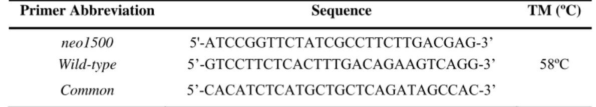
PCR strategy relied on the amplification of the junction of the inserted cassette with the target gene flanking sequences, as in Figure 4. For the detection of both mutated and Wt alleles, two independent pairs of primers, neo1500 and Wild-type, were respectively used (Table 1).
Table 1: Primers sequence for mice genotyping.
Primer Abbreviation Sequence TM (ºC)
neo1500 5'-ATCCGGTTCTATCGCCTTCTTGACGAG-3’
Wild-type 5’-GTCCTTCTCACTTTGACAGAAGTCAGG-3’ 58ºC Common 5’-CACATCTCATGCTGCTCAGATAGCCAC-3’
Figure 4: Schematic representation of the genotype strategy for LCN2-null mice. The exons are
numbered and identified in light grey. Primers Common and Wild-type were used to amplify the Wt allele, whereas neo1500 in conjunction with the Common primer was used to identify the mutated allele, both with 500 bp.
The Common primer is specific for the Lcn2 gene and, in combination with either neo1500 or Wild-type primers allows to distinguish the LCN2-null mice from the Wt, respectively. Two independent PCR mixtures were required since the product size in both sets of primers is of 500 bp. For each PCR reaction, 1 μl of DNA (100 ng/μl) was used for a final volume of 20 μl of PCR reaction, using the mix and reaction conditions as described in Table 2:
Table 2: PCR Mix composition and reaction conditions.
Master Mix (20 µl) Volume
(µl)
DNA Template PCR conditions
H2O (Milli_Q) 14.5
1 µl
(100 ng/ µl)
Taq Buffer (with (NH4)2SO4)1 2.0 95ºC 2’
MgCl2 (25 mM) 1.2 95ºC 1’ dNTPs (10 mM) 0.4 58ºC 45” 30 cycles Common (10 µM) 0.4 72ºC 1’ neo1500 (10 µM) 0.4 72ºC 5’ Wild-type (10 µM) 0.4 4ºC ∞ Taq Polymerase (5 U/ µl)1 0.1
1Fermentas Life Science, California, USA
The amplified PCR products were separated in a 2% agarose gel stained with ethidium bromide.
2.3 Behavioral assessment
To explore the role of LCN2 in the normal CNS development and function, we evaluated the impact of the absence of this protein through a behavioral characterization both during postnatal development and in adulthood.
One cohort of animals from two independent litters was submitted to a set of behavioral paradigms through the first 21 days of life, to evaluate the acquisition of neurological hallmarks during brain development, the called developmental milestones protocol. Other set of animals from three independent group of litters was assessed for their behavior in adulthood (2-3 months old), which included general evaluation of motor, emotion and cognitive dimensions.
At the end of the experiments, all animals were sacrificed. Both protocols were replicated at least twice.
2.3.1 Developmental milestones
Assessment of neurobehavioral neonatal development included the execution of a range of well-described tests used to evaluate neurologic parameters such as motor, reflexes and strength/coordination development (Hill et al., 2008; Lim et al., 2008). This procedure was designed to allow a fast throughput so that several litters can be examined daily within a relatively short period of time (Hill et al., 2008; Lim et al., 2008).
The day of birth was considered as postnatal day (PND) 0, in which pups were weighed and inspected for any gross malformations or particular traits. From that day on, mice were daily examined for the acquisition of developmental milestones and weight gain until PND 21, the weaning day. To allow for a fast identification of each mouse in a litter, on the first days after birth, pups were marked with a respective number and on PND 3, toe clipping was done not only for the identification of each mouse in the litter but also for proposes of genotyping.
On the day of testing, the home cage was moved into the testing room and left to habituate for at least 30 min. During the experimental execution, the pups were left in the same room as the mother and the time of separation was minimized as possible. The experimenter was always the same and identified the mouse by its marked number, being blind for the genotype. The execution of each test was random, as well as the animals order to perform them each day.
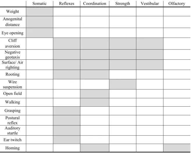
Each test has a range of time period during the 21 days to be performed (Table 3). The outcomes of the tests focus on the time to accurately perform, or respond to, a stimulus or posture.
Table 3: Data sheet for developmental milestones. Neonatal mouse pups were daily examined from PND
0 to 21 for performance on a battery of developmental tests assessing strength, coordination and the appearance of reflexes. The shaded areas identify the tests to be performed on each day.
Milestones test Postnatal day 0 1 2 3 4 5 6 7 8 9 10 11 12 13 14 15 16 17 18 19 20 21 Weight Anogenital distance Surface righting Negative geotaxis Cliff aversion Rooting Postural reflex Wire suspension Walking Grasping Auditory startle Ear twitch Open field Air righting Eye opening Homing
Therefore, the latency of time (in seconds, s) for the animal to execute the test was registered and afterwards converted to dichotomic scores, as indicated in Table 4; this allows for a quantitative measurement of the behavior. The animal is considered to exhibit a mature response on a specific test when the highest score attributed is observed for two consecutive days (Hill et al., 2008; Lim et al., 2008).
Table 4: Conversion of time intervals registered for each test into dichotomic scores [adapted from (Hill et al., 2008)]. Score 0 1 2 3 Surface righting no response within
30 s slowly (10-25 s) rights itself but
rights itself but it takes up to 10 s to do it rights itself immediately in less than 1 s Negative geotaxis
does not move at all within the 30 s
period
turns its body 180º to the ‘head up’ position in a period time inferior to 30 s
Cliff aversion
freezes/does not respond within the
30 s test-period
turns very slowly back to the surface
(10-25 s)
avoids the cliff, but it still takes some time to turn, up to
10 s
turns back in less than 1 s
Rooting
does not move the head toward the
filament
moves the head toward the filament
Wire
suspension falls immediately
grasp the bar with all four limbs (maximum time set
of 30 s)
Auditory
startle does not jump jump
Ear twitch not present present
Open field does not respond in 30 s
moves out of the circle in less than
30 s
Walking no locomotion
pivoting – moving
around with the help of the head and forelimbs, but not using the
hind limbs
crawling – moving
on all four limbs, dragging the belly
over the surface
walking – mature
locomotion with the body supported
completely by the four limbs
Eye opening both closed one open both open
Air righting lands on its back lands on the surface with all four paws
Grasping no grasping
places its paw on the cotton bud, but it does not hold on
firmly
places its paw on the cotton bud with
more force, but when the cotton bud is pulled, cannot hold it
grasps the cotton bud very firmly
Postural
reflex not present present
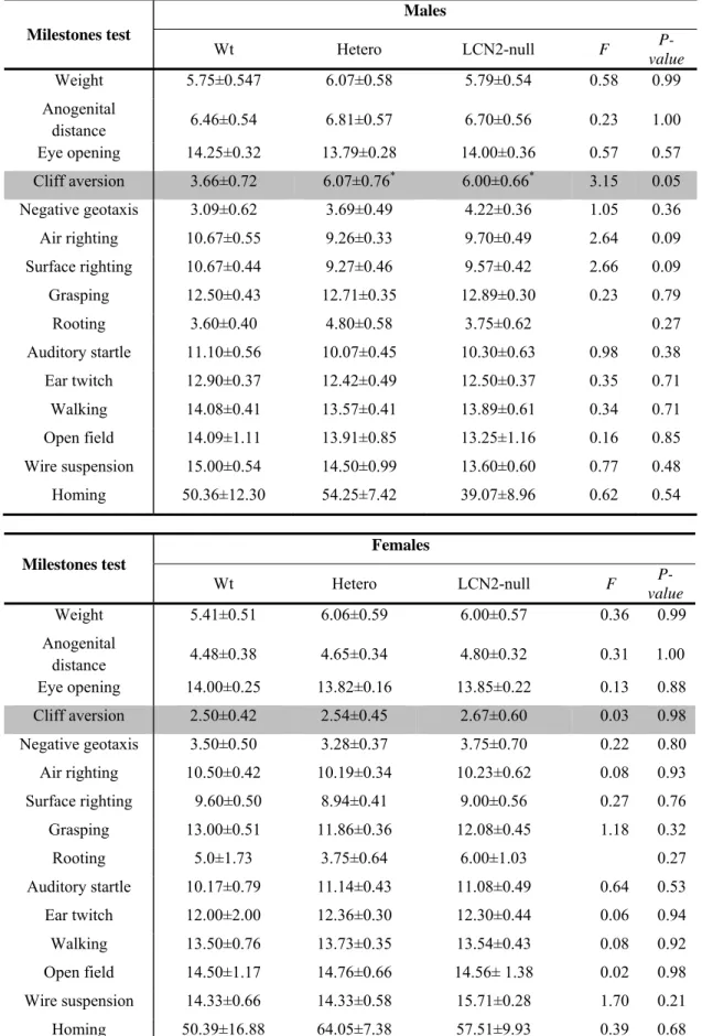
Two independent groups of litters were used for the milestones evaluation. A first group of mating produced 5 litters each composed of between 5 to 12 pups, with a total of 35 pups (8 Wt, 16 Hetero and 11 LCN2-null offspring). All animals, both males and females, were submitted to the milestones protocol, and at the weaning day, they were housed in group of 5 animals in each cage and left to grow.
A second set of mating gave rise to 5 litters with a total of 42 pups (10 Wt, 20 Hetero and 12 LCN2-null). Again, all animals, males and females, were submitted to the milestones protocol but instead, at the PND 21, animals were sacrificed.
Each test and the time period of execution as well as the respective behavioral dimension that it evaluates, will be next described in detail (Hill et al., 2008; Lim et al., 2008) (summarized in Table 5).
Table 5: Summary of each test analyzed and the respective dimension that it evaluates.
Somatic Reflexes Coordination Strength Vestibular Olfactory
Weight Anogenital distance Eye opening Cliff aversion Negative geotaxis Surface/ Air righting Rooting Wire suspension Open field Walking Grasping Postural reflex Auditory startle Ear twitch Homing Somatic parameters
As a measure for morphological development, animals were daily weighed and the anogenital distance (ANGD - distance between the opening of the anus and the opening of the genitalia) was measured. Eyes opening were also daily analyzed and a score was attributed as indicated in Table 4.
Neurological reflexes
Cliff aversion, PND1-14 (Labyrinthine reflex and body righting mechanisms, strength, and coordination)
In this test, the mouse pup was positioned on the edge of a small box with a smooth surface with the digits of the forepaws and the snout hanging over the edge of the box. The time for the pup to turn and begin to crawl away from the edge was registered. If the pup lost footing and slipped off the box, the test was repeated once more. If the mouse did not respond within 30 s, the test was terminated. The test was repeated daily until it was performed correctly in fewer than 30 s for two consecutive days.
Negative geotaxis, PND1–14 (Labyrinthine reflex and body righting mechanisms, strength, and coordination)
The animal was placed head down on a square of screen set at an angle of 45°. The time for the pup to turn 180° to the “head up” position was recorded. If the pup lost footing and slipped on the screen, the test was repeated once more. If the mouse did not respond within 30 s, the test was finished. The mature response was considered to be achieved when pups were able to perform correctly in fewer than 30 s for two consecutive days.
Surface righting, PND1-13 (Labyrinthine reflex and body righting mechanisms, strength, and coordination)
The mouse pup was held gently on its back and released. The time for the pup to flip over onto its abdomen with all four paws touching the surface of the table was registered. If the mouse did not respond within 30 s, the test was ended. The mature response was considered to be achieved when pups were able to right itself in less than 1 s for two consecutive days.
Air righting, PND8–21 (Labyrinthine reflex and body righting mechanisms, strength, and coordination)
The mouse pup held upside down was released from a height of approximately 60 cm and its landing position was examined. The first day that the pup landed right side up on all four paws was recorded. The test was repeated daily until the pup lands on all four paws for two consecutive days.
Rooting, PND1–12 (Tactile reflex and motor coordination)
A fine filament of cotton was used to perform this test. The filament was slowly and gently stroked three times from front to back along the side of the pup’s head. Rooting occurred if the pup moved its head toward the filament. If there was no response to the brushing filament on one side of the head, the test was repeated once on the other side. Testing continued daily until the pup responded correctly for two consecutive days.
Wire suspension, PND4–14 (Strength)
The pup, hold with its forepaws in a 3-mm diameter metal wire suspended at 5 cm above a soft surface was released, and the length of time the mouse remains grasping the bar was measured. If the pup failed off immediately, the test was repeated once again. The test was repeated daily until the mature response was achieved when the animal was able to grasp the bar holding with all four limbs.
Open field, PND8–21 (Locomotive coordination and extinguishing of pivoting behavior)
In this test, a mouse pup is placed in the center of a circle with 13 cm in diameter and the time taken to move out was recorded. If the mouse did not respond in 30 s, the test was finished. The mouse was tested daily until it responded in fewer than 30 s for two consecutive days.
Walking, PND5-21 (Locomotive coordination and muscular strength)
In this test, the animals were able to freely move on the bench for 60 s. The first day the pups were able to move with the body completely supported by the four limbs was registered.
Grasping, PND5-21 (Freeing reflex)
The mouse pup palm forelimb was stimulated with a cotton swab. The day when the pup flexed the paw to grasp the object was recorded.
Postural reflex, PND5-21 (Reflex)
Animals placed in a small plastic box were gently shaken up and down and left and right. Acquisition of a mature postural reflex was considered when the animal was able to maintain its original position in the box by extending all four limbs.
Auditory startle, PND7–18 (Auditory reflex)
The mouse pup was placed on the laboratory bench, and its reaction to a handclap at a distance of 10 cm was recorded. The test was repeated daily until the pup responded correctly with a quick involuntary jump for two consecutive days.
Ear twitch, PND7–15 (Tactile reflex)
A fine filament twisted of a cotton swab was gently brushed against the tip of the ear three times. The pup was considered to achieve a mature response when it responded by flattening the ear against the side of the head for two consecutive days.
Homing, PND12 (Locomotion and olfactory capabilities)
Individual pups were placed in the center of a standard cage containing 1/3 of bedding from the home cage and the remaining 2/3 of the cage with new clean bedding. The latency to turn and move onto the home cage bedding was assessed in three separate trials. The pup was considered to have successfully ‘homed’ when all four paws had crossed onto the home cage bedding and the pup remained there for at least 10 s. Each trial lasted a maximum of 120 s with an inter-trial interval of approximately 10 s.
2.3.2 Adult behavior
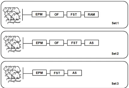
Unraveling the role of LCN2 in the brain function included the use of a battery of standard tests that evaluate motor and locomotion, emotion (anxiety and depressive-like states) and cognition at adulthood. With that purpose, we submitted young adult (2-3 months old) LCN2-null male mice and their respective littermates to a set of behavioral paradigms (Figure 5).
Three independent set of animals from 3 different groups of matings, with a total of 28 Wt, 34 Hetero and 28 LCN2-null males animals were used. Not all sets were submitted to the same behavior tests, although some are common across them and when performed, the same timeline was used in the 3 independent experiences (Figure 5).
Figure 5: Schematic representation of the timeline used for adult behavior. For this assessment, 3
independent set of animals were used in 3 independent experiences. The order of tests presented corresponds to the order of performance done in each set. AS, Acoustic Startle; EPM, Elevated Plus Maze; FST; Forced Swim Test; OF, Open Field; RAM, Radial Arm Maze.
By the time of weaning, animals were housed 5 per cage and only handled in the week before the behavior protocol started in order to minimize the stress that could be caused by the experimenter manipulation. Tests were performed always in the light day period, since physiological and biochemical parameters change throughout the day (Wotjak, 2004). Before each test, animals were transferred to the test room for habituation for 30 min.
The detailed description of each test used will now be specified.
i) Elevated Plus Maze (EPM)
The EPM apparatus (ENV-560; MedAssociates Inc, St. Albans, VT, USA) was used to assess anxious-like behaviors and it consisted of two opposite open arms (50.8×10.2 cm) and two enclosed arms (50.8×10.2×40.6 cm) elevated 72.4 cm above the floor. The junction center area between the four arms measured 10×10 cm.
Mice were placed individually in the center of the maze facing a closed arm and were allowed to freely explore it for 5 min. Behavioral parameters were recorded with the use of an infra-red photobeam system connected to a computer with specific software (MedPCIV, MedAssociates Inc). The time spent in the closed arms was used to calculate the percentage of time in the closed arms which was taken as an index of anxiety-like behavior: the more time spent in the closed arms meant an anxious-like
state. The total number of entries in the closed arms was used as a measure of general locomotor activity.
ii) Open Field (OF)
The OF arena measures a combination of both exploratory and locomotor activities and aspects of anxiety. The OF apparatus consisted of a brightly illuminated square arena of 43.2×43.2 cm equipped with infrared beams located at the level of the arena floor on both X and Y axes (to monitor the central and peripheral horizontal activities) and above the floor on two sides of the chamber (to monitor vertical activity) (Med Associates Inc.). Mice were individually placed in the center of the arena and allowed to freely explore it for 5 min. The resulting data was analyzed using the Activity Monitor software (Med Associates, Inc.), considering two previously defined areas: a central and an outer area. Activity parameters such as total distance travelled, vertical activity and time spent in each predefined area, as well as the average velocity, were analyzed. Also, the ratio between the time spent in the center and in the periphery of the open field arena was used as an indicative of anxiety-like state (Prut and Belzung, 2003).
iii) Forced Swim Test (FST)
Learned helplessness was evaluated in the FST for the assessment of depressive-like behaviors. The test was conducted using a slightly modified method described by Porsolt and colleagues (1977) (Porsolt et al., 1977). Briefly, each animal was individually placed in glass cylinders filled with water (25°C; depth 30 cm) for 5 min. Test sessions were videotaped and manually scored as the immobility time and latency to immobility using the Etholog V. 2.2 software, always by the same experimenter (Ottoni, 2000) blind to the animals genotype under assessment. The minimal duration for first bout of immobility was set at 1 s. A mouse was judged immobile when it ceased all active behaviors (i.e. struggling, swimming and jumping) and remained passively floating or making minimal movements necessary to maintain the nostrils above water (Castagne et al., 2009). Learned helplessness behavior was defined as an increase in time of immobility and a decrease in both latency to immobility and swimming time.