Mónica Filipa dos Santos Mendes
Master of Science in Biomedical Engineering
Probing Radiosensitisers in Electron
Transfer Experiments
Thesis submitted in partial fulfilment of the requirements for the degree of
Doctor of Philosophy in
Radiation Biology and Biophysics Applied Atomic and Molecular Physics
Supervisor: Prof. Paulo Limão-Vieira, Full Professor
Universidade Nova de Lisboa
Co-supervisor: Prof. Gustavo García Gómez-Tejedor, Full Professor
Consejo Superior de Investigaciones Científicas
Examination Committee Chairperson: Prof. João Carlos da Palma Goes
Rapporteurs: Prof. Luís Paulo da Mota Capitão Lemos Alves Prof. Alexander Dorn
Members: Prof. Pedro António de Brito Tavares Prof. Gustavo García Gómez-Tejedor
Probing Radiosensitisers in Electron Transfer Experiments
Copyright © Mónica Filipa dos Santos Mendes, Faculdade de Ciências e Tecnologia, Universidade NOVA de Lisboa.
A Faculdade de Ciências e Tecnologia e a Universidade NOVA de Lisboa têm o direito, perpétuo e sem limites geográficos, de arquivar e publicar esta dissertação através de exemplares impressos reproduzidos em papel ou de forma digital, ou por qualquer outro meio conhecido ou que venha a ser inventado, e de a divulgar através de repositórios científicos e de admitir a sua cópia e distribuição com objetivos educacionais ou de investigação, não comerciais, desde que seja dado crédito ao autor e editor.
“Nothing in life is to be feared, it is only to be
understood. Now is the time to understand
more, so that we may fear less”
ACKNOWLEDGEMENTS
I feel very grateful for the opportunity I had to start my PhD 4 years ago. During this time I have learned such important and interesting things that made me grow both professionally but, especially as a person. Do a PhD is like a roller coaster with funny moments, frustrating days and motivating days but it is worthy in the end. All of these moments could not be lived and achieved without the best people by my side. So, I would like to thank…
To the financial support from the Portuguese National Funding Agency FCT-MCTES through PD/BD/106038/2015 scholarship. This work was also supported by Radiation Biology and Biophysics Doctoral Training Programme (RaBBiT, PD/00193/2012); UID/Multi/04378/2013 (UCIBIO).
To Prof. Dr. Paulo Limão-Vieira for all the support, supervision and motivation throughout the course of this work, as well as for the opportunity to visit other international groups and attend scientific meetings. Thank you for believing in my work!
To Prof. Dr. Gustavo García for support, supervision and for giving me the opportunity to be part of his research group and performed part of my work in his lab, as well as for the opportunity to visit other international groups and attend scientific meetings. These two years in Madrid were truly important and unforgettable.
To Prof. Dr. Filipe Ferreira da Silva for his friendship, support and scientific conversations that help me so much.
All of the remaining members of Molecular Physics and Applications research group, particularly Ana Cruz and Afonso Moutinho for their willingness to help at any moment.
To the Department of Physics of NOVA University of Lisbon and CEFITEC through UID/FIS/00068/2019 and PTDC/FIS-AQM/31281/2017.
To the Instituto de Fisica Fundamental of Consejo Superior de Investigaciones Cientifícas (CSIC) in Madrid for giving me all the support, supervision and working conditions to perform the work of this thesis.
To all my colleagues of CEFITEC, Emanuele Lange, André Rebelo, João Ameixa, Alexandra Loupas, Tiago Cunha, Guilherme Meneses, Telma Santos, Sarvesh Kumar, João Silva, José Romero, Rodrigo Rodrigues, Gonçalo Barreto and Diego Farago Pastega. Thank you for all the good moments and conversations that made everything easier.
To Filipa Pires for her friendship and tremendous support during these years. I have no doubt that without you it would be much more difficult.
To Alessandra de Souza Barbosa for her friendship and good times we spent together. I am glad that you decided to visit our lab one day.
To all my colleagues of CSIC, Lilian Ellis-Gibbings, Ali Traoré, Alexey Verkhovtsev, Carlos Guerra, Filipe Costa and Lidia Álvarez. In particular, to Ana Lozano for her kindness, motivation and friendship that encouraged me to pursue my work. I am really grateful for having meet you! To my dear friends Mariana Baptista and Filipa Costa for all the support and long conversations about science. Thank you for your motivation and to believe in me! You were my first friends in science, and you will be with me forever. I have no doubt about that.
To my lifelong friends that have been with me in almost all the important and memorable moments of my life, even when we are separated by miles and miles. So, thank you to Carina, Luís, Hélder, Beto, Jorge, Lisandra, Sandrine, Bruno, Ana Rita, Lígia, Licas, Carla, Raquel, Daniela, Vanessa, Cláudia and Cíntia.
I would like to give a special thanks to my dear friend Susana. You are amazing and I am a such lucky person to have you in my life throughout the last 23 years.
To Ricardo for your love, patience, to believe in me and being by my side no matter what. Thank you! You are my best!
In finally, I would like to dedicate this thesis to my dearest family, my brother Miguel, my sister Lili, my grandmother “Avó” São, my grandmother “Avó” Laurinda (who is watching me wherever she is) and my love parents. Everything was because of them. Thank you for your unconditional love and support. You always believed in me and gave me confidence to go through all the obstacles in my life. I own you everything! Thank you so much! Love you!
ABSTRACT
The impact of ionising radiation in the living systems is being investigated for decades, because its capability to induce damage in tissues and cells, compromising the DNA molecule integrity, resulting in mutations and eventually cells death. Considering this, ionising radiation can be very useful in different fields, especially in radiation therapy. However, it is necessary to guarantee that the effects of radiation in normal tissues during a radiation treatment are minimised. Many efforts have been made to improve the radiotherapy protocols, namely by the application of radiosensitisers which enhance the effect of radiation. Recent research investigations have demonstrated the role of secondary low-energy electrons as the main damaging agents in DNA. These secondary electrons can interact directly or indirectly with molecules, producing highly reactive species (ions and radicals). Moreover, it is also known that electrons do not exist freely in the physiological medium, but rather in solvated and/or in pre-solvated states. Therefore, studies on electron transfer between atoms/ions and biomolecules seems crucial to better understand the molecular mechanism of radiation interaction.
The work presented in this thesis consists on the study of electron transfer collisions of atoms/ions in molecules of biological relevance. Initially, neutral potassium collisions in imidazole, nitroimidazoles (4-nitroimidazole and 2-nitroimidazole) and methylated compounds (1-methyl-4-nitroimidazole and 1-methyl-5-(1-methyl-4-nitroimidazole) were investigated by time-of-flight (TOF) mass spectrometry in a crossed beam experiment comprising a neutral potassium beam and a molecular effusive beam. In these experiments the anionic fragmentation patterns and yields were obtained. These results present some differences from the dissociative electron attachment (free electrons) results, highlighting the importance of charge transfer studies in understanding the molecular reactions upon radiation. The second part of the work was performed in a novel crossed beam setup where collisions between oxygen anions and molecules as nitrogen, water and pyridine were investigated by measuring positive and negative fragmentation patterns through TOF mass spectrometry. From these studies we obtained for the first time experimental electron detachment cross-section of O2− in water and pyridine.
Keywords: Electron transfer, radiosensitisers, time-of-flight mass spectrometry, atom/ion molecules
RESUMO
A interação da radiação ionizante com os sistemas biológicos tem despertado o interesse da comunidade científica nas últimas décadas, principalmente devido à capacidade de induzir dano ao nível dos tecidos e células, comprometendo as biomoléculas, nomeadamente a integridade da molécula de ADN. Considerando estes efeitos, a radiação ionizante pode ser muito útil em diferentes áreas, nomeadamente em medicina através do seu uso em terapia oncológica. No entanto, é necessário garantir que, durante os tratamentos, os tecidos normais adjacentes ao tecido tumoral sejam preservados evitando posteriores danos a longo prazo. Neste sentido, têm sido feitos esforços no sentido de otimizar os protocolos de radioterapia, nomeadamente através de compostos moleculares radiossensibilizadores. Descobertas recentes demonstraram que os eletrões secundários de baixa energia que se formam após irradiação do meio biológico desempenham um papel fulcral nos danos ao nível do ADN. Estes eletrões secundários podem interagir direta ou indiretamente com as moléculas produzindo espécies altamente reativas, tais como iões e radicais livres. Sabe-se ainda que os eletrões não estão presentes de forma livre no meio fisiológico, mas solvatados ou pré-solvatados em molécula de água. Desta forma, estudos de transferência de eletrão entre átomos/iões e biomoléculas são crucias para um melhor conhecimento ao nível dos mecanismos moleculares envolvidos na interação com a radiação.
O trabalho apresentado nesta tese consiste no estudo de transferência de eletrão em colisões de átomos/iões com moléculas de relevância biológica. Inicialmente, foram investigadas colisões de potássio com a molécula imidazol, nitroimidazóis (4-nitroimidazol e 2-nitroimidazol) e compostos metilados (1-metil-4-nitroimidazol e 1-metil-5-nitroimidazol) através de estudos de espectrometria de massa do tipo tempo-de-voo. Para tal usou-se um equipamento de feixes cruzados onde um feixe de átomos neutros de potássio colide com um feixe molecular em fase gasosa. Durante as medidas experimentais foram obtidos os padrões de fragmentação para as moléculas em estudo, que foram comparados com estudos de captura eletrónica dissociativa. Verificou-se que existem alguma diferenças entre estes dois métodos, o que realça a importância de estudo de transferência de carga para melhor compreender as reações moleculares após irradiação. A segunda parte deste trabalho foi desenvolvida num equipamento inovador de feixes cruzados com o qual se podem realizar estudos de colisões entre iões negativos de oxigénio e moléculas, tais como azoto molecular, água e piridina. Através de espectrometria do tipo tempo de voo foram analisados os padrões de fragmentação para iões positivos e negativos, e obtidos experimentalmente pela primeira vez secções eficazes de dissociação eletrónica em colisões envolvendo O2− e moléculas como água e piridina.
CONTENTS
ACKNOWLEDGEMENTS ... VII ABSTRACT ... IX RESUMO ... XI CONTENTS ... XIII LIST OF FIGURES ... XVII LIST OF TABLES ... XXI ACRONYMS AND SYMBOLS ... XXIII
CHAPTER 1 ... 1
INTRODUCTION ... 1
1.1 Motivation ... 1
1.1.1 Cancer ... 1
1.1.2 Effects of ionising radiation in biological systems ... 2
1.1.3 Impact of low-energy electrons in biomolecules ... 4
1.1.3.1 Direct effects: dissociative electron attachment ... 5
1.1.3.2 Indirect effects: production of highly reactive species ... 7
1.1.4 Electron transfer processes in biomolecules damage ... 9
1.1.5 Radiosensitisers ... 11
1.2 Outline of the Thesis... 13
CHAPTER 2 ... 15
COLLISION THEORY IN ELECTRON TRANSFER PROCESSES ... 15
2.1 Two-Particles Collision ... 15
2.2 Atom−Molecule Collisions ... 20
2.3 Ion−Molecule Collisions ... 22
CHAPTER 3 ... 25
EXPERIMENTAL SETUPS ... 25
3.1 Potassium−Molecule Collision Crossed Beam Experimental Setup ... 25
3.1.1 Overview ... 25
CONTENTS
3.1.3 Langmuir-Taylor detector ... 28
3.1.4 Molecular Target Oven ... 28
3.1.5 Time-of-Flight Mass Spectrometer ... 29
3.1.5.1. TOF spectrometry introduction... 29
3.1.5.2. Dual-Stage linear TOF ... 30
3.1.5.3. Dual-stage Reflectron TOF ... 32
3.1.6 Hemispherical Energy Analyser (HEA) ... 33
3.1.7 Vacuum system ... 33
3.2 O2− − Molecule Collision Crossed Beam Experimental Setup ... 35
3.2.1 Overview ... 35
3.2.2 Anionic projectile beam ... 36
3.2.3 TOF mass spectrometers ... 37
3.2.3.1. Post-collisional negative and positive ions detection ... 37
3.2.3.2. Post-collisional primary beam detection ... 38
3.2.4 Data acquisition methodology ... 39
3.2.4.1. Total electron detachment cross-sections ... 39
3.2.4.2. Positive and negative ions detection ... 41
3.2.5 Optimisation procedures ... 42
3.2.6 Vacuum system ... 44
CHAPTER 4 ... 47
ELECTRON TRANSFER EXPERIMENTS IN K−MOLECULES COLLISIONS ... 47
4.1 Selective Bond Excision of Nitroimidazoles in Electron Transfer Experiments... 47
4.1.1 Experimental Section ... 54
4.1.2 Theoretical Section... 54
4.2 Dynamics of Negative Ions in Potassium Collisions with Imidazole, Nitroimidazoles and Methylated Compounds ... 56
4.2.1 Introduction ... 57
4.2.2 Experimental Methods ... 59
4.2.3 Theoretical Method ... 59
4.2.4 Results and Discussion ... 59
4.2.5 Conclusions ... 71
4.3 Ion-Pair Formation in Neutral Potassium-Neutral Pyrimidine Collisions: Electron Transfer Experiments ... 72
4.3.1 Introduction ... 73
4.3.2 Experimental Methods ... 74
4.3.3 Theoretical Method ... 75
4.3.4 Results and Discussion ... 76
4.3.5 Conclusions ... 81
CHAPTER 5 ... 83
CONTENTS
Experimental Electron Detachment Cross-sections for Collisions of O2− with N2
Molecules in the Energy Range 50-7000 eV ... 83
5.1.1 Introduction ... 84
5.1.2 Results and Discussion ... 85
5.1.3 Conclusions ... 88
Experimental Electron Detachment Cross-sections for Collisions of O2− from Water and Pyridine Molecules in the Energy Range 10−4000 eV ... 90
5.2.1 Introduction ... 91
5.2.2 Results and Discussion ... 93
5.2.3 Conclusions ... 99
CHAPTER 6 ... 101
CONCLUSIONS ... 101
6.1 Concluding Remarks ... 101
6.1.1 Electron Transfer in K − Molecule Collisions ... 101
6.1.2 Electron Transfer in O2− − Molecule Collisions ... 103
6.2 Future Work ... 103
LIST OF FIGURES
Figure 1.1. Chronological diagram of radiation-induced damage. ... 3 Figure 1.2. Single electron tracks simulation in liquid water. (a) Electrons with 10 keV incident energies are slowed down by successive interactions with matter (e.g. elastic scattering ●, rotational excitation ●, vibrational excitation ●, electronic excitation ●, neutral dissociation ●, ionisation ●, and electron attachment ●). Image taken from ref. [32]... 4 Figure 1.3. Formation of a temporary negative ion in the DNA molecule after the capture of low-energy electrons that which leads to DNA strand breaks. Taken from ref. [36]. ... 6 Figure 1.4. Measured quantum yields, per incident electron (3 to 20 eV), for the induction of DSBs (A), SSBs (B), and loss of the supercoiled DNA form (C), in DNA films. Taken from ref. [37]. .... 7 Figure 1.5. DNA damage caused by indirect effects (water radiolysis). Taken from ref. [60]. ... 9 Figure 2.1. Schematics of adiabatic and diabatic potential energy curves for an atom-atom collision. E1 and E2 represent the adiabatic states (full curves). The dashed lines represent the diabatic states: H11 (covalent) and H22 (ionic). Rc is the crossing radius. Adapted from ref. [62]. ... 19 Figure 2.2. Schematics of atom-atom scattering representing the four possible pathways considering the impact parameter b and two different crossing radii. Adapted from ref. [62]. ... 20 Figure 3.1. Schematics of the linear TOF experimental apparatus: a) potassium oven; b) charge-exchange chamber; c) cationic potassium source; d) deflecting plates; e) Langmuir-Taylor detector; f) molecular target oven; g) collision/extraction region; h) TOF mass spectrometer; and i) channeltron detector. Adapted from ref. [62]. ... 26 Figure 3.2. Schematics of the new reflectron TOF apparatus and the hemispherical analyser: a) stack of laser cut electrodes (reflectron); b) deflecting plates; c) lens elements; d) hemispherical analyser; e) Einzel lens at the entrance of analyser; f) channeltron detector; and g) microchannel plate (MCP) detector. Taken from ref. [120]. ... 27 Figure 3.3. Schematics of the charge exchange hyperthermal neutral potassium beam formation.. 27 Figure 3.4. Langmuir-Taylor detector. Taken from ref. [120]. ... 29 Figure 3.5. Schematics of the implemented linear TOF mass spectrometer. ... 31 Figure 3.6. Electrical connections of the TOF extraction system. ... 31 Figure 3.7. Schematic representation of the basic principle of a Reflectron TOF mass spectrometer showing two ions with the same m/z produced in the extraction region with different velocity distributions. ... 32 Figure 3.8. Vacuum system schematics: 1) Rotary pump; 2) Electro-magnetic valve; 3) Membrane valve; 4) Diffusion pump; 5) Baffle; 6) Gate valve; 7) Potassium chamber; 8) Collision chamber; 9) Turbomolecular pump; 10) Flexible tube; 11) Vacuum gauge control unit with dial indicator; 12) Penning gauge; 13) Vacuum gauge control unit with digital indicator; 14) TOF mass spectrometer; 15) Pirani gauge. Taken from ref. [120]. ... 34
LIST OF FIGURES
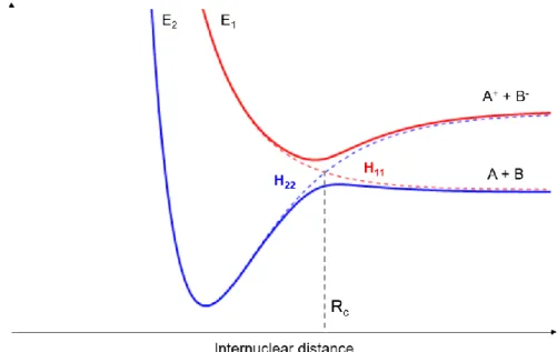
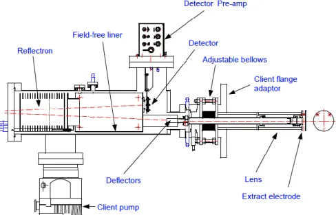
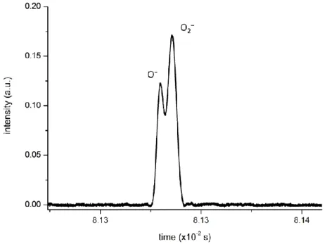
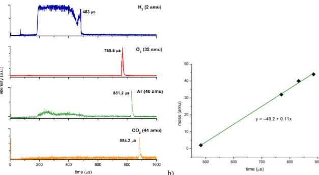
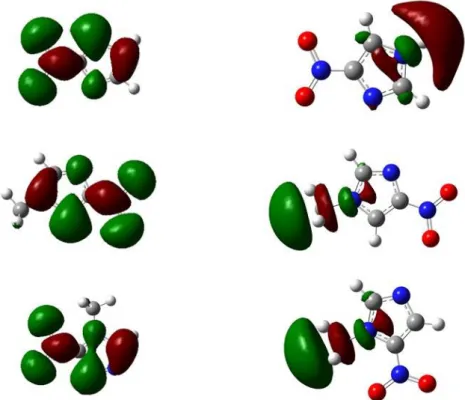
Figure 3.9. Reflectron TOF instrument overview recently installed from Kore Technology, UK. Image courtesy of © Kore Technology, Ltd. ... 34 Figure 3.10. Schematics of the experimental setup with: V, pulsed supersonic valve; C, hollow cathode discharge; A1 and A2, anodes; L1, L2 and L3, Einzel lenses; D1, D2, D3 and D4, deflecting plates; M1 and M2, magnets; E1, E2, E3 and E4, extraction plates; G2, focusing/attenuation grids; EG, electron gun; GC, gas cell; MCP 1 and MCP 2, multichannel plate detectors; QMS, quadrupole mass spectrometer; SEM, secondary electron multiplier detector; P1, P2, P3 and P4, turbomolecular pumps. ... 35 Figure 3.11. Electrical schematics of the anion beam source. V, pulsed supersonic valve; A1 and A2, anodes; C, hollow cathode discharge; L1, Einzel lens; VD, discharge voltage; VF, focusing voltage. ... 37 Figure 3.12. Schematics of the implemented TOF spectrometer with: a) the first commercial extraction system implemented into the chamber; b) the modified extraction system with a homemade cylindrical scattering chamber. E1 and E2 are the extraction plates and F3 is an acceleration grid. ... 38 Figure 3.13. Schematics of the second extraction system implemented to detect and analyse the anionic primary beam. ... 39 Figure 3.14. Typical Time-of-flight mass spectrum of the projectile beam (O2−/O−) at 200 eV in laboratory frame. ... 41 Figure 3.15. Example of a time-of-flight mass spectrum of N2 at 560 eV where: the black line is the extraction pulse at E1, the pink line is the anionic incident beam and the green line is the collision resultant positive ions. ... 42 Figure 3.16. a) time-of-flight spectra of different discharge precursor gases: hydrogen (blue line), oxygen (red line), argon (green line) and carbon dioxide (yellow line); b) calibration curve time x mass. ... 43 Figure 3.17. Time-of-flight mass spectrum of positive ions detection in O2− + CH3NO2 at 1300 eV (lab frame). ... 44 Figure 3.18. O2 precursor discharge gas analysis in four different situations. ... 44 Figure 4.1. Molecular structures of 4-nitroimidazole (4NI), 2-nitroimidazole (2NI), 1-methyl-4-nitroimidazole (Me4NI) and 1-methyl-4-1-methyl-4-nitroimidazole (Me5NI). ... 49 Figure 4.2. Time-of-flight mass spectra of negative ions from electron transfer experiments at 100 eV lab frame energy for 4(5)NI, 2NI, Me4NI and Me5NI. ... 50 Figure 4.3. Time-of-flight mass spectra showing anions produced in electron transfer experiments at 10 eV lab frame energy for 4(5)NI, 2NI, Me4NI and Me5NI. The metastable parent anion M− is visible for all cases while the loss of a OH• radical is only operative in 4(5)NI and 2NI, the former also showing CN– formation. ... 51 Figure 4.4. Left column: M06-2X/aug-cc-pvtz level of theory optimized structures of radical anions and their SOMOs; Right column: lowest σ* orbitals from HF/D95VH level of theory, for 4(5)NI, Me4NI and Me5NI. C N, and O atoms are represented in white, grey, blue and red colours, respectively. Positive and negative values of the wave function have a red and green colours, respectively. ... 53
LIST OF FIGURES
Figure 4.5. Molecular structure of 4-nitroimidazole, 2-nitroimidazole, methyl-4-nitroimidazole, 1-methyl-5-nitroimidazole and imidazole. ... 59 Figure 4.6. Time-of-flight negative ion mass spectra in potassium collisions with 4-nitroimidazole (4NI), 2-nitroimidazole (2NI), 1-methyl-4-nitroimidazole (Me4NI), 1-methyl-5-nitroimidazole (Me5NI) at 30eV lab frame energy (15.7 and 16.3 eV available energy in the centre-of-mass, respectively). See text for details. ... 66 Figure 4.7. 4-nitroimidazole (4NI) branching ratios (fragment anion yield/total anion yield) of the main negative ions formed as a function of the collision energy in the centre-of-mass frame. See text for details. ... 66 Figure 4.8. Time-of-flight negative ion mass spectra in potassium collisions with 4-nitroimidazole (4NI), 2-nitroimidazole (2NI), 1-methyl-4-nitroimidazole (Me4NI), 1-methyl-5-nitroimidazole (Me5NI) at 100 eV lab frame energy (62.6 and 64.5 eV available energy in the centre-of-mass, respectively). See text for details. ... 67 Figure 4.9. 1-methyl-4-nitroimidazole (Me4NI) branching ratios (fragment anion yield/total anion yield) of the main negative ions formed as a function of the collision energy in the centre-of-mass frame. See text for details. ... 67 Figure 4.10. Time-of-flight negative ion mass spectra in potassium collisions with 4-nitroimidazole (4NI), 2-nitroimidazole (2NI), 1-methyl-4-nitroimidazole (Me4NI), 1-methyl-5-nitroimidazole (Me5NI) at 500 eV lab frame (330 and 340 eV available energy in the centre-of-mass, respectively). See text for details. ... 68 Figure 4.11. 1-methyl-5-nitroimidazole (Me5NI) branching ratios (fragment anion yield/total anion yield) of the main negative ions formed as a function of the collision energy in the centre-of-mass frame. See text for details. ... 68 Figure 4.12. Time-of-flight negative ion mass spectra in potassium-imidazole (IMI) collisions at 30, 100 and 500 eV lab frame energy (12.8, 52.9 and 282 eV available energy in the centre-of-mass, respectively). See text for details. ... 70 Figure 4.13. Imidazole (IMI) branching ratios (fragment anion yield/total anion yield) of the main negative ions formed as a function of the collision energy in the centre-of-mass frame. See text for details. ... 70 Figure 4.14. Molecular structure of pyrimidine (Pyr). ... 74 Figure 4.15. Time-of-flight negative ion mass spectra in potassium-pyrimidine (Pyr) collisions at 30, 100 and 700 eV lab frame energy (13.8, 56.2 and 419.3 eV available energy in the centre-of-mass, respectively). See text for details. ... 79 Figure 4.16. Energy loss spectrum of K+ ions measured in the forward direction in collisions of potassium atoms with pyrimidine (Pyr) at 111 eV lab frame energy (67.2 eV in the centre-of-mass system). See text for details... 80 Figure 4.17. Pyrimidine (Pyr) branching ratios (fragment anion yield/total anion yield) of the main negative ions formed as a function of the collision energy in the centre-of-mass frame. See text for details. ... 82 Figure 5.1. Electron detachment cross-sections in the 50-7000 eV energy range for O2− collisions with N2. ... 87
LIST OF FIGURES
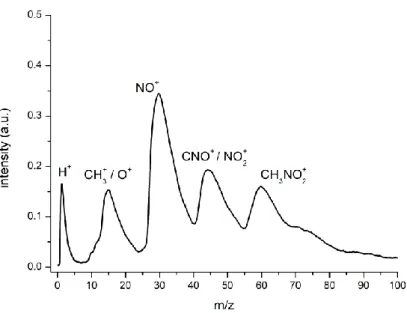
Figure 5.2. Total electron detachment cross-sections for O2− on N2 (black squares) compared with previously published experimental results from Jalbert et al. (blue triangles) and Bennett et al. (violet circles), and a theoretical model from Jalbert et al. (green dash line). The red circles represent the total ionization cross-sections for the formation of N2+ and N+. See also the legend on the plot for other symbols. ... 89 Figure 5.3. Molecular structure of water (H2O) and pyridine (C5H5N)... 93 Figure 5.4. Electron detachment cross-sections in the 10−4000 eV energy range for O2− collisions with water (cyan diamonds) and pyridine (purple squares). ... 94 Figure 5.5. Comparison between electron detachment section values and the relative cross-section for positive and negative ions formation in O2− + H2O collisions. The values are normalized to the maximum value. ... 96 Figure 5.6. Comparison between electron detachment section values and the relative cross-section for positive and negative ions formation in O2− + C5H5N collisions. The values are normalized to the maximum value. ... 96 Figure 5.7. Time-of-flight positive and negative ion mass spectra from O2−-pyridine collisions at 500 eV lab frame energy. ... 98 Figure 5.8. Time-of-flight positive and negative ion mass spectra from O2−-pyridine collisions at 1000 eV lab frame energy. ... 99
LIST OF TABLES
Table 4.1. Assignment of the negative ions formed in potassium collisions with 4-nitroimidazole (4NI), 2-nitroimidazole (2NI), 1-methyl-4-nitroimidazole (Me4NI), 1-methyl-5-nitroimidazole (Me5NI), and imidazole (IMI). ... 64 Table 4.2. Calculated dipole moments obtained with HF/aug-cc-pvtz level of theory and vertical electron affinities (VEAs) obtained with HF/D95V level of theory, for 4-nitroimidazole, 1-methyl-4-nitroimidazole, 1-methyl-5-nitroimidazole and imidazole. ... 65 Table 4.3. Calculated highest occupied molecular orbitals (HOMOs) and lowest unoccupied molecular orbitals (LUMOs) for 4-nitroimidazole (4NI), methyl-4-nitroimidazole (Me4NI), 1-methyl-5-nitroimidazole (Me5NI) and imidazole (IMI) anions obtained at the M06-2X/aug-cc-pvtz level of theory. ... 65 Table 4.4. Calculated lowest unoccupied molecular orbitals (LUMOs) for pyrimidine (Pyr) and pyrimidine (Pyr) in the presence of a potassium atom in the perpendicular geometry inside the pyrimidine ring. Energies in eV. ... 78 Table 4.5. Negative ions formed in potassium collisions with pyrimidine (Pyr). ... 82 Table 5.1. Present experimental results of total electron detachment cross-sections for N2 in collisions with O2−. ... 88 Table 5.2. Experimental electron detachment cross-sections for O2− collisions with water and pyridine. ... 95 Table 5.3. Tentative assignment of negative and positive ions formed in O2− collisions with water (H2O). ... 97 Table 5.4. Tentative assignment of negative and positive ions formed in O2− collisions with pyridine (C5H5N). ... 98
ACRONYMS AND SYMBOLS
AD Auto-detachment
amu Atomic mass unit
b Impact parameter
BEIR Biologic Effects of Ionising Radiation
BRs Branching ratios
CEC Charge exchange chamber
CEFITEC Centre of Physics and Technological Research
CSIC Consejo Superior de Investigaciones Científicas
CT Constant transmission
CTSR Charge-transfer to shape resonances
d Distance
DBS Dipole bound state
DCSs Differential cross-sections
DD Direct detachment
DEA Dissociative electron attachment
DFT Density functional theory
DNA Deoxyribonucleic acid
DSB Double strand breaks
e− Singleelectron
E0 Pass energy
EA Electron affinity
EAad Adiabatic electron affinity
Eav Available energy
ECM Energy of the centre-of-mass
Ek Kinetic energy
Elab Kinetic energy in the laboratory frame
FWHM Full-width at half-maximum
GC Gas cell
Ĥ Hamiltonian operator
H● Hydrogen radical
H0 Non-perturbated Hamiltonian
H11 Diabatic potential curves
H12 Coupling factor
H22 Diabatic potential curves
H2O●+ Water cation
HCE Hollow cathode effect
ACRONYMS AND SYMBOLS
HOMO Highest occupied molecular orbital I Transmitted anion signal
I0 Initial anion beam intensity
ICRP International Commission on Radiological Protection
IE Ionisation energy IMI Imidazole k Boltzmann constant k Electronic state K+ Cationic potassium K+
hyp Hyperthermal cationic potassium
K0 Neutral potassium
K0
hyp Hyperthermal neutral potassium
l Effective path length
LCAM Atomic and Molecular Collisions Llaboratory
LEE Low-energy electrons
LNT Linear no-threshold
LUMO Lowest unoccupied molecular orbital
LZ Landau-Zener method
m Mass
M–# Anionic transient state
MCP Microchannel plate detector MDSB Multiple double strand breaks
Me4NI 1-methyl-4-nitroimidazole
Me5NI 1-methyl-5-nitroimidiazole
mK Mass of potassium
mm Mass of the molecular target
MO Molecular orbital
NO• Nitric oxide radical
O2●− Superoxide anion
OH● Hydroxyl radical
P Gas pressure
p Landau-Zener non-adiabatic transition probability
PEPIPICO Photoelectron-photoion-photoion coincidence spectroscopy
PET Positron emission tomography
Pyr Pyrimidine
QMS Quadrupole mass spectrometer
r Spatial coordinates of the electron
R Spatial coordinates of the nuclei
Rc Crossing radius
ACRONYMS AND SYMBOLS
ROS Reactive oxygen species
SC Scattering chamber
SCF Self-Consistent Field
SFS Sector field sweep
SMC Schwinger multichannel
SSB Single strand breaks T Temperature
t Total flight time
Tc Scattering chamber temperature
tcol Collision time
Te Kinetic energy operator of the electrons
THF Tetrahydrofuran
Tm Operation temperature of Baratron gauge
Tn Kinetic energy operator of the nuclei TNI Transient negative ion
TOF Time-of-flight
tvib Vibrational time
U Potential energy
UV Ultra-violet V sum of potential
V Electric field
v Relative velocity
VAE Vertical attachment energy
VEA Vertical electron affinity
VFRs Vibrational Feshbach resonances
Vp Voltage applied between analyser hemispheres
VUV Vacuum ultra-violet
XPS X-ray photoelectron spectroscopy z Charge of the particle
α Experimental correction factor
νr Radial velocity
π* Antibonding orbital
σ* Antibonding orbital
σexp Electron detachment cross-sections
Φ(r; R) Adiabatic electronic wavefunctions
Ψ(r, R) Total wavefunction
Ωk(R) The nuclear wave function
∆E endoergicity
2NI 2-nitroimidazole
CHAPTER 1
INTRODUCTION
1.1 Motivation
1.1.1 Cancer
Cancer represents a leading cause of death in developed and developing countries [1,2]. According to the more recent statistics it was estimated that 18.1 million people were diagnosed with a cancer disease and 9.6 million died from cancer in 2018, meaning that about 1 in 6 deaths worldwide is due to cancer. The World Health Organization and the International Agency for Cancer Research predicts an increase up to 29.5 million of new cancer cases in 2040 [3], which makes it one of the most important obstacles to life expectancy around the world in the 21st century [2]. The numbers also reveal that around 70% of deaths from cancer occur in less economically developed countries. In Portugal, the incidence of new malignant tumour cases has increased around 3% per year [4]. The most recent data reported in 2010 show an incidence rate of 441.9/100 000, with 67% of cancers diagnosed at ages greater than 60 years old [4,5].
Briefly, this noncommunicable disease is characterized by an uncontrolled proliferation of abnormal cells (tumour cells), which have suffered several mutations resulting into a malignant tumour. These tumour cells can also migrate to different parts of the body through the blood flow or lymph in a process named as metastasizing. Metastases become more difficult to control and are considered a major cause of death from cancer. The reasons for cancer occurrence are many and varied, and some of which are still unknown. However, in the last decades, some efforts have been made in order to understand the major risks factors for cancer, especially through epidemiologic studies. The possibilities of developing a neoplasm disease are related to a combination of genetic and external factors. The external factors can be classified in three different groups: physical
CHAPTER 1.INTRODUCTION
carcinogens (such ionising radiation (e.g. UV)), chemical carcinogens (such as asbestos, tobacco smoke, food and water contaminants) and biological carcinogens (such as infections by virus and bacteria) [3,6]. It is now identified that around 90-95% of the most cancer cases are associated with lifestyle and environment [7]. About 30% of cancer deaths are due to behavioural and dietary risks, such as, high body mass index, low fruit and vegetable intake, lack of physical activity, tobacco and alcohol use [6,8]. Therefore, it becomes urgent to search and find strategies to fight against this epidemic disease, either through early diagnosis or new treatment methods.
In the next subchapters we will focus on the role of radiation in living organisms especially due its capacity to induce at the molecular and atomic levels alterations in biomolecules.
1.1.2 Effects of ionising radiation in biological systems
The biological organisms are continually being exposed to electromagnetic radiation. This interaction with radiation can result from natural sources, such as food (potassium-40 is a radioactive isotope present in bananas), sun, cosmic radiation, minerals and materials found in the ground (e.g. uranium-238, radon-222, radium-226) and even living organisms (e.g. radioactive carbon-14 used to dating organic material). Besides this natural exposure, we can also be a target to man-made radiation which comprises essentially the radiation used for medical diagnosis and treatment, atmospheric nuclear testing, wars, nuclear power production, and finally accidental nuclear disasters as Chernobyl and Fukushima [9].
The radiation sources can be divided in two main groups according to the energy and the effects they produce when interacting with matter. If we look at the electromagnetic spectrum, for lower energies we find radiation sources like microwaves, infrared and visible radiation which are considered as non-ionising radiation. At higher energies (shorter wavelengths) we find UV radiation, X-ray and gamma radiation, termed ionising radiation. Other particles like alpha, electrons, neutrons and heavy ions can also be treated as ionising radiation given the spectral energy ranges they are commonly used. Ionising radiation is capable, when interacting with matter, to ionise it through direct and indirect processes producing several species along the ionisation track [10]. This occurs when the energy of incident radiation transferred to the target is higher than the ionisation energy of atoms and molecules. The ionisation energy is the minimum energy necessary to ionise an atom and ranges from few eV for alkali atoms to 24.6 eV for helium (noble gas) [11,12]. Ionising radiation is very important and useful in different fields, such as industry, agriculture and in medicine. In medical applications it can be used for diagnosis (diagnostic radiology and nuclear medicine) and treatment of diseases particularly cancer-related diseases (radiotherapy, brachytherapy, among others).
Besides the benefits and positive effects about the use of ionising radiation, especially in medical applications, it can also cause adverse health effects, even when we are dealing with low-dose ionising radiation [13,14]. According to the US National Academy of Sciences BEIR VII Committee
1.1 MOTIVATION
[15], approximately 50% of cancer patients are treated with ionising radiation. Yet, the most important effect in humans resulting from low doses of radiation is the possibility to induce cancer [16]. So, we are facing an antagonistic effect of using ionising radiation. Therefore, recommendations from the dedicated Committees is the determination of the effects of low-dose ionising radiation on DNA damage, especially the long-term effects. Epidemiological studies using the survivors of Hiroshima and Nagasaki atomic bombs in 1945 have shown that there is a relationship between cancer incidence and radiation doses in organs. Using this data is possible to extrapolate the effects of low-dose radiation from exposure to high-doses based on a linear no-threshold (LNT) model [16,17]. This model has been used to define numerous international rules and standards of radiation protection (ICRP) [18]. Other nuclear accidents, like Chernobyl (April 1986) and Fukushima (March 2011) were also used to associate the risk of developing cancer diseases (as leukaemia, lymphoma, thyroid cancers, skin cancers, sarcomas, lung and breast carcinomas) in long-term after exposure to high doses of radiation [19]. Many other studies regarding the secondary effects of ionising radiation after the use in some medical treatments and even, after some diagnostic scans, were carried out in order to determine the main causes produced by exposure to very low radiation doses. These are extremely important to assess because during these procedures not only the tumour tissues are affected but also healthy tissue [18,20–22].
Figure 1.1. Chronological diagram of radiation-induced damage.
Briefly we can describe the ionising radiation interactions with living organisms on a time scale. At the time a primary high-energy ionising radiation interacts with biomolecules and tissues
CHAPTER 1.INTRODUCTION
generates multiple sequences of physical and chemical reactions, within the early stages of irradiation (~10-16 s), resulting mainly in electronic excitation and ionisation of the molecular constituents, with subsequent bond breaks creating large amounts of secondary electrons and radicals. The damage in DNA molecule created by these secondary species are characterized by single strand breaks (SSB), double strand breaks (DSB), clustered damage, base damage and loss of the supercoiled helix integrity. The consequences of these injuries in long-term (biological stage) can lead to several mutations, chromosome aberrations, cell inactivation and other effects that compromise the genome integrity which may result in cancer [23–26] (figure 1.1). Given this complexity, a complete understanding of these processes represents a challenging task.
1.1.3 Impact of low-energy electrons in biomolecules
The impact of high-energy ionising radiation with biosystems generates a large amount of different secondary species along the radiation track. As referred before, the ionisation processes triggered by the primary beam create secondary electrons in abundance which interact with biological tissue causing damage in biomolecules, especially in DNA. In fact, it is well-established that low-energy electrons (LEE) and the radicals originated by them are the most responsible for the damage in DNA and other cell molecular components. Typically, 5×10⁴ secondary electrons (below 20 eV) are produced per MeV of primary radiation [27–29]. Along the track and due to successive inelastic interactions (see figure 1.2) with the medium, these secondary electrons transfer their kinetic energy until they reach a near-zero energy (thermalisation) undergoing solvation [30,31]. At this energy range, different electron induced processes can happen yielding different levels of molecular alterations, either producing ionisations (energies typically above 7 eV), bond rupture or resonant attachment, among several others.
Figure 1.2. Single electron tracks simulation in liquid water. (a) Electrons with 10 keV incident energies are slowed down by successive interactions with matter (e.g. elastic scattering ●, rotational excitation ●, vibrational excitation ●, electronic excitation ●, neutral dissociation ●, ionisation ●, and electron attachment ●). Image taken from ref. [32].
1.1 MOTIVATION
1.1.3.1 Direct effects: dissociative electron attachment
Electron attachment can occur even at very low-energies as close to 0 eV. It is defined as a direct electron capture by a target molecule leading to formation of a transient negative ion (TNI) state with a lifetime of ~10-16 s [33]. The process is considered as a resonant scattering, which means that the energy of the incident electron must be exactly the energy necessary to reach a quasibound state of the molecular target (resonance) [34]. If there is no energy lost during the interaction, which means that the energy of the incident electron is roughly speaking the same of the scattered electron (excluding rotational excitation), the process is termed elastic. However, the scattering mechanism is considered inelastic where the energy of the incident electron is higher than the energy of the scattered electron. In the latter case, the formation of a TNI represents a transition from the electronic ground state of the neutral molecule (ABC + e-) to the potential energy surface of the molecular anion (ABC-)and can be described following the Franck-Condon transitions. The TNI has a very short lifetime which depends on the size of the molecule as well as the width of the resonance according to the Heisenberg’s uncertainty principle Γ~ℏ
𝜏 , where Γ is the energy width of the resonant anionic
state, ћ is the Plank’s constant/2π and τ is the lifetime of the anionic state. As the TNI is very unstable it can decay through three different channels: radiative stabilization (equation 1.1), auto-detachment (AD) (equation 1.2) and dissociative electron attachment (DEA) (equation 1.3).
e−+ ABC ⟶ (ABC)−#⟶ (ABC)−+ hν (1.1)
e−+ ABC ⟶ (ABC)−∗⟶ (ABC)#+ e− (1.2)
e−+ ABC ⟶ (ABC)−∗⟶ AC + B− (1.3)
(ABC)−# means TNI with an excess of internal energy. The formation of a stable molecular anion via radiative stabilization is much less likely and happens for time scales much longer than the other two processes (10-12 s) which means it is not a competitive channel. Auto-detachment consists on the ejection of an extra electron from the TNI without dissociation. In the case of an inelastic mechanism the incoming electron may leave the target molecule in an electronic or vibrational excited state. This is a competitive process to DEA, which occurs when the TNI decay resulting in dissociation with production of anions and one (or more) neutral species (equation 1.3). DEA typically occurs in the timescale of 10-14 s to 10-12 s [34]. Briefly, the TNI dissociates through the instability created by the extra electron, which is initially captured into an unoccupied anti-bonding molecular orbital, has the capability to change the intramolecular potential and adds an excess of internal energy (figure 1.3). DEA depends on the energy of the incoming electron and can also be site and bond selective, i.e. this process can selectivity occur whether the electron attaches to specific sites in the atoms and bonds of a molecule [35].
CHAPTER 1.INTRODUCTION
Figure 1.3. Formation of a temporary negative ion in the DNA molecule after the capture of low-energy electrons that which leads to DNA strand breaks. Taken from ref. [36].
During the last decades much effort has been given to the investigation of LEE impact with molecules, with considerable interest to biomolecules (e.g. DNA bases and amino acids). In 2000, a pioneering study performed by Boudaïffa and co-workers demonstrated that electrons with energies up to 20 eV are capable to induce SSB and DSB in plasmid DNA in ultra-high vacuum conditions (see figure 1.4). These authors have also observed that DNA damage is highly dependent on the initial kinetic energy of the incident electron, particularly below 15 eV, and even that SSB and DSB yields in the region of 7−10 eV incident electrons are one to two orders of magnitude larger than those for 10 to 25 eV photons. Therefore, the mechanisms of DNA damage depend not only on the quantum of energy absorbed but also on the nature of the particle that deposits the energy [37]. Further to these findings, Huels et al. reported that besides SSB and DSB also multiple double strand breaks (MDSB) occur from interactions with electrons below 100 eV, showing a strong monotonic increase above 30 eV, however less intense than in SSBs and in DSBs [38]. These authors concluded that MDSB are related with strand breaks clusters (nano-clusters) involving multiple successive interactions of one single electron in different sites of the supercoiled DNA. After these studies, several investigations were and are being performed in order to better understand the DEA processes operative in both gas and condensed-phases.
Studies using DNA sugars [39,40] and bases [35,41,42] showed that electrons with very low energies (< 3 eV) attack the molecular target decomposing it, especially by the loss of H atoms, leading to formation of the dehydrogenated parent anion. It is being reported that a simple bond cleavage can be related to the triggering of other bond cleavages resulting in degradation of an entire cyclic structure. In most of the cases, it has been reported that this decomposition is remarkably bond and site selective [35,39,42,43]. In some studies, in order to investigate how certain dissociation channels may be quenched as a function of the electron energy, deuterated and/or methylated compounds specific sites in molecules have been used. Additionally, studies using slow electrons as a tool to control chemical reactions in condensed phase has also been reported. Balog and Illenberger performed studies in thin films of 1,2-C2F4Cl2 molecules by setting the electron beam energy to values below 3 eV which resulted in a complete modification into molecular chlorine and other products. This effect is based on both selectivity and energy dependence and is triggered by a DEA
1.1 MOTIVATION
process [44]. Electron irradiation of thymine and halogen-substituted nonamers films [45] and pyrimidine [46] have shown that the TNI contributes significantly via DEA to molecular dissociation at low energies.
Besides the extremely importance to investigate DEA processes in DNA and cell components, other molecules have attracted the interest of the scientific community because their electron scavenging properties (e.g. halogenated compounds) make them very useful as radiosensitisers in medical treatments. This will be discussed in Section 1.1.5.
Figure 1.4. Measured quantum yields, per incident electron (3 to 20 eV), for the induction of DSBs (A), SSBs (B), and loss of the supercoiled DNA form (C), in DNA films. Taken from ref. [37].
1.1.3.2 Indirect effects: production of highly reactive species
As described in the previous paragraphs, secondary electrons are capable not only to induce directly alterations in the structure of biomolecules, with focus on DNA constituents, but are responsible to induce changes through indirect effects. The indirect effects of secondary electrons are related to the production of free radicals, atoms or molecules with unpaired electrons, after interaction with other cell components (DNA, water, oxygen and proteins). Most of these radicals are created from the interaction of the incoming radiation with the water molecule (water radiolysis), since around 80% of the cell is water [26]. These species are highly reactive and are responsible to permanent damage of the target molecule. It is assumed that one third of the damage in the genome is caused by direct effects whereas two thirds by indirect effects [36].
Thus, investigating the indirect mechanisms by LEE is essential to a comprehensive knowledge of the damage produced at the molecular level. Experiments were carried out using biomolecules
CHAPTER 1.INTRODUCTION
surrounded by water [31] and thin films of short single DNA strands embedded in a water medium [47,48]. Such studies have proved that the presence of water around DNA changes the decomposition channels and the SSB and DSB yield functions. Furthermore, theoretical studies have demonstrated that DNA solvation leads to an enhancement in electron capture with energies near zero eV or lower via the modification of adiabatic electron affinity of solvated DNA bases in water [49,50]. As result of water radiolysis, several reactive species are formed, such as hydroxyl radicals (OH●), hydrogen radicals (H●), as well as electrons (known as pre-hydrated electrons) [51], schematically represented in figure 1.5. It is assumed that these species are responsible for most of the damage induce in cells genome, leading to severe injuries, essentially because they have high electron affinities and can produce the excision of hydrogen atoms from the biomolecules [30,52]. When high-energy ionising radiation interacts with water molecules, ionisation processes are initiated producing water radical cations and electrons (equation 1.4). This reaction rapidly transfers electrons to DNA causing rupture and production of transient anions. As H2O●+ is a strong acid it immediately reacts with the medium components (equation 1.5) or can migrate over distances of a few molecular diameters by resonant electron transfer and produce hydroxyl radicals and solvated electrons (equation 1.6). Moreover, electronically excited states of water can also be produced and dissociate into H and OH radicals (equation 1.7) [53][54]. H2O rad → H2O•++ e− (1.4) H2O•++ H2O ⟶ H3O++OH• (1.5) 2H2O ⟶ H3O++OH•+ eaq− (1.6) H2O∗⟶ OH•+ H• (1.7)
These are some examples of the interaction of radiation with water but there are also several other channels related to the indirect effects of LEEs. One of the most important reactions is the production of oxygen-derived free radicals (the so-called reactive oxygen species: ROS), which are short-lived and highly reactive. The hydroxyl radical (OH●), the superoxide anion (O2●-) and the hydrogen peroxide (H2O2) are some of the ROS [55]. At low levels, the ROS generation happens under normal physiologic conditions to guarantee the good cellular functioning by regulating the expression of specific genes and acting in the maintenance of redox balance of the organism. However, if a burst occurs in the normal functioning of cells, through high-energy irradiation or in cancer for instance, abnormal high levels of ROS are generated which can result in oxidative stress. Excess cellular levels of ROS can cause damage to proteins, nucleic acids, lipids, membranes and organelles such as mitochondria with severe consequences for organism [56,57]. Therefore, the high levels of ROS in cellular microenvironment can be harmful to healthy cells or can be used as intermediate species in tumour treatments using radiation [58,59].
1.1 MOTIVATION
Figure 1.5. DNA damage caused by indirect effects (water radiolysis). Taken from ref. [60].
1.1.4 Electron transfer processes in biomolecules damage
In the previous sections it was briefly discussed the role of direct electron attachment and the consequent dissociation processes of molecules. However, many elementary collisional processes are not direct electron impact but rather depend upon electron transfer, since free electrons have very limited lifetimes, losing their energies in successive inelastic collisions and reacting with or being solvated by surrounding molecules. In this context, the studies on molecular damage by free electron attachment processes must be complemented with studies on electronic capture of “bound” electrons (as in atom-molecule and ion-molecule collisions).
In atom-molecule collisions the electron is not free but weakly bounded to a neutral atom, which acts as an electron donor (A). This represents a better approximation to what may happen in the physiological environment. In this type of collision, the presence of an electron donor makes the collision system a three-body system, where the projectile significantly changes the fragmentation pathways of the target molecules (BC). Briefly, it is a collision system composed by a projectile which transfers a valence electron to the molecular target when reaching a given distance from the target producing a cationic donor and a transient molecular anion [61–63]. Thereupon, several channels can be opened as described in the following equations:
A + BC ⟶ A++ (BC)−#⟶ A++ BC− (non-dissociative ionisation) (1.8)
A + BC ⟶ A++ (BC−)# ⟶ A++ B−+ C (dissociative ionisation) (1.9) A + BC ⟶ A++ (BC−)# ⟶ AB++ C− (associative chemionisation) (1.10)
CHAPTER 1.INTRODUCTION
These processes can be characterised by two physical properties: the ionisation energy (IE) of the electron donor atom (A) and the electron affinity (EA) of the target molecule (BC). EA is defined as the energy difference between the neutral (BC) and the anion (BC−), termed vertical electron affinity (VEA) [34] and if it is related to their respective ground states, then is termed EAad, adiabatic electron affinity. In this case it termed adiabatic electron affinity. It is also important to mention the endoergicity (∆E) of a reaction, which is (at large atom-molecule collisions) the difference between the IE of the donor and the EA of the target, as shown in equation 1.12. If the ionisation energy is higher that the electron affinity, the endoergicity is positive, meaning that the process is endothermic.
∆E = IE(A) − EA(BC) (1.12)
For large atom-molecule distances the ionic potential energy surface lies above the covalent. Due to the Coulomb potential there is a crossing point at small distances at which both potential energy surfaces have the same value. The crossing distance Rc is inversely proportional to the endoergicity of the reaction. Consequently, in a reaction where it is formed a cation and an anion (ion-pair formation) only the lowest ionic state will be involved. On the other hand, it should be noted that in these processes the electron transfer is only possible between configurations of the same symmetry and multiplicity [63–65]. Indeed, the presence of a third body (cationic donor projectile post-collision) in the collisional mechanism may yield a different fragmentation pattern after the TNI decays in contrast to DEA experiments. In fact, this was verified by Antunes et al. [66] in a study using nitromethane as a molecular target, the formation of a parent anion (CH3NO2−) which is not detected in DEA experiments. Other studies developed in the Lisbon group have demonstrated the ability to change the probability of certain reaction channels and to induce the formation of new dissociation pathways by electron transfer in potassium-molecule collisions. Ferreira da Silva et al. performed studies on different amino acids describing the side chain in the fragmentation pattern comparing the ionic yields. In some of them the dehydrogenated parent anion is one the most intense fragments and possibly leading to the formation of metastable fragments (at higher collision energies) [67,68]. Dissociative ion-pair formation in collisions of fast potassium atoms with benzene and fluorobenzene was also studied by Limão-Vieira et al. [69]. Many studies performed by Almeida et al. have shown the importance of a third body in the “stabilisation” of the TNI in the collisional system. By tuning the collision energy of the hyperthermal neutral potassium beam in collisions with methylated and deuterated pyrimidine molecules Almeida et al. have showed that H− loss is site and bond-selective [70,71]. Furthermore, they also studied D-Ribose [72], THF [73] and uridine [74] sugar where major enhancements in the formation of OH− and O− were observed compared with DEA experiments. Investigation on thymine and uracil molecules by Almeida et al. [75] also provide information about the dissociation channels and the fragmentation pattern by electron transfer as well as CNO− formation as the major ring breaking anionic product for both molecules.
1.1 MOTIVATION
Following these studies, the focus of this thesis is the study of electron transfer processes making use of a crossed molecular beam setup where the electron donor is a neutral alkali atom (potassium atom) due to its low ionisation energy (IE(K) = 4.34 eV). Although neutral potassium does not exist in the physiological environment, it is a good candidate to mimic the charge transfer mechanism in DNA damage caused by LEEs. A set of measurements were performed for molecular targets of the nitroimidazoles family, including methylated compounds and imidazole. A comprehensive comparison was also established between DEA results and the results are thoroughly presented in this thesis. These studies were fully performed in the Atomic and Molecular Collisions Laboratory (LCAM) in Lisbon.
Furthermore, and considering the importance of what has been described in Section 1.1.3.2, ion-molecule collisions were also performed at Laboratorio de Interaccion Radiación-Materia at CSIC in Madrid. In order to study such processes, we have developed and improved a novel crossed beam experimental setup to investigate negative and positive ion formation as well as cross-sections from charge transfer processes between the superoxide anion (O2−) as electron donor and isolated molecules, N2, water and pyrimidine as targets.
1.1.5 Radiosensitisers
Many cancer diseases are treated using ionising radiation through radiotherapy. The microenvironment of some solid tumours is characterised by regions of low oxygen (hypoxia), which plays a fundamental role in tumour progression. Tumour hypoxia arises from the high rate of tumour growth that cannot be sustained by a limited oxygen supply (the so-called “oxygen effect”). Hypoxia is directly related with aggressiveness of the tumour, and resistance to all available sorts of cancer treatment, including chemotherapy, radiotherapy and indirectly surgery [76,77]. The radiosensitisers used in radiotherapy to enhance tumour control of radioresistant hypoxic tumours are called electron-affinic molecules. However, the detailed molecular mechanism of actual radiosensitisation is still unknown. A working hypothesis is that these molecules undergo redox reactions inside the cells that are deficient in oxygen, and that the nitroimidazole ring facilitates reduction via the formation of radical anions [78]. In particular, the 5-nitroimidazole, nimorazole was shown to be effective in several clinical trials and it has been used in routine treatments of head and neck cancer in Denmark [79,80].
Feketeová and co-workers have reported on the formation of radical ions from radiosensitisers using different spectroscopy techniques [43,81–84]. They have concluded that low−energy electrons (0−8 eV) effectively decompose 4-nitroimidazole and two methylated isomers via DEA. The observation that neutral and radical anions are formed via DEA with high efficiency already at threshold (0 eV), shows the significant importance of the study of the molecular mechanisms involved in these reactions and emphasises the implications of that for the development of
CHAPTER 1.INTRODUCTION
nitroimidazole-based radiosensitisers in tumour radiation therapy. Yu and Bernstein demonstrated that the decomposition of three nitroimidazole model molecules following electronic excitation generates NO as an initial decomposition product at the nanosecond laser excitation wavelengths, with vibrational warm and rotational cold distributions of the NO product, which are independent of excitation wavelengths [85]. Some other studies using halogen-modified nucleic acids (such as 5-bromouridine, 5-fluoroacil, 5-chlorouracil, etc.) have been suggested as the most promising radiosensitisers for targeted therapies [86–89].
1.2OUTLINE OF THE THESIS
1.2 Outline of the Thesis
The present doctoral thesis describes the studies on interaction of low-energy electrons via charge transfer processes with different molecular targets. To pursue this objective, two different cross beam setups were used:
(1) To investigate atom-molecule collisions an hyperthermal neutral potassium (K) beam was used as electron donor. The molecular targets studied were 4-nitroimidazole and methylated-derived, imidazole and pyrimidine.
(2) To study ion-molecule collisions, the projectile used was a superoxide (O2−) anion. The molecular targets studied were nitrogen molecule, water and pyridine.
This thesis is organized in six chapters as follows:
• Chapter 1: It is an introduction to the thesis presenting the main motivation and a state-of-the art for state-of-the presented scientific research.
• Chapter 2: It discussed some fundamental aspects of atom and ion-molecule collision theory used to support the analysis of the results.
• Chapter 3: It is presented and comprehensively described the experimental apparatus used to perform electron transfer experiments in both laboratories, Lisbon and Madrid.
• Chapter 4: It contains the experimental results obtained in electron transfer experiments between neutral potassium and 4-nitroimidazole and methylated compounds, imidazole and pyrimidine, as well as a comprehensive literature review, analysis and discussion results. • Chapter 5: It contains the experimental results obtained in electron transfer experiments
between the negative oxygen ion and the nitrogen molecule, water and pyridine as well as a comprehensive literature review, analysis and discussion of results.
• Chapter 6: It is a conclusion summarizing the results and some ideas and suggestions for future work that can complement the present research.
C
HAPTER
2
COLLISION THEORY IN ELECTRON
TRANSFER PROCESSES
In this chapter it will be introduced the principal theoretical aspects about electron transfer processes. Firstly, it will be discussed the generic fundaments about the simplest case in collisions between two particles referring the Born-Oppenheimer approximation. Ion-pair formation will be also explained based on the Landau-Zener formalism. Thereafter, atom-molecule collisions will be explained focusing on diatomic molecules. Finally, some important notions will be expounded regarding collisions between ions and molecules, which constitutes the second part of this thesis dedicated to the experimental results. It is important to point out that the molecules studied in this thesis are more complex than the cases presented in this chapter. However, theoretical descriptions given in the next sections represent useful guiding concepts to analyse and explained most of the experimental results obtained in the thesis.
2.1 Two-Particles Collision
In collisions between two particles in their ground states two main processes can take place: the elastic and inelastic scattering. Let us consider as simplification the atom-atom collision case. The elastic scattering occurs when kinetic energy is transferred from an atom to the other yielding the same two atoms in their original electronic states. The inelastic scattering refers to the collision resulting in differences in final electronic states of the atoms. Here it will be discussed the case of electron transfer interactions, which comprises an electron transfer from a donor atom to an acceptor
CHAPTER 2.COLLISION THEORY IN ELECTRON TRANSFER PROCESSES
atom with possible electronic excitation of the target. Equation 2.1 represents schematically this process.
A + B ⟶ A++ (B)∗− (2.1)
Where A is the donor atom, B is the acceptor atom and * means electronic excited. Equation 2.1 represents an ion-pair formation process, where after the interaction a cation and an anion are produced. The interaction between two atoms in a collision can be described through the time-dependent Schrödinger equation [61,62], as in equation 2.2:
𝐻̂𝛹(𝑟, 𝑅) = 𝑖ℏ (𝑑𝛹(𝑟,𝑅)
𝑑𝑡 ) (2.2)
In this equation, Ĥ is the Hamiltonian operator, Ψ(r, R) is the total wavefunction of the atom-atom system, r is the spatial coordinates of the electron and R of the nuclei. The Hamiltonian can be described through three components, the Tn (kinetic energy operator of the nuclei), Te (kinetic energy
operator of the electrons) and V is the sum of potential between the particles involved in the system that depends parametrically on the nuclear coordinates, as described in equation 2.3.
𝐻̂ = 𝑇𝑛+ 𝑇𝑒+ 𝑉 (2.3)
Since the collisional systems can be much more complex than just two single particles interacting, it is necessary to find proper approximations to the solution of the Hamiltonian. In the next paragraphs it will be discussed the Born-Oppenheimer approximation. This approximation considers the nuclear motion and the electronic motion as two independent parameters. Therefore, the total wave function
Ψ(r, R) can be expressed as a complete, orthogonal set of adiabatic electronic wavefunctions Φk(r; R) that depend parametrically on the nuclear coordinates R. In other words, separating this
wavefunction into a nuclear and electronic wavefunction [61,63,90,91]. The total wavefunction can now be defined according to equation 2.4.
Ψ(𝑟, 𝑅) = ∑ Φ𝑘 𝑘(𝑟; 𝑅). Ω𝑘(𝑅) (2.4)
in which Ωk(R) is the nuclear wave function associated with each electronic state k. Consequently, the Born-Oppenheimer approximation comprises two main assumptions: the nuclear motion can be considered as a classical trajectory R(t) which considers that the nuclei is moving as a function of the final state of the electrons, and it is possible to state that the nuclei are fixed relative to the electrons’ motion because they are much more massive. Considering this, the Hamiltonian can be re-written just as an electronic Hamiltonian ignoring the Tn parameter as follows:

![Figure 1.5. DNA damage caused by indirect effects (water radiolysis). Taken from ref. [60]](https://thumb-eu.123doks.com/thumbv2/123dok_br/15583432.1049558/35.892.209.703.106.437/figure-damage-caused-indirect-effects-water-radiolysis-taken.webp)