Proteolysis of ovine and caprine
caseins in solution by enzymatic
extracts from flowers of
Cynara cardunculus
M. Jose´ Sousa and F. Xavier Malcata
Escola Superior de Biotecnologia, Universidade Cato´lica Portuguesa, Porto, Portugal
Primary proteolysis of ovine and caprine Na-caseinate at 30°C in phosphate buffer at pH 6.5 or 5.5 in the absence of NaCl and at pH 5.2 with 5% (w/v) NaCl by cardosins in aqueous extracts of Cynara cardunculus flowers was investigated using urea-polyacrylamide gel electrophoresis and reversed-phase high performance liquid chromatography. Caprine caseinate underwent more extensive degradation than ovine caseinate under the same conditions (pH 6.5 and pH 5.5); proteolysis ofb- and as-caseins in ovine and, to a lesser extent, in caprine caseinates was reduced in the presence of 5% (w/v) NaCl. Peptide profiles of the pH 4.6-soluble extract had different patterns throughout ripening arising from the different specificity of cardosins toward ovine and caprine Na-caseinates. The major cleavage sites in ovine (caprine) caseinate were Phe105-Met106 (Lys116-Thr117) for k-casein, Leu127-Thr128 and Tyr191 (Glu100-Thr101, Leu127-Thr128, Leu136-Pro137 and Leu190-Tyr191) for b-casein, Phe23-Val24 (Phe23-Val24, Trp164-Tyr165 and Tyr173-Thr174) for as1-casein and Phe88-Tyr89 (Ser9-Ser10, Phe88-Tyr89 and Tyr179-Leu180) foras2-casein. © 1998 Elsevier Science Inc.
Keywords: plant rennet, cheese ripening, proteases, enzyme specificity
Introduction
One of the most successful rennets of plant origin, as apparent from long term use in Portugal for the manufacture of farm cheeses from ovine milk, is obtained from the flowers of the thistle, Cynara cardunculus L.1
An acid proteinase was first isolated from dried thistle flowers by Faro et al.2and shown to induce milk clotting via
cleavage of the sensitive bond Phe105-Met106 in bovine k-casein.3,4The aqueous extract of flowers of C.
cardun-culus was further shown to possess three active proteinases (once termed cynarases or cyprosins, and currently termed cardosins) which have been isolated, purified and partly characterized in terms of activity5– 8 and specificity4,9,10
toward pure bovine caseins. More recently, two additional aspartic proteinases were isolated from the fresh stigmae of a standard variety of C. cardunculus L. Based on the structural and kinetic properties of these enzymes, it was
concluded that they result from different genes and are different from the previously reported proteinases of the same plant; hence, they were named cardosin A and cardosin B.11,12Each cardosin consists of two subunits with
apparent molecular weights of 31 and 15 kDa for cardosin A and 34 and 14 kDa for cardosin B.12,13 Specificity and
kinetic studies were performed using the b-chain of oxi-dized insulin.3 Taking advantage of the derived
chro-mophoric peptides, it was reported that cardosin A is similar to chymosin whereas cardosin B resembles pepsin.13,14
Although considerably less susceptible than the Phe105-Met106 bond of k-casein, peptide bonds of as1-,as2-, and
b-caseins are also hydrolyzed by cardosins (and by chymo-sin) if subject to appropriate environmental conditions. The susceptibility of bovine Na-caseinate to proteolysis is strongly influenced by the state of aggregation of the substrate15 which is in turn affected by pH and NaCl.
Fundamental information on the action of cardosins on ovine and caprine milks is scarce, and straightforward extrapolation of conclusions obtained with pure bovine caseins to ovine and caprine counterparts is risky. In fact, unlike bovine caseins, ovine caseins exhibit two groups of electrophoretic bands. The group with lower mobility con-tains b-casein and is divided into two variants, b1- and
Address reprint requests to Dr. F. X. Malcata, Universidade Catolica Portuguesa, Escola Superior de Biotecnologia, Rua Dr. Antonio Bernar-dino de Almeida, P– 4200 Porto, Portugal
b2-casein, which differ in the level of phosphorylation (6
and 5, respectively),16although levels ranging continuously
from 1–7 have also been reported.17The group with higher
mobility consists of three bands (as1-,as2- andas3-caseins),
designated as a whole as as-casein region despite the
microheterogeneity arising from variations in the degree of glycosylation and/or phosphorylation coupled with genetic polymorphism.18 –21
The scope of this work was to experimentally monitor the evolution with time of the breakdown patterns of ovine and caprine caseins by cardosins in crude aqueous extracts of C. cardunculus using experimental conditions that par-allel milk (pH 6.5) and cheese at early stages of ripening in the absence of salting (pH 5.5) or at early stages of ripening with 5% NaCl (w/v) (pH 5.2) with the goal of furthering knowledge on the nature of action of this plant rennet in cheesemaking and ripening.
Materials and methods
Enzyme
Dried flowers of the wild thistle (C. cardunculus L.) were obtained in the Serra da Estrela region (Portugal). The crude extract was prepared by grinding the stigmae of the flowers for 36 s, homog-enizing in 0.1 m citrate buffer at pH 5.9, and centrifuging at 12,000 g for 5 min. The crude extract (0.1 g of ground flowers ml21 buffer) had a protein concentration of 4.54 mg ml21as determined by the Coomassie spectrophotometric method using bovine serum albumin as standard.
Milk clotting activity
Rennet clotting activity was measured using low-heat skim milk powder NILAC™
(NIZO, Ede, The Netherlands); the milk sub-strate was prepared by dissolving 12 g in 100 ml of 1022m CaCl2
(pH 6.5) at 30°C and 2 ml of this substrate were mixed with 0.2 ml of crude enzyme extract. One rennet unit (RU) was defined as the amount of crude enzyme extract needed to coagulate 10 ml of milk substrate at 30°C in 100 s.
Substrate
Whole ovine and caprine caseins were prepared via isoelectric precipitation of ovine and caprine milks, respectively, by acidifi-cation to pH 4.2– 4.3 with 6.0 m HCl, heating to 37°C for 30 min and centrifugation at 6,000 g for 10 min. The precipitate was recovered by filtration and washed several times with distilled water. The caseins were dispersed in distilled water, the pH adjusted to 7.0 with NaOH and the system allowed to equilibrate for at least 3 h at 4°C before lyophilization.
Hydrolysis of caseins
Ovine and caprine Na-caseinates were dissolved in 100 mm phosphate buffer at pH 6.5 or pH 5.5, or at pH 5.2 in the presence of 5% NaCl, to a final concentration of 10 mg ml21. The crude enzyme extract (0.270 RU ml21) was then added at a ratio of 0.526 ml to 10 ml of caseinate (v/v) and the experimental solutions (together with appropriate controls at each pH and salt concentra-tion) were held at 30°C in a thermostated water bath. Aliquots were taken at 1 min, 30 min, 1 h, 3 h, 6 h, 12 h and 24 h. Samples for urea-polyacrylamide gel electrophoresis were mixed with the sample buffer containing mercaptoethanol and urea to inactivate the enzyme and unfold the protein, whereas those for
reversed-phase high performance liquid chromatography were heated at 85°C for 30 min to inactivate the enzyme.
Urea polyacrylamide gel electrophoresis (Urea-PAGE)
Samples (0.75 ml) of hydrolysates, obtained and quenched as described above, were prepared for urea-PAGE by adding an equal volume of double-concentrated sample buffer.22Urea-PAGE was
performed using a Protean II XI vertical slab-gel unit (Bio-Rad Laboratories, Watford, UK) according to the method of Andrews36
(12.5% T-acrylamide plus bisacrylamide, 4% C-bisacrylamide as a percentage of T) at pH 8.9, with modifications;23the gels were
stained with Coomasie Blue G-250 (Bio-Rad, Richmond CA) using the method of Blakesley and Boezi.24 Quantification of
intact b1-,b2-, as1-, as2- and as3-caseins was by densitometry
using a model CD60 densitometer (Desaga, Sarstedt-Gruppe, Germany).
Reversed-phase high performance liquid chromatography (RP-HPLC)
Samples of hydrolysates (2 ml), obtained and quenched as de-scribed above, were adjusted to pH 4.6 by addition of 60 ml of 33.3% (w/v) acetic acid, held at room temperature for 10 min and then 60ml of 3.33 m sodium acetate added.25The samples were
centrifuged at 6,000 g for 10 min and the supernatants recovered for analysis. RP-HPLC was performed by the method of Singh et al.26using a Beckman system (Beckman Instruments, San Ramon
CA). A Lichrosorb RP-8 (5mm) 250 3 4 mm column (Merck, Darmstadt, Germany) was employed with an Lichrocart 4-4 guard column (Merck). Elution was at 30°C using a mobile phase of two solvents: A (0.1% trifluoroacetic acid in water) and B (0.1% trifluoroacetic acid in acetonitrile) at a flow rate of 1.0 ml min21 starting with 100% A for 5 min and then a linear gradient for 55 min up to 50% B, holding at 50% B for 6 min, a linear gradient for 4 min up to 60% B and finally holding at 60% B for 3 min. Absorbance of the eluate was read at 214 nm. Samples (10 mg ml21) were dissolved in a mixture of solvents A and B (1: 0.01, v/v), filtered through 0.22 mm cellulose acetate filter and an aliquot (75ml) of the permeate was injected.
Isolation of peptides
Peptides were isolated by RP-HPLC as described above or by electroblotting from urea-PAGE gels using a mini Trans-Blot™
electrophoretic transfer cell (Bio-Rad, Hercules CA). In the latter case, transfer of peptides was at 100 V for 30 min in 10 mm CAPS (3-cyclohexylamino-1-propanesulfonic acid) buffer with 10% methanol onto polyvinylidene difluoride membranes (PVDF) with a pore size of 0.22 mm (Bio-Rad). Membranes were rinsed with water and stained in 0.2% Ponceau S in 1% acetic acid for 1 min followed by destaining in water. Bands corresponding to peptides were excised from the membranes and sequenced (5–10 cycles).
Sequence analysis
Peptides were sequenced at the National Food Biotechnology Centre, University College Cork via Edman degradation on an automated pulsed liquid-phase protein-peptide sequencer (Applied Biosystems model 477A, Foster City CA). Liberated amino acids were detected as their phenylthiohydantoin derivatives.
Results and discussion
Experimental data
Urea-PAGE electrophoregrams of ovine and caprine Na-caseinates hydrolyzed by extracts of C. cardunculus (0.0142 RU ml21) at pH 6.5, pH 5.5, and pH 5.2 with 5% NaCl (w/v) throughout the hydrolysis time are shown in Figures 1a and 1b. As expected, the extent of degradation ofb- and
as-caseins in both ovine and caprine Na-caseinates
de-pended on ionic strength and pH. Ovine Na-caseinate at pH 6.5 or 5.5 (Figure 1a, lanes 2– 8) showed similar rates of hydrolysis ofb-caseins (b1- andb2-casein) andas-caseins
for 3 h of hydrolysis; however, b-casein was almost completely degraded in all cases whereasas-caseins showed
only extensive degradation at pH 5.5 by 6 h of hydrolysis (Figure 1a, lane 9). Caprine Na-caseinate exhibited more
Figure 1 Urea-PAGE electrophoregrams of ovine Na-caseinate (a) and caprine Na-caseinate (b) hydrolyzed by extracts of C. cardunculus at 30°C and pH 6.5 (lanes 2–5), pH 5.5 (lanes 6 –9), and pH 5.2 with 5% (w/v) NaCl (lanes 10 –13) for 1 min, 1 h, 3 h and 6 h, respectively. Bovine Na-caseinate was included as standard in lane 1 of both (a) and (b)
extensive degradation than ovine Na-caseinate under the same conditions (pH 6.5 and pH 5.5); b-casein was com-pletely degraded at pH 6.5 by 3 h whereas complete degradation was observed by 1 h at pH 5.5. Although as-caseins were not completely broken down even after 6 h
at pH 5.2 in the presence of 5% (w/v) NaCl (Figures 1a and 1b, lanes 10 –13), as-caseins were hydrolyzed faster than
b-caseins in ovine and caprine Na-caseinates.
Under all conditions tested, b-casein in ovine and ca-prine Na-caseinates was hydrolyzed by cardosins to yield a pair of bands of higher electrophoretic mobility, comparable
to that of bovine b-I-casein. This is in agreement with previous reports pertaining to ovine casein hydrolyzed by calf chymosin.21 Bovine and ovine b-caseins have very
similar amino acid sequences. The cleavage sites are prob-ably conserved; the corresponding bonds in ovineb-casein susceptible to chymosin are Leu190-Tyr191 and Ala187-Phe188.21In bovine and ovine milk cheeses,b-caseins (b
1
-and b2-casein) were degraded to about the same extent
(approximately 33%), but in caprine milk cheesesb-casein was degraded to a greater extent (approximately 44%) by proteinases of C. cardunculus.27 The bonds in bovine Figure 2 RP-HPLC profiles of pH 4.6-soluble peptides from ovine Na-caseinate (a) and caprine Na-caseinate (b) hydrolyzed for 12 h by extracts ofC. cardunculus at pH 6.5 (A), pH 5.5 (B) and pH 5.2 with 5% (w/v) NaCl (C)
b-casein most susceptible to action by chymosin and pro-teinases from flowers of C. cardunculus are Leu192-Tyr193 and Ala189-Phe190,9,28 the hydrolysis of which yields
b-I-casein.28In studies of proteolysis of caprineb-casein by
calf rennet, Marcos et al.29for several varieties of cheeses
and Carretero et al.30 for Montsec cheese, the existence of
bands accounted for byb-I- and b-II-caseins was reported. Ovine and caprineas-caseins were hydrolyzed initially to a
set of bands of higher electrophoretic mobility which may correspond to bovineas1-I-casein. Two bands with greater
electrophoretic mobility than the as1-I-caseins, tentatively
denoted as A, were apparent at pH 6.5 and 5.5 after 1 h of
hydrolysis in ovine Na-caseinate, and at pH 6.5, 5.5 and 5.2 with 5% (w/v) NaCl after 1 h of hydrolysis in caprine Na-caseinate. Such bands are fragments from as-I-caseins
(f 24-*) because they apparently contain the same sequence at the N-terminus. Bands with similar electrophoretic mo-bility and the same corresponding N-terminal sequence were found in solutions of bovine as1-casein when
incu-bated with extracts from flowers of C. cardunculus.28
Further degradation products with higher electrophoretic mobility and poorly stained (short or medium peptides) could be seen but were not well resolved due to limitations of the analytical techniques employed.
Proteolysis ofb-casein and as-caseins in ovine and, to a
lesser extent, in caprine Na-caseinates (Figures 1a and 1b) by extracts of C. cardunculus was reduced in the presence of 5% (w/v) NaCl at pH 5.2. Sousa28reported that
proteol-ysis of bovineb-casein by extracts of C. cardunculus at pH 6.5 was increasingly inhibited as NaCl concentration was increased up to 20%. It is also known that proteolysis of bovine b-casein is dependent on pH, ionic strength and
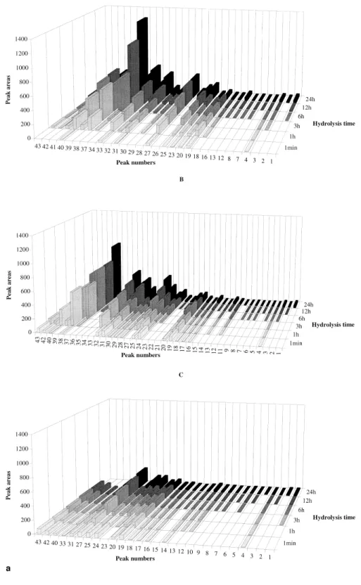
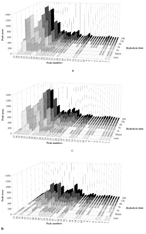
Figure 3 Changes in the area of the RP-HPLC peaks representing the major peptides produced by hydrolysis of ovine Na-caseinate (a) and caprine Na-caseinate (b) catalyzed by extracts ofC. cardunculus at 30°C, and pH 6.5 (A), pH 5.5 (B) and pH 5.2 with 5% (w/v) NaCl (C) for various times
temperature.31 Optimum formation of the bovineb-casein
peptidesb-I, b-II and b-III by calf chymosin occurs at pH 6.4, 4.6 and below 4.6, respectively32,33; however, Mulvihill
and Fox33 claimed that inhibition decreased as pH was
decreased from 7.0 to 4.6.b-III-casein was not produced in
the presence of 2.5% NaCl at pH values below 3.4, but other peptides (e.g., b-IV-casein and b-V-casein) were formed under these conditions (b-IV-casein was also produced in the absence of NaCl at pH 3.4 but only at high enzyme/ substrate ratios). Trujillo et al.34 found that caprine b-I-Figure 3 Continued
casein was produced by calf rennet at all pH values and the corresponding electrophoretic band increased in intensity as pH was increased. At low pH values (i.e., not above 4.6), the polypeptideb-II-casein was the main breakdown prod-uct detected andb-III-casein was not detected at all at any pH value in the presence of 5% NaCl. Although definitive evidence is lacking, it has been suggested that NaCl exerts an influence on the hydrolysis ofb-casein via modification (folding or aggregation) of the substrate rather than of the enzyme.33Less marked influence of NaCl on proteolysis of
bovine as-caseins by extracts of C. cardunculus and by
chymosin28 supports this view.
The RP-HPLC peptide profiles in the pH 4.6-soluble fraction of ovine and caprine Na-caseinate hydrolyzed during 12 h at 30°C and pH 6.5, pH 5.5 or pH 5.2 with 5% (w/v) NaCl are shown in Figures 2a and 2b. The peptide profiles in the pH 4.6-soluble fraction of ovine caseinate were virtually similar to those of caprine caseinate after 1 min of hydrolysis (data not shown), but after 12 h of hydrolysis, caprine caseinate (Figure 2b) exhibited a higher number (except at pH 5.5) and a higher concentration of peptides than ovine caseinate (Figure 2a). This observation may be due to changes in specificity of cardosins on ovine and caprine milk caseins under different ionic strength conditions. Despite the fact that caprine caseinate showed a higher concentration of peptides at pH 6.5 and pH 5.5 than at pH 5.2 with 5% NaCl (Figure 2b), the number of peptides produced in caprine caseinate was not too different from that in ovine caseinate. The isolated peptides were se-quenced by the Edman procedure from their n-terminus in an attempt to identify them. Ovinek-casein was cleaved by cardosins at Phe105-Met106 (peptide 19) whereas caprine k-casein was cleaved by cardosins at Lys116-Thr117 (pep-tide 14). In bovinek-casein, the most susceptible bond was Phe105-Met1064and it was also shown that such cleavage
was able to induce milk clotting as typically happens with acid proteinases. The N-terminal sequences of the peptides designated as 33 [b-(f191-*)], 37 [b-(f1-*)], 38 [b-(f128-*)] and 40 [b-(f128-*)] [b-(f191-*)] (Figure 2a) indicated that ovineb-casein was cleaved by cardosins at bonds Leu-190-Tyr191 and Leu127-Thr128, so possible fragments formed are b-(f1/127), b-(f128/192), b-(f128/207) and b-(f191/ 207); cardosins showed, on the other hand, ab-(f191/207) broader specificity toward caprine b-casein, such as at bonds Glu100-Thr101, Leu127-Thr128, Pro136-Leu137, and Leu192-Tyr193 as indicated by the peptides designated as Leu190-Tyr191 19 [b-(f137-*)], 20 [b-(f128-*)], 28 [b-(f191-*)], 29 [b-(f101-*)], 30 [b-(f128-*)], 31 [b-(f1-*)] and 32 [b-(f128-*)] (Figure 2b). Chymosin cleaves bovine b-casein in solution at seven sites which, in decreasing order of rate of attack, are Leu192-Tyr193, Ala189-Phe190, Leu165-Ser166, Gln167-Ser168, Leu163-Ser164, Leu139-Leu140 and Leu127-Thr128 (Visser and Slangen, 1977). Under the experimental conditions used by Charles and Ribadeau-Dumas (1984), only the bonds Ala189-Phe190 and Leu192-Tyr193 of bovine b-casein can be cleaved. Cardosins cleave six bonds over a similar time frame and the relative susceptibility to attack is, in decreasing order, Leu192-Tyr193, Leu191-Leu192, Leu165-Ser166, Phe190-Leu191, Ala189-Phe190 and Leu127-Thr128.35 Cardosins
thus seem to cleave more bonds than chymosin in the bulky,
hydrophobic segment Ala189-Phe-Leu-Leu-Tyr193 of bo-vineb-casein.
The N-terminal sequence of ovine and caprineas1-casein
was identified in peptides designated as 27 (Figure 2a) and 19 (Figure 2b), respectively, as complementary to the peptide associated with the band identified in urea-PAGE as as-I-casein (Figures 1a and 1b); hence, the bond cleaved in
as1-casein was likely Phe23-Val24. Caprineas1-casein was
cleaved by cardosins at the bonds Phe23-Val24, Trp164-Tyr165 and Tyr173-Thr174 as derived from the N-terminal sequences of peptides 24 [as1-(f165-*)], 28 [as1-(f24-*)]
and 28 [as1-(f174-*)] (Figure 2b). The most susceptible
bond to the action of chymosin and cardosins in bovine as1-casein is Phe23-Phe24.9,28Its hydrolysis yieldsas1
-(f1-23) and (f24-199);22the corresponding susceptible bond in
ovineas-casein with respect to chymosin21and to cardosins
is Phe23-Val24, so the electrophoretic band designated as as1-I-casein in ovine and caprine caseins (Figures 1a and
1b) is probably the peptide Val24-Trp199. Macedo et al.35
reported that cardosins can, in addition, cleave the eight bonds Tyr153-Tyr154, Trp164-Tyr165, Tyr165-Tyr166, Tyr166-Val167, Phe145-Tyr146, Leu149-Phe150, Leu156-Asp157 and Ala163-Trp164 whereas chymosin can only cleave the Phe23-Phe24 bond of bovine as1-casein.
Mc-Sweeney et al.22 identified at pH 6.5 and 5% NaCl the
following seven cleavage sites of bovine as1-casein by
chymosin: Phe28-Pro29, Leu40-Ser41, Leu149-Phe150, Phe153-Tyr154, Leu156-Asp157, Tyr159-Pro160 and Trp164-Tyr165. These peptide bonds were also cleaved at pH 5.2 in the presence of 5% NaCl. In addition, Leu11-Pro12, Phe32-Gly33, Leu101-Lys102, Leu142-Ala143 and Phe179-Ser180 could also be cleaved. Five of these bonds were also cleaved by cardosins at pH 6.5,22 i.e.,
Phe23-Phe24, Leu149-Phe150, Phe153-Tyr154, Leu156-Asp157 and Trp164-Tyr165, but cardosins could not cleave the two bonds containing a prolyl residue at the C-terminal side.
Ovine as2-casein was cleaved by cardosins at
Phe88-Tyr89 as derived from the N-terminal sequence of the peptide denoted as 31 [as2-(f89-*)] (Figure 2a), and caprine
as2-casein was cleaved by cardosins at Ser9-Ser10,
Phe88-Tyr89 and Tyr179-Leu180, thus leading to peptides 19 [as2-(f180-*)], 20 [as2-(f89-*)], 25 [as2-(f89-*)] and 28
[as2-(f10-*)] (Figure 2b); in bovine as2-casein, cardosins
catalyzed the hydrolysis of two peptide bonds between rather hydrophobic amino acids (such as Phe88-Tyr89 and Tyr95-Leu96), thus releasing the heptapeptide Tyr89-Tyr95.35Grappin et al. claimed that bovinea
s2-casein was
resistant to catalytic action by chymosin, but McSweeney et al. argued that chymosin could act upon both Phe88-Tyr89 and Tyr95-Leu96.
Changes in the relative peak area of the major peptides produced by cardosins after 1 min, 1 h, 3 h, 6 h, 12 h and 24 h are represented in Figures 3a and 3b for ovine and caprine caseinates. The peptide profiles of the ovine case-inate hydrolysates were similar to one another, although those peptide peaks denoted as 26 and 41 were present only at pH 6.5, and those peptide peaks denoted as 11, 21, 22, 35 and 36 were present only at pH 5.5. At pH 6.5 after 1 min of hydrolysis (Figure 3a-A), the primary peptides apparent were those denoted as 4, 19, 20, 23, 25, 27, 31, 33 and 40. The peptides denoted as 23, 27 [as1-(f1-*)], 31 [as2
-(f89-*)] and 33 [b-(f191-*)] showed the highest specific formation rates, such as 8.02, 4.77, 1.313 10 and 1.30 3 10 UA (units of absorbance) min21RU21, respectively. At the same pH but after 1 h of hydrolysis (Figure 3a-A), the secondary peptides denoted as 32, 37 [b-(f1-*)] and 38 [b-(f128-*)] were those that exibited the highest formation rates, such as 1.04 3 10, 4.70 3 10 and 1.05 3 10 UA min21 RU21, respectively. They did not reach a plateau during the whole period of study (24 h). At pH 5.5 after 1 min of hydrolysis (Figure 3a-B), the primary peptides apparent were those denoted as 4, 16 –20, 24, 25, 28 –32, 38, 40, 42 and 43. Peptides denoted as 16, 18, 20, 24, 25, 29, 30 and 32 displayed the highest formation rates, such as 3.21, 1.42, 2.63, 1.57, 3.79, 2.49, 7.96 and 6.45 UA min21RU21. The secondary peptides denoted as 27, 33 and 37 (Figure 3a-B) showed the highest formation rates by 1 h, such as 8.58, 7.30 and 2.19 3 10 UA min21RU21, respectively. The peptides produced at pH 5.2 with 5% (w/v) NaCl after 1 min of hydrolysis (Figure 3a-C) were, with the exception of peptide denoted as 10, also produced at either pH 6.5 or pH 5.5. The major primary and secondary peptides were those denoted as 24 and 23, respectively, with formation rates of 4.75 and 3.38 UA min21RU21, respectively. The peptide profiles of the caprine hydrolysates at pHs 6.5, 5.5 and 5.2 with 5% (w/v) NaCl (Figure 3b, A-C) had a number of peptides in common. Hydrolysates from caprine casein-ate produced at pH 6.5 after 1 min (Figure 3b-A) included as primary peptides those denoted as 1, 2, 13, 14, 20, 22–28 and 31–36. The highest formation rates were observed for those peptides denoted as 20 [b-(f128-*) 1 as2-(f89-*)], 24
[as1-(f165-*)], 26, 28 [as1-(f24-*) 1 as1-(f174-*) 1
b-(f191-*) 1 as2-(f10-*)], 31 [b-(f1-*)] and 32
[b-(f128-*)], such as 7.87, 4.85, 9.99, 2.20 3 10, 5.37 3 10 and 4.133 10 UA min21RU21, respectively. At pH 5.5 after 1 min of hydrolysis (Figure 3b-A), the primary peptides were those denoted as 1– 4, 9, 10, 13–15, 19 –29, 31, 32 and 34 –37 whereas the ones that showed the highest formation rates were 19 –23, 28, 29 and 31, such as 1.343 10, 9.18, 1.643 10, 7.86, 1.26 3 10, 8.37, 1.14 3 10 and 4.85 3 10 UA min21 RU21, respectively. The secondary peptides produced from caprine caseinate at pH 6.5 that showed the highest formation rates were 19 [as1-(f1-*)1 as2-(f180-*)
1 b-(f137-*)], 21 and 29 [b-(f101-*)], such as 1.18 3 10, 8.95 and 1.973 10 UA min21RU21, respectively, but at pH 5.5 no secondary peptide was found that exhibited a significant rate of formation. All peptides produced at pH 5.2 and 5% (w/v) NaCl (Figure 3b-C) were also produced either at pH 6.5 or pH 5.5. The major primary and secondary peptides were denoted as 28 and 21 with corresponding formation rates of 1.48 3 10 and 2.44 3 10 UA min21 RU21, respectively.
Acknowledgments
Financial support for author Sousa was provided by a Ph.D. fellowship issued by program PRAXIS XXI (BD-2763/93), Portugal. Partial funding for this research work was pro-vided through grants within program PRAXIS XXI (project IMPACTO: Investigac¸a˜o dirigida ao melhoramento do processo de produc¸a˜o de queijo da Serra por integraca˜o de abordagens cientı´ficas e tecnolo´gicas), Portugal and
PAMAF-IED (project PROTOLACTIS: produc¸a˜o, por tec-nologias optimizadas, de lacticı´nios tradicionais certifica-dos), Portugal.
References
1. Macedo, A., Malcata, F. X. and Oliveira, J. C. The technology, chemistry and microbiology of Serra cheese: A review. J. Dairy Sci. 1993, 76, 1725–1739
2. Faro, C. J., Alface, J. S. and Pires, E. V. Purification of a protease from the flowers of Cynara cardunculus L. Cieˆn. Biol. 1987, 12, 201
3. Faro, C. J., Moir, A. J. and Pires, E. V. Specificity of a milk-clotting enzyme extracted from the thistle Cynara cardunculus L.: action on oxidized insulin andk-casein. Biotecnol. Lett. 1992, 14, 841–846 4. Macedo, I., Faro, C. J. and Pires, E. M. Specificity and kinetics of
the milk-clotting enzyme from cardoon (Cynara cardunculus L.) toward bovinek-casein. J. Agri. Food Chem. 1993, 41, 1537–1547 5. Heimgartener, U., Pietrzak, M., Geertsen, R., Brodelius, P., Silva Figueiredo, A. C., and Pais, M. S. S. Purification and partial characterization of milk clotting proteases from flowers of Cynara
cardunculus. Phytochemistry 1990, 29, 1405–1410
6. Campos, R., Guerra, R., Aguiar, M., Ventura, O. and Camacho, L. Chemical characterization of proteases extracted from wild thistle (Cynara cardunculus.). Food Chem. 1990, 35, 89 –97
7. Faro, C. Purificac¸a˜o e caracterizac¸a˜o fı´sico-quı´mica da protease de
Cynara cardunculus L. Ph.D. thesis, Universidade de Coimbra,
Portugal, 1991
8. Cordeiro, M. C., Jakob, E., Puhan, Z., Pais, M. S. and Brodelius, P. E. Milk clotting and proteolytic activities of purified cynarases from Cynara cardunculus—a comparison to chymosin.
Milchwis-senschaft 1992, 47, 683–700
9. Macedo, I. Especificidade caseinolı´tica da protease de Cynara
cardunculus L. Ph.D. thesis, Universidade de Coimbra, Portugal,
1993
10. Pires, E., Faro, C., Macedo, I., Esteves, C., Morgado, J., Verı´ssimo, P., Pereira, D. and Gomes, D. Flor de cardo versus quimosina no fabrico de queijos artesanais. Quı´mica 1994, 54, 66 – 68
11. Faro, C. J., Verı´ssimo, P., Lin, Y., Tang, J. and Pires, E. V. In:
Aspartic proteinases: Structure, Function, Biology and Biomedical Implications. Takahashi, K. (ed.). Plenum Press, New York, USA.
1995, 373–377
12. Verı´ssimo, P., Faro, C., Moir, A. J., Lin, Y., Tang, J. and Pires, E. Purification, characterization and partial amino acid sequencing of two new aspartic proteinases from fresh flowers of Cynara
cardun-culus L. Eur. J. Biochem. 1996, 235, 762–768
13. Verı´ssimo, P., Esteves, C., Faro, C. and Pires, E. The vegetable rennet of Cynara cardunculus L. contains two proteinases with chymosin and pepsin-like specificities. Biotechnol. Lett. 1995, 17, 621– 626
14. Ramalho-Santos, M., Verı´ssimo, P., Faro, C. and Pires, E. Action on bovineas1-casein of cardosins A and B, aspartic proteinases from the flowers of the cardoon Cynra cardunculus L. Biochim. Biophys.
Acta 1996, 1297, 83– 89
15. Fox, P. F. Influence of aggregation on the susceptibility of casein to proteolysis. J. Dairy Res. 1970, 37, 173–180
16. Richardson, B. C. and Mercier, J. C. The primary structure of the ovineb-caseins. Eur. J. Biochem. 1979, 99, 285–297
17. Chianese, L., Garro, G., Ferranti, P., Malorni, A., Addeo, F., Rabasco, A., and Pons, P. M. Discrete phosphorylation generates the electrophoretic heterogeneity of ovineb-casein. J. Dairy Res. 1995, 62, 89 –100
18. Richardson, B. C. and Creamer, L. K. Comparative micelle struc-ture. V. The isolation and characterization of the major ovine caseins. N. Z. J. Dairy Sci. Technol. 1976, 11, 46 –53
19. Boisnard, M. and Petrissant, G. Complete sequence of ovine-casein messenger RNA. Biochimie 1985, 67, 1043–1051
20. Mercier, J. C., Gaye, P., Soulier, S., Hue-Delahaie, D. and Vilotte, J. L. Construction and identification of recombinant plasmids carrying cDNAs coding for ovine as1, as2-, b, k-casein and
b-lactoglobulin. Nucleotide sequence of as1-casein cDNA.
Bio-chimie 1985, 67, 959 –971
calf chymosin. M.Sc. thesis, National University of Ireland, Cork, Ireland, 1995
22. McSweeney, P. L. H., Olson, N. F., Fox, P. F., Healy, A., and Hojrup, P. Proteolytic specificity of chymosin on bovineas1-casein.
J. Dairy Res. 1993, 60, 401– 412
23. Shalabi, S. I. and Fox, P. F. Electrophoretic analysis of cheese: Comparison of methods. Ir. J. Food Sci. Technol. 1987, 11, 135–151 24. Blakesley, R. W. And Boezi J. A. A new staining technique for proteins in polyacrylamide gels using Coomassie Brillant Blue G-250. Anal. Biochem. 1977, 82, 580 –581
25. McGann, T. C. A., Mathiassen, A. and O’Connell, J. A. Applica-tions of the Pro-Milk Mk II. Part III. Rapid estimation of casein in milk and protein in whey. Lab. Prac. 1972, 21, 628 – 631, 650 26. Singh, T. K., Fox, P. F. and Healy, A. Water-soluble peptides in
Cheddar cheese: isolation and identification of peptides in the diafiltration retentate of the water-soluble fraction. J. Dairy Res. 1995, 62, 629 – 640
27. Sousa, M. J. and Malcata F. X. Proteolysis in bovine, ovine and caprine cheeses manufactured with a plant rennet (Cynara
cardun-culus), Proceedings of ‘‘X Congresso Nacional de Bioquı´mica’’,
Braga, Portugal: October 31–November 2, 1996, pp 86, P9 –29 28. Sousa, M. J. C. F. Plant rennet substitute from flowers of Cynara
cardunculus. M.Sc. thesis, National University of Ireland, Cork,
Ireland, 1993
29. Marcos, A., Esteban, M. A., Leo´n, F. and Ferna´ndez-Salguero, J. Electrophoretic patterns of European cheeses: Comparison and quantification. J. Dairy Sci. 1979, 62, 892–900
30. Carretero, C., Trujillo, A. J., Mor-Mur, M. and Guamis, B. Electro-phoretic study of casein breakdown during ripening of goat’s milk cheese. J. Agric. Food Chem. 1994, 421, 1456 –16550
31. Dalgleish, D. G. The enzymatic coagulation of milk. In:
Develop-ments in Dairy Chemistry Vol 1 (Fox, P. F., Ed). Applied Science
Publishers, London, 1992, 157–187
32. Guiney, J. A. A study of various factors influencing b-casein hydrolysis by rennet. M. Sc. thesis, National University of Ireland, Cork, 1973
33. Mulvihill, D. M. and Fox, P. F. Proteolysis of bovineb-casein by chymosin: Influence of pH, urea, and sodium chloride. Ir. J. Food
Sci. Technol. 1978, 2, 135–139
34. Trujillo, A. J., Guamis, B. and Carretero, C. J. Proteolysis of goat
b-casein by calf rennet under various factors affecting the cheese
ripening process. J. Agric. Food Chem. 1995, 43, 1472–1478 35. Macedo, I., Faro, C. J. and Pires, E. M. Caseinolytic specificity of
cardosin, an aspartic protease from the cardoon Cynara cardunculus L.: Action on bovine as- and b-casein and comparison with chymosin. J. Agri. Food Chem. 1996, 44, 42– 47
36. Andrews, A. T. J. Proteinases in normal bovine milk and their action on caseins. J. Dairy Res. 1983, 50, 45–55
37. Carles, C. and Ribadeau-Dumas, B. Kinetics of action of chymosin (rennin) on some peptide bonds of bovineb-casein. Bio/Chemistry 1984, 23, 6839 – 6843
38. Grappin, R., Rank, T. and Olson, N. Primary proteolysis of cheese proteins during ripening. A review. J. Dairy Sci. 1994, 68, 531–540 39. McSweeney, P. L. H., Olson, N. F., Fox, P. F. and Healy, A. Proteolysis of bovineas2-casein by chymosin. Z. Lebensm. Unters.
Forsch. 1994, 199, 429 – 432
40. Visser, S. and Slangen, K. J. On the specificity of chymosin (rennin) in its action on bovine b-casein. Neth. Milk Dairy J. 1977, 31, 16 –30