i
DEVELOPMENT AND CHARACTERIZATION OF
A CO-CULTURE TWO-DIMENSIONAL
BLOOD-BRAIN BARRIER FOR THE STUDY OF
NANOPARTICLE PERMEATION
O
SUBTÍTULO
BÁRBARA BRUNA DA SILVA MENDES
DISSERTAÇÃO DE MESTRADO APRESENTADAÀ FACULDADE DE ENGENHARIA DA UNIVERSIDADE DO PORTO EM ENGENHARIA BIOMÉDICA
iii
Faculdade de Engenharia da Universidade do Porto
Development and characterization of a co-culture
two-dimensional blood-brain barrier for the study of
nanoparticle permeation
Bárbara Bruna da Silva Mendes
Dissertação realizada no âmbito do
Mestrado em Engenharia Biomédica
Orientador: Prof. Dr. Bruno Sarmento
Co-orientador: Prof. Dr. Domingos Ferreira
v
Agradecimentos
O resultado contido nesta tese não seria possível sem um conjunto especial de pessoas que disponibilizaram o seu tempo e o seu conhecimento, a quem desejo ser capaz de agradecer da forma que merecem.
Em primeiro lugar queria deixar o meu obrigado ao professor Bruno Sarmento pela dedicação e tempo disponibilizados na leitura deste documento escrito e nos conselhos partilhados ao longo deste ano. Ao seu grupo de investigação que nas longas reuniões permitiram discussões científicas e troca de conhecimento e dúvidas. Um especial obrigado à Carla por toda a ajuda nas técnicas realizadas no INEB, bem como no apoio no desenvolvimento de diversos protocolos.
Ao professor Domingos e Doutora Cláudia Marques pelo acolhimento recebido no Departamento de Tecnologia Farmacêutica e pelo apoio e partilha de conhecimento na parte laboratorial na fase de aprendizagem. Ao professor Paulo Costa por toda a ajuda na compreensão do uso do software e no princípio teórico dos diversos equipamentos associados ao departamento e a posterior ajuda na discussão de resultados. Ao Joel que, sem dúvida, cedeu o seu tempo e conhecimentos dedicado sempre a ajudar novos alunos, bem como a sua imprescindível ajuda com a logística associada a todo o departamento, na requisição de material bem como na marcação das diferentes técnicas usadas. Um especial obrigado ao laboratório de Química Aplicada, que me ajudaram tanto na parte inicial do uso de equipamentos, bem como na cedência de materiais e reagentes que foram fundamentais para o progresso do meu trabalho laboratorial. Um especial obrigado, à Joana Queiroz, Catarina Moura e Doutora Sofia Costa Lima pela incansável simpatia.
A toda a minha família que nunca duvidou de mim e sempre me deram um incentivo extra para sempre acreditar. À minha querida avó São que é uma das grandes inspirações da minha vida e que me fez gostar tanto de trabalhar na área de Neurociências. Ao meu querido Mário por todo o carinho e confiança que deposita em mim todos os dias, foi um caminho nem sempre fácil mas o teu apoio nunca faltou em nenhuma circunstância.
vii
Abstract
The most crucial limitation in diagnosis and treatment of the tumor brain is the unique and complicated environment imposed by the central nervous system barriers, mainly due to blood-brain barrier. It can be said that blood-brain barrier is a sort of sanctuary site with unique structural and biochemical properties. In the last decades, nanotechnology has been studied to solve this problem, since nanoparticles present a small size and a large surface area which improves the characteristics of drugs. Specifically, in drug encapsulation and in functionalization for a specific target which minimizes unwanted effects and maximize therapeutic effects.
Therefore, the development of a faithful in vitro cell system, which would reflect as many relevant in vivo BBB properties as possible, it is an important first step in the evaluation of new drugs and new drug delivery systems to cross this barrier. Here, it is purpose a human cell model of the blood brain barrier for use as tool for screening nanoparticles interactions, with emphasis to camptothecin loaded in solid lipid nanoparticles.
In this system, a triple co-culture was established. Endothelial cells were grown in the luminal side of the semi-permeable filter, astrocytes on the inverted side of the insert and a glioma cell line on the bottom of the abluminal side. First, permeability to three different well-known compound showed that endothelial monolayer, besides the lower trans-endothelial electrical resistance values, mimic a highly restrictive barrier. Also, immunocytochemistry and scanning electron images showed a confluent endothelial monolayer at 7th day and
4,6x10⁴ cells/cm2 initial concentration. On the same day, astrocytes were co-cultured and on
2nd a glioma cell line at the same endothelial cell proportion was added. When glioma cell
line was added to the in vitro model, endothelial cells co-cultured with astrocytes, it is clear the barrier disruption, by decreasing of trans-endothelial electrical resistance values and for scanning electron images it is possible analyze the loss of tight junctions. Finally, the addition of astrocytes is inconclusive because of cellular concentration limitation, however the
viii
astrocytes on scanning electron images seems can influence endothelial monolayer by mechanical forces.
In parallel, it were developed camptothecin loaded in solid lipid nanoparticles as an anti-cancer model drug. The pharmacokinetic properties of camptothecin are not favorable to its free administration, since the compound has low water solubility and is chemically unstable, only at acidic pH has anti-cancer activity. So, incorporation of camptothecin within the hydrophobic matrix is essential to protect the drug from degradation, to increase the therapeutic effect.
The work done before for our group it was accurate and it was established new conditions to produce and characterize these nanoparticles. Solid lipid nanoparticles loaded with camptothecin were produced and characterized. The size around 200 nm, the charge slightly negative and association efficiency values are suitable for cell membrane passage and uptake under normal physiological conditions. Then, the nanoparticles were tested on the three different cells used on the in vitro model. Camptothecin loaded in solid lipid nanoparticles consistently showed higher potency as compared to the free camptothecin and low cytotoxicity. Also, it showed more biocompability with endothelial cells that with the astrocytoma cell line.
Therefore, it is necessary analyze nanoparticles permeation on the in vitro model and compare those results with the in vivo. However, it is expected that the triple co-culture model purposed is a good alternative to screening nanoparticles formulations and can be a new insight to study blood brain barrier structure and mechanisms.
ix
Resumo
A baixa eficácia no diagnóstico e no tratamento do tumor cerebral tem como principal razão os complexos mecanismos presentes no sistema nervoso central, especificamente presentes na barreira hemato-encefálica. Atravessar esta barreira torna-se numa tarefa quase impossível principalmente devido às células presentes e aos fatores químicos e biológicos envolvidos. Recentemente, a nanotecnologia tem sido uma área de investigação de grande interesse, uma vez que as nanopartículas apresentam um tamanho reduzido e uma grande área superfície-volume, o que pode melhorar a eficácia terapêutica.
Desta forma, o desenvolvimento de um sistema celular in vitro, que apresente o maior número de propriedades in vivo, sem dúvida é o primeiro passo para a avaliação de novos medicamentos que necessitam de atravessar a barreira hemato-encefálica. Com o objetivo de aumentar a compreensão das interações presentes nesta barreira e os efeitos das células cerebrais com as nanopartículas é proposto o desenvolvimento de um modelo celular humano da barreira hemato-encefálica e o estudo da camptotecina encapsulada em nanopartículas lipídicas sólidas.
Neste modelo, um sistema de tripla co cultura é estabelecido. As células endoteliais foram colocadas no lado apical da membrana, os astrócitos no lado contrário da membrana no lado basolateral e o glioma no fundo da placa também no lado basolateral. Estudos de permeabilidade com três diferentes moléculas demonstram que a monocamada endotelial, apesar dos baixos valores de resistência elétrica trans endotelial, consegue mimetizar uma barreira selectiva como acontece na situação in vivo. Também, as imagens de imunocitoquímica e de microscopia electrónica é possível observar uma monocamada confluente ao sétimo dia e com uma concentração inicial de 4,6x10⁴ cells/cm2. Quando, a
linha cellular de glioma é adicionada ao modelo torna-se clara a disrupção da barreira pela dimuição dos valores de resistência elétrica trans endotelial e pelas imagens de microscopia electrónica onde é possível observar a perda das ligações características das células
x
endoteliais. Por último, aquando da adição dos astrócitos os resultados foram inconclusivos devido à limitação celular das células primários, porém através da microscopia electrónica torna-se evidente uma influência mecânica dos astrócitos com as células endoteliais.
Em paralelo foram desenvolvidas nanopartículas incorporados com camptotecina como fármaco anti-cancerígena modelo. As propriedades farmacocinéticas da camptotecina não são favoráveis à sua administração em fármaco livre, uma vez que o fármaco apresenta uma baixa soubilidade em água e apresenta uma estrutura química instável, só a pH ácido é que apresenta actividade anti-cancerígena. Desta forma, a incorporação do fármaco numa matriz hidrofóbica é essencial para proteger a camptotecina da degradação e assim aumentar a eficácia terapêutica.
O trabalho desenvolvido anteriormente pelo nosso grupo com este fármaco foi melhorado e novas condições de produção e caracterização foram desenvolvidas. As nanopartículas com o fármaco incorporado apresentaram tamanhos cerca dos 200nm e carga ligeiramente negativa, o que está de acordo com passage da membrane cellular e a incorporação em condições fisiológicas normais. Posteriormente, as nanopartículas foram testadas nas células usados no modelo cellular. As nanopartículas sem fármaco em todas as condições apresentam baixa citotoxicidade. Por outro lado, a camptotecina livre apresenta a citotoxicidade mais elevada em todas as condições. A camptotecina incorporada nas nanopartículas apresenta uma maior eficácia em comparação com o fármaco livre, e apresenta uma maior biocompatibilidade nas células endoteliais do que com a linha celular de glioma.
Assim sendo, é necessário analisar a permeabilidade das nanopartículas no modelo in vitro e comparar os resultados com modelos in vivo. Porém, é esperado que o modelo proposto é uma boa alternativa para avaliar novos fármacos incorporados em diferentes sistemas e pode ser uma nova estratégia para estudar a estrutura e mecanismos associados à barreira hemato-encefálica.
Palavras-chave: Modelo in vitro, Barreira hemato-encefálica, Camptotecina, Glioma
xi
Contents
Chapter 1 ... 1
Introduction ... 1
1. Blood Brain Barrier ... 2
2. Transports across the BBB ... 5
3. Drug targeting to the brain ... 10
4. BBB models ... 13
Chapter 2 ... 19
Aim... ... 19
Chapter 3 ... 21
Materials & Methods ... 21
Materials ... 21
Methods ... 22
3.1. Cell Culture ... 22
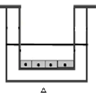
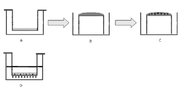
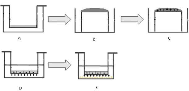
3.2 In vitro models ... 24
3.3 Camptothecin loaded Solid Lipid Nanoparticles ... 31
3.4. In vitro studies ... 35
Chapter 4 ... 37
Results & Discussion ... 37
4.1. Characterization of the in vitro mouse model ... 38
4.2. Characterization of the in vitro human BBB model on monoculture ... 39
4.3. The influence of different cells on the in vitro BBB model ... 45
4.4. Nanoparticles characterization ... 52 4.5. In vitro studies ... 54
Chapter 5 ... 57
Conclusion ... 57Chapter 6 ... 59
Future work ... 59References ... 61
xiii
List of Figures
Figure 1.1 The cell associations at the BBB [5].. ... 3 Figure 1.2 Simplified explanation of the molecular composition of endothelial TJ at the
BBB are shown [17].. ... 4 Figure 1.3 Different type of blood brain barrier (BBB) transporters adapted from [8]... ... 5 Figure 1.4 Mechanisms of transport across BBB [2].. ... 9 Figure 1.5 Overview of different strategies for brain targeting of drugs adapted from [23]. . 10 Figure 3.1 Schematic illustration of [A] in vitro BBB model and [B] in vivo BBB.. ... 24 Figure 3.2 The experimental procedure for monoculturing the bEnd3 cell line. ... 25 Figure 3.3 The experimental procedure for monoculturing the endothelial cells.. ... 26 Figure 3.4 The experimental procedure for co-culturing the endothelial cells and U87 cell
line.. ... 26 Figure 3.5 The experimental procedure for coculturing the endothelial cells and
astrocytes on different sides of the semi permeable filter.. ... 27 Figure 3.6 The experimental procedure for co-culturing the endothelial cells, astrocytes
and U87 cell line on different sides of the semi permeable filter.. ... 28 Figure 3.7 Schematic representation of SLN production by high shear homogenisation
followed by ultrasonication adapted from[40]. ... 32 Figure 4.1 TEER measurements in different cell mouuse densities on monoculture.. ... 38 Figure 4.2 TEER measurements in different cell human densities on monoculture... 39 Figure 4.5 Cellular gate and fluorescence histogram from hCMEC/D3 flow cytometry
experiment using VE-Cadherin-FITC.. ... 42 Figure 4.7 Permeability assay.. ... 44 Figure 4.8 TEER measurements in co-culture, endothelial cells and U87 cell line. ... 46 Figure 4.9 TEER measurements in co-culture, endothelial cells and primary astrocytes
xiv
Figure 4.10 TEER measurements in endothelial cells cultured with U87 cell line and
primary astrocytes cells.. ... 47 Figure 4.11 Permeability assay on 7th day. Permeability experiment using the FD4
molecule.. ... 47 Figure 4.12 Fluorescence histogram from hCMEC/D3 co-cultured with U87 cell line using
VE-Cadherin-FITC.. ... 49 Figure 4.13 SEM images of surface endothelial cells co-cultured with astrocytes and U87
cell line on 7th day.. ... 49
Figure 4.14 SEM images of endothelial cells surface co cultured with astrocytes and U87 cell line in on 7th day.. ... 50
Figure 4.15 SEM images of endothelial cells, astrocytes and U87 cell line in culture on 7th
day.. ... 51 Figure 4.16 Volume density (%) analysis of Empty and CPT-loaded SLN particle size.. ... 52 Figure 4.17 Calibration curve to extrapolate CPT concentration values using HPLC
method. ... 53 Figure 4.18 Reduction of Alarmar blue reagent on endothelial cells, on glioma cells and
primary astrocytes.. ... 54 Figure 4.19 Cellular viability of hCMEC/D3, human astrocytes and U87 cell line using the
xv
Acronyms
ABC Adenosine triphosphate-Binding Cassete AET Active Efflux Transporter
AMT Absorptive-Mediated Trancytosis BBB Blood-Brain Barrier
BCRP Breast Cancer Resistance Protein bEnd3 Immortalized mouse Endothelial cells bFGF basic Fibroblast Growth Factor BSA Bovine Serum Albumin
CMT Carrier-Mediated Transport CNS Central Nervous System
CPT Camptothecin
CPT-SLN Camptothecin-loaded SLN
DIV-BBB Dynamic In vitro Blood-Brain Barrier DLS Dynamic Light Scattering
DMEM Dulbecco’s Modified Eagle’s Medium DMSO DimethylSulfOxide
DPBS Dulbecco’s Phosphate Buffered Saline EC Endothelial Cells
EDTA EthyleneDiamineTetraacetic Acid FBS Fetal Bovine Serum
FD Fluorescein isothiocyanate – Dextran hCMEC/D3 human Capillary Endothelial cells
HPLC High Performance Liquid Chromatography HPLC High Performance Liquid Chromatography JAMS Junctional Adhesion Molecules
MDR Multidrug Resistance Proteins
MRP Multidrug Resistance-associated Proteins NVU NeuroVascular Unit
xvi
RMT Receptor-Mediated Transcytosis SEM Scanning Electron Microscopy SLN Solid Lipid Nanoparticles
TEER Trans-Endothelial Electrical Resistance TJ Tight Junctions
WGA Wheat Germ Agglutinin
1
Chapter 1
Introduction
The most crucial limitation in diagnosis and treatment of the neuronal diseases is the unique and highly controlled microenvironment of the Central Nervous System (CNS) barriers [1-4]. There are three key interfaces at which cells form barriers between the blood and the CNS, namely the Blood-Brain Barrier (BBB), blood cerebrospinal fluid barrier and the arachnoid barrier [5], being BBB the most important. This interface is formed by specialized endothelial cells (EC) in close association with basement membrane and neighboring cell types within the neurovascular unit [6]. The microvascular endothelium at the BBB is characterized by the presence of tight junctions between adjacent EC, lack of fenestrations, and minimal pinocytotic vesicles [7].
Besides the delivery of drugs to CNS through BBB being really poor and the treatment inefficient, the drugs used present many limitations such as side effects and bioavailabitity. Therefore, it is necessary to develop drug carriers to improve the effects in diagnostics, therapy and theragnostics [8]. The development of a close in vitro cell system is a difficult task and an important first step in the evaluation of new drugs and drug delivery systems to cross the BBB [9]. However, at present, no in vitro model can faithfully reproduce all the properties and characteristics of the in vivo BBB model.
This chapter will focus on BBB structure and mechanisms. Firstly, its importance functional and morphological it will be analyzed. A brief description of the different types of transporters it will be discussed, as the different techniques to perform brain drug targeting and the current strategies. Finally, it will be present various in vitro models divided into static and dynamic ones.
2
1.
Blood Brain Barrier
In 1885, more than 120 years ago, Paul Ehrlich was the first to demonstrate the presence of a barrier between blood and brain. He found that intravenously injected dyes, rapidly contrast all organs except the brain [10]. A few years later, his student Edwin Goldman made other experience where he injected these dyes into the cerebrospinal fluid. He found that this route had free access to neural tissue, however not of the peripheral organs. So, these dyes were prevented from directly entering the blood supply of the brain [2]. Since this crucial discovery, our understanding of the BBB molecular structure, physiological processes and our knowledge in its transporters increased [1].
BBB is regarded as an active, dynamic and extremely complex interface between the blood and the CNS which has specific structural and biochemical properties [9]. It is clear that BBB is very important in the protection of neurons from fluctuations in the plasma component and it was the main factor that leads to its development. This interface controls the rate of influx and efflux of biological substances needed for the brain metabolic processes and neuronal function. Because, of its selectivity, the BBB plays a crucial role to regulate the trafficking between blood and CNS and the determination of neuroimmunology and neuropathology [11]. So, BBB provides protection against many toxic compounds and pathogens. For this reason, it is of paramount importance in regulating the constancy of the brain internal environment [9]. It also contributes to ion homeostasis function which keeps the ionic composition optimal for synaptic signaling and preserves neural connectivity. It allows immune surveillance and responses to minimal inflammation and cell damage [5, 12].
Morphologically, the BBB is formed by specialized EC, paving the luminal side (the blood capillary side) in close association with basement membrane and neighboring cell types, which include perivascular pericytes, astrocytes, neurons and microglia in the abluminal membrane. These various cell types and basal lamina collectively constitute the neurovascular unit (NVU), Figure 1 [13]. Other important characteristic of in vivo BBB is the shear stress over the surface of the cells which is a tangential force generated by the blood flow [14]. Shear stress promotes the differentiation process and maintenance of BBB phenotype [15].
Specifically, the basement membrane of the cerebral endothelium is constituted by three apposed layers, made of different types of extracellular matrix classes of molecules such as collagen IV [16], glycoproteins, proteoglycans, laminin and various types of matrix adhesion receptors. In adults his membrane is about 30-40 nm thick and separates ECs and pericytes from the surrounding extracellular space [17]. Their interconnections produce a complex matrix which anchors cells and establishes the support for neighboring cells [18]. Also, the neighboring cells play an important role improving the barrier functions. It is a fact that ECs
3
are the major cellular constituent of the brain. The principal features associated with BBB ECs which differ these cells from the other ones in the rest of the body are the lack of fenestrations, low level of pinocytic vesicles, a high mitochondrial content, the presence of more extensive tight junctions (TJ) with electrical resistance as high as 8000 Ohmcm2 and
the expression of various transporters that influence molecule transport to the brain [19]. Concerning to astrocytes, they constitute nearly half of brain cells [20] and encircle 90% of the BBB endothelium on the abluminal side. Astrocytes have a key feature on the induction and maintenance of BBB integrity, namely by the secretion of factors such as transforming growth factor-β, glial-derived neurotrophic factor, basic fibroblast growth factor (bFGF) and angiopoetin 1 into the medium; alter the expression of drug transporters such as P-glycoprotein (P-gp) and induce tighter TJ [21]. Although this importance has been documented since two decades, the molecular pathways still remains unclear [5, 18]. As referred to pericytes, also known as vascular smooth muscle cells, they have a physical association with the endothelium. The release of ECs factors can induce migration of pericytes and affect the maintenance of the integrity of the vessel. Pericytes are able to control the capillary diameter, due to their contractile characteristics, then modulate the cerebral blood flow [22]. Regarding to microglia cells, the exact mechanisms of how microglia influences BBB properties are still unknown, however it is clear that they are playing an important role in immune response and consequently in the BBB integrity [22]. So far, very few are known about the precise role that neurons play on the BBB phenotype. Although, there are some evidence that neurons affect cerebral blood flow and can regulate the function of blood vessels [7].
Figure 1.1 The cell associations at the BBB [5]. The NVU is a complex cellular system that includes highly specialized endothelial cells, a high concentration of pericytes embedded in the endothelial cell basement membrane; astrocytic endfeet associated parenchymal basement membrane, neurons and immune cells. Considering all cellular interactions presents on neurovascular unit, it can be said that BBB presents unique structural and biochemical properties.
4
TJ consist of an extreme complex of integral proteins spanning the intercellular cleft (occludin and claudins), junctional adhesion molecules (JAMs) and cytoplasmic accessory proteins (zonula occludens (ZO) -1, -2, -3 and cingulin) bound to the actin cytoskeleton, Figure 2. Specifically, claudins form the primary seal of TJ forming dimmers and bind homotypically to claudins on adjacent cells, the level of claudin expression determines TJ integrity. Occludins are the dynamic regulatory protein responsible on TJ regulation, to enhance the transendothelial electrical resitance (TEER) and restrict the paracellular permeability. Together, claudins and occludins form the extracellular component of TJs and are both required for formation of the BBB. And JAMs can regulate the leukocyte migration and it is involved in cell-to-cell adhesion [21]. Basically, ZO proteins serve as recognition proteins for TJ placement and as support structure for signal transduction proteins [17]. In case of adherens juntions, they are located near the basolateral side of ECs. Cadherin proteins span the intercellular cleft and are linked into the cell cytoplasm. The principal function of these junctions is holding the cells together giving the tissue structural support [13]. TJ and AJ components are known to interact and influence TJ assembly [19].
Figure 1.2 Simplified explanation of the molecular composition of endothelial TJ at the BBB are shown [17]. Three integral proteins form the TJ structure: claudins, occludins and JAM. Claudins produce the primary seal of TJ. Occludin function as a dynamic regulatory protein and JAMs are important to regulate leukocyte migration. TJ consists of accessory proteins such as ZO. All this mechanisms are important to improve the BBB tightness and to reduce the compounds that can cross the BBB.
5
2.
Transports across the BBB
With regard to BBB, it can be said that it is a sort of sanctuary site as it strictly controls the exchanges between the blood and brain compartments [8]. As far as small molecule drugs are concerned, more than 98% cannot enter the brain [23]. Consequently, crossing the BBB is a great challenge.
Nonetheless several transporters have been established by means of which solute molecules move across BBB. The various systems that mediate the transport across BBB can be divided into three categories - small molecule, large molecule and efflux transporters (figure 1.3 and 1.4). Within the small molecule transporters there are two possibilities, the diffusion transport and the carrier-mediated transport (CMT). In the first, the passage of molecules across the EC of the BBB can occur between adjacent cells (the paracellular pathway) or through the cells (the transcellular pathway) [7]. Active efflux transporter (AET) is another type of route. Among them the most extensively characterized is adenosine triphosphate-binding cassete (ABC) transporter family. Macromolecule transporters include receptor-mediated transcytosis (RMT), absorptive-receptor-mediated transcytosis (AMT) and cell-receptor-mediated- cell-mediated-transcytosis. This last one refers only to immune cells transport [12].
Figure 1.3 Different type of blood brain barrier (BBB) transporters adapted from [8]. The scheme is divided into three large groups. The first group is about small molecule transporters. The second group is about macromolecule transporters. And the last group is about active efflux transporters (AET). Each group is divided in various small groups which have different biologic and physical characteristics – diffusion and carrier-mediated transport (CMT); receptor-mediated transcytosis (RMT); adsorptive-mediated transcytosis (AMT) and cell-adsorptive-mediated transytosis (CMT); adenosine triphosphate-binding cassete (ABC) transporters.
2.1. Small molecule transporters
Within the small molecule transporters there are two possibilities, the diffusion transport – either simply diffusion or facilitated transport across aqueous channels – and the active transport which is mediated by a carrier such as proteins. In the first, the passage of
6
molecules across the ECs of the BBB can occur between adjacent cells (the paracellular pathway) or through the cells (the transcellular pathway) [8].
With regard to transcellular transport, Lipinski and co-researchers developed five rules that determine if a compound is more likely to be membrane permeable and easily absorbed by the body [24]. In order to achieve his goal, Lipinski analyzed the physicochemical properties of more than 2000 drugs and candidate drugs in clinical trials. His work resulted in the establishment of five criteria that must be fulfilled by the compounds. These are: no more than five hydrogen bond donors (nitrogen or oxygen atoms with one or more hydrogen atoms); no more than ten hydrogen bond acceptors (nitrogen or oxygen atoms); a molecular mass lower than 500 Da; an compound's lipophilicity, expressed as a quantity known as logP (the logarithm of the partition coefficient between water and 1-octanol) lower than 5, and compound classes that are substrates of biological transporters are exceptions to the rule [24]. It is important to state that the rule of five applies only to absorption by passive diffusion of compounds through cell membranes; compounds that are actively transported through cell membranes by transporter proteins are exceptions to the rule. Therefore, it is of limited significance nowadays [23]. However, it is clear that if the molecular weight of a drug molecule is higher than 400 Da or the drug forms more than eight hydrogen bonds, the probability of crossing the blood-brain barrier via passive diffusion in pharmacologically significant amounts is very low [12].
Even though the paracellular transport of hydrophilic substances is virtually absent due to the unique properties of the TJs, small lipid soluble substances, like alcohol and steroid hormones, penetrate transcellularly by dissolving in their lipid plasma membrane [13]. In brief, the paracellular transport is a passive movement of a molecule through the aqueous route of the intercellular cleft between EC via small pores or flaws in the tight junctions. It represents a central functional component of BBB regulation [7].
The relationship between the paracellular and transcellular permeability is the key feature in the regulation of overall trans-endothelial permeability in the endothelium [25].
Concerning to CMT, the active transport is an important transporter that carriers essential polar nutrients to cross the brain namely as glucose, amino acids, and purine bases. This type of route uses carriers, that is to say, membrane-restricted systems commonly involved in the transport of small molecules with a specific size and a molecular weight smaller than 600 Da [8]. The solute carriers may be bi-directional, in which case the direction of net transport is determined by the substrate concentration gradient; unidirectional either into or out of the cell; or involve an exchange of one substrate for another; or be driven by an ion gradient depending on electrochemical gradients. It is also a fact that CMT is substrate selective, considering that the transport rate depends on the degree occupation of the carrier.
7
Considering that glucose is the main energy source of the brain, glucose transporter-1 plays a vital role within the transporters. Since the density of glucose transporter-1 at the abluminal membrane is higher than at the luminal, there is a homeostatic control for glucose influx into the brain [13, 25].
2.2. Macromolecules transporters
Transcytosis of macromolecules across the BBB via endocytotic mechanisms provides the main route by which large molecular weight solutes such as proteins and peptides can enter the CNS intact. Macromolecule transporters include RMT, AMT and CMT.
Extensive studies of RMT have revealed that it offers selective uptake of many different types of ligands, namely plasma proteins, enzymes and growth factors [8], RMT occurs in three steps, first the endocytosis of macromolecules specially bound to a receptor on the endothelial surface of BBB, followed by diffusion across the endothelium, and exocytosis on the opposite site. ECs comprise different receptors, such as transferrin receptor [26], insulin receptor [27], lipoprotein receptors [28], and insulin-like growth factors [27]. Regarding insulin molecules, the ligand first binds to the receptor present at specialized areas of plasma membranes called coated pits. Once bound to ligand, these coated pits invaginate into the cytoplasm and form coated vesicles. The ligand is dissociated from receptor by acidification of endosome and crosses to the other side of the membrane [23].
AMT, also known as pinocytosis, is mediated by electrostatic interaction between positively charged substrates and the negatively charged plasma membrane (i.e. heparin sulphate proteoglycans) [12]. It is important to state that this process does not involve specific plasma membrane receptors. Normally, endocytosis occurs upon binding of the cationic compound to the plasma membrane. In order to protect the brain from nonspecific exposure to polycationic compounds, this vesicular transport is actively downregulated in the BBB [23].
Recently, a new transporter was identified which is based on transport of immune cells (like monocytes or macrophages) to cross the intact BBB [23]. CMT can transport any type of molecules or materials and particulate carrier systems, whereas other mechanisms normally permit only solute molecules with specific properties.
2.3. Active efflux transporters
As opposed to the above described influx routes, the efflux transporters play a different role. It is a fact that they can be considered a "first line of defense", since it is up to them to remove xenobiotic molecules and brain potentially neurotoxic endogenous from the brain tissue back into the circulation. In addition, they can significantly restrict the entry of substrate into brain parenchyma. Up until now, the most extensively characterized efflux transporter proteins at BBB is the ABC transporter family [29, 30]. In humans they are a
8
superfamily of proteins containing 48 members which are grouped into 7 sub-families, ABC A to G. Among the large number of ABC transporters, only three of them are expressed at the blood-brain barrier. The ABCB sub-family contains the multidrug resistance proteins (MDR) of which P-glycoprotein (P-gp) is the best-known representative, the ABCC subfamily contains the multidrug resistance-associated proteins (MRP) and ABCG sub-family contains breast cancer resistance protein (BCRP) [31].
Regarding to the P-gp, it is a membrane-bound protein (170 kDa) which is present at high concentrations in the luminal membrane of the blood-brain barrier endothelium. P-gp has a high affinity for a wide range of cationic and lipophilic compounds and therefore limits the transport of many drugs, including cytotoxic anticancer drugs, antibiotics, hormones, and HIV protease inhibitors. At present, P-glycoprotein is considered the most prominent element of selective barrier function that limits xenobiotics from entering the brain [31].
MRP transports mainly organic anions, glutathione, glucuronide- or sulfateconjugated compounds, as well as various nucleoside analogs. Thus it acts as an organic anion transporter while it also transports neutral organic drugs. MRP transporters have five isoforms present in BBB and BCSFB.
Another BBB efflux transporter is BCRP, which appears to be expressed in the luminal membrane of the cerebral EC in a similar manner to p-glycoprotein. Recent studies suggest some cooperation between BCRP and p-glycoprotein inasmuch as they limit xenobiotics from entering the brain and compensate one another [23]. Four vital reasons justify the need to understand the regulation of these transporters. First of all, it is not known how barrier properties can be altered through environmental factors. Second, inflammatory and oxidative stress seems to affect ABC transporter expression in other barrier and excretory tissues. Third, it is crucial to find out how specific CNS diseases alter transporter function. Finally, it is necessary to discover how ABC transporter-specific inhibitors can improve drug delivery [31].
9
Figure 1.4 Mechanisms of transport across BBB [22]. This figure explains the involved mechanism of each BBB transporter.
10
3. Drug targeting to the brain
As said before, the most limiting factor in the development of new strategies of diagnosis and therapeutics is the crossing of the BBB. To overcome these limitation, innumerous strategies has been studied to improve the pharmacological quantity that it is able to improve therapeutic efficiency.
Firstly, it will be discuss various routes including direct delivery to CNS and direct systemic delivery. Then, it will be focus in noninvasive approach, namely it will be discussed physiological strategies such as transporter mediated delivery; chemical ones using as example cationic proteins; conjugation of drugs with antibodies is a biological strategy and the various colloidal carrier systems. Figure 1.5 shows a systematic classification of various approaches.
Figure 1.5 Overview of different strategies for brain targeting of drugs adapted from[23]. Various strategies can be used to improve diagnostic and therapeutic – such as invasive ones (systemic and local drug delivery), and drug delivery strategies which are modified according to the purpose.
3.1. Novel drug delivery systems
In this field nanotechnology has been a key feature since it improves the characteristics of different agents [32]. Nanotechnology is the creation and use of functional materials with at least one characteristic dimension measured in nanometers (scale =10-9). In this technology
unique phenomena enable novel applications because nanosystems have new properties such as large surface-volume ratio, surface charge, small and controlled size. The small size of the nanoparticles enables them to penetrate the BBB and facilitates drug delivery across the barrier [33]. They have large surface-volume ratio resulting in an increase of local interaction
11
and thereby increasing the rate of dissolution. At the same time, it is possible to do surface functionalization (to improve shelf-life) and to use them as drug carriers (to increase drug bioavailability). They allow for controlled and slow drug release in the brain, while decreasing peripheral toxicity and side effects [23].
Several colloidal system have been studied to improve the BBB crossing such as immunolipossomes [34], Solid Lipid Nanopaticles (SLN) [35], poly(butyl cyanoacrylate) (PBCA) nanoparticles, chitosan nanoparticles, albumin nanoparticles [36].
Functionalization of nanocarriers is one of the most important steps or challenges in formulating nanocarriers for drug delivery. There are two forms the passive or/and active targeting. The passive targeting depending of tissue characteristics like the enhanced permeability and retention on many types of tumors [36]. However, in case of human brain diseases has not been shown to be effective. The advantage of active targeting is the increase of the amount of drug in the target tissue, thereby increasing the pharmacological response and reducing systemic side effects [8]. For example, in the case to increase time of nanoparticles in the organism and drug bioavailability is necessary use active targeting by coating the surface with polyethylene glycol or surfactants like polysorbate 80. Because, if the nanoparticles are unmodified, they rapidly are adsorb, mainly by opsonins, and eliminate for the organism by the macrophages of the reticuloendothelial system [36].
The blood-to-brain transport system is of considerable interest in drug delivery for targeting of drug molecules into brain whereas peptides and small molecules may use specific transporters expressed on EC [8]. Active physiological or disease-induced drug targeting strategies use modified drugs to take advantage of native BBB nutrient transport systems or by conjugation to ligands that recognize receptors expressed at the BBB [37]. So, only drugs that closely mimic the endogenous carrier substrates will be transported into the brain. Nowadays, the research of nanoparticles to target BBB is in RMT, AMT and P-gp mechanisms, mainly [37].
Accordingly to AMT, the most limitation of this approach is lack of tissue selectivity, which can potentially cause side effects [38]. Its approach is based on SynB vectors, penetratin and Tat which are various examples of cell penetrating peptides [12]. Cell penetrating peptides have an enormous potential for diagnostic and therapeutic applications because their low cytotoxicity and the tremendous variety of cargo that can be loaded [8]. Recently, Liu and co-workers made a polymer core-shell NPs and on its surface is an anchored Tat molecule. The results shown that the surface with TAT improved their uptake cellular by EC [39].
RMT has been successful in transporting large drug molecules, drug carrying lipossomes, nanoparticles and polymeric complexes even without it [37]. As referred before, RMT has
12
different types of receptors and each receptor has its specific ligands and approaches [8]. In the case of diptheria receptor, it is strongly up-regulated in inflammatory conditions, which occur in CNS diseases. Recently, Boer and his group worked in CRM197 which is a non-toxic mutant of diphtheria toxin and applied it as a targeting vector for drug delivery to the brain in diagnostic and therapeutic applications [8].
At present, P-gp is considered the most prominent element of selective because it limits xenobiotics from entering and accumulates in the brain. So, one of the strategy is inhibits efflux transport systems by coating the nanoparticle surface with polaxamer 188 and polysorbate 80, for example. Polysorbate 80 is an important component because it is cleared adsorb apolyprotein E or B and followed by endocytosis and trancytosis.
3.2. Administration strategies
Regarding to direct delivery to CNS, there are different approaches. Accordingly to intracerebral (intraparenchymal) delivery, drugs are delivered directly into the parenchymal space of the brain. They can be injected by intrathecal catheters, by controlled release matrices and by microencapsulated chemicals or recombinant cells [3]. Unfortunately, access to the parenchyma is minimal so that a larger dose is required [8]. Alternatively, convection enhanced diffusion is used to increase drug uptake by bulk flow. Furthermore, brain implants (biodegradable/non-biodegradable polymeric materials encapsulating drugs) can be used for the local delivery, too [37]. Another possible route is the intraventricular route, which it is also used for drugs (small or large molecules) that do not cross the BBB and where no BBB drug delivery is available [40]. Lastly, the intrathecal route involves delivery of drugs into the cistern magna of the brain. In this delivery there is a chance of drugs spreading along the distal space of spinal canal. For this reason it is best used to treat spinal diseases [3].
Another possible to administer drug-loaded is the direct systemic delivery. In intravenous delivery, the most commonly used route to administrate larger doses of drugs into the body [41], the drug is deliver directly into the general circulation by avoiding its first-pass metabolism and has great potential to deliver drugs to almost all neurons in the brain. Unfortunately, drug availability is affected by its exposure to peripheral organs and rapid clearance. Consequently, there is only a little accumulation of the drug in the brain. Similarly, intra-arterial administration allows drugs to access the brain vasculature, before they enter peripheral tissue and it is possible to avoid first pass metabolism. By using BBB disrupting agents it is possible to increase the effects of this route [3]. The intranasal route is based on the principle that drugs exit the submucosa space of the nose into the brain CSF compartment. The nasal epithelium has many advantages such as high permeability, avoidance of first-pass metabolism, small doses and self-administration. However, this administration damages the nasal mucosa and decreases the quantity of drug available [23].
13
3.3. Chemical Stimuli
Parallel to last strategies, there has been a significant effort in delivering drugs to the brain with BBB disruption [63]. In this approach the substances are directly delivered to the CNS by using certain chemical substance or by applying energy (ultrasonic waves or electromagnetic radiations). However, the BBB disruption exposes the brain to infection and damage from toxins [6].
4. BBB models
It is, indisputable that in vivo models are the best candidates to study the permeability phenomena at BBB. However, these resources, typically rats or mice, are scarce, expensive, and difficult to study both in detail and real-time. Conversely, ex vivo and in vitro models are good alternatives due to their simplicity and controlled environment [11]. Nevertheless, the research community also recognizes that reproducing the physiology and the functional response of the BBB in vitro is a challenging task. In vitro BBB models started to emerge in early 1990s and offer a number of desirable advantages such as cost effectiveness, versatility enable controlled, repeatable and non-invasive tests like permeability assays, resistance measurements and microscopy. Moreover, they can alter multiple BBB determining factors namely: use different cell isolation procedures, cell culture conditions, configuration of cells on culture and the cell types (origin and species) [9, 17]; use different systems, such as static or dynamic. Nonetheless, in vivo validation is still required [9]. In the follow paragraphs, it will be discuss different factors and/or variables that can improve the BBB model and the different approaches that have been used to reproduce in vivo BBB.
4.1. Criteria for in vitro models of the BBB
At present no in vitro model can faithfully reproduce all the properties and characteristics of the in vivo BBB model since the latter has several types of cells and junctions that give rise to unique properties [7]. Considering that a single different factor is enough to change the BBB fundamental properties, there are several requirements that an ideal in vitro BBB model should meet. These include:
The ability to enable the expression of TJ between adjacent EC which directly facilitate the formation of a selective barrier [9, 42];
In vivo-like asymmetric distribution of relevant transporters which confers polarization of the EC [9];
Mechanotransductive effects of shear stress from fluid flow on EC which determines cell differentiation and tight junction formation [42];
14
The ability to discriminate the permeability of substances according to their molecular weight [9, 42];
Maintenance of high electrical resistance that represents the maturity and soundness of the structures [43];
The ability to reproduce the effects of a large variety of hemodynamic and systematic/inflammatory insults on the BBB [9];
Availability, convenience, predictability and reproducibility [17].
An ideal BBB model should be able to reproduce all these characteristics. Unfortunately, the techniques available at present do not allow the monitoring of all these features. Therefore, new artificial systems such as bioreactors will have to incorporate a number of controlled parameters namely control/adjust oxygen and carbon dioxide levels in the culture medium, real-time monitoring of BBB integrity, medium sampling, among others. They will also have to include an array of computer-controlled sensors to monitor a variety of physiological parameters (e.g. glucose, lactate). Advances in this field will only be made possible with the introduction of new cell culture apparatus [9].
4.2. Overview of current in vitro BBB models
In the last decades we have witnessed the development of cell culture techniques in which cells are immersed in a homogeneous culture medium [44]. The main advantage of cell culture is that it allows us to select the stimuli that cells are exposed to, something that would be very difficult to reproduce in vivo. Besides, using different types of BBB cells, it will be possible use various apparatus which are possible to distinguish into two main groups: static and dynamic systems. The main difference is that dynamic models include fluid flow. However, most often the final choice of the BBB model is determined by the researcher's needs as well as the characteristics of the laboratory, namely time, cost and to what extent the model has to be able to reproduce in vivo conditions. Accordingly, either one or the other model can be more advantageous depending on the purpose of the investigation [43]. In the following sections current in vitro models of the BBB are analyzed; for improve understand of each model it will be referred to the corresponding key publications.
4.2.1. Cell types
Nowadays, most of the current successful BBB in vitro models are based on primary cell cultures [4] due to their high TEER values and low passage brain EC retain which closely resemble in vivo models, although the several passages of initial cultures entail the down-regulation or even the loss of many features [17]. Moreover, there may be a limitation in the availability of the primary cells as a result of the accessibility of the animals, while these cells are also more susceptible to internal and external contaminations than cell lines. In
15
addition, this approach has high costs and requires time-consuming and special skills for the isolation of brain EC. Primary cultures can use mammals such as rats, mice, pigs and bovines. Rodent models are advantageous in that they are available and it is possible to use them as transgenic animals [17]. However, their small size and the relatively low amounts of EC that can be obtained from them leads to the use of other models, namely pigs or bovines [45]. Not only it is possible to obtain large quantities of EC (up to 200 million as opposed to 1-2 million cells per rat brain) [7], but they also offer good permeability properties, more closely to the human BBB. On the other hand, their availability is restricted and they are not so well characterized regarding their biochemical or molecular composition [5]. Finally, the use of human primary cells is equally restricted by the unavailability of experimental material. The source material is usually acquired either from autopsies or biopsies, so this tissue often cannot be considered as a healthy resource [4]. Therefore, it was necessary to develop immortalized several cell lines [46].
Immortalized cell lines offer a considerable number of advantages. They are reliable (using trusted well-established sources), consistent (cell source is controlled and consistent), long-lasting (important cell features do not disappear over time), accessible (cells are available to be purchased at any time) and preparation time and cost are reduced [44]. Despite lacking certain BBB features and having low TEER, there is no doubt that immortalized cell lines are an emergent solution for BBB models. Immortalized cell lines are available from many species, although the most frequently used models derive from rats [47]. Other models of cell lines are the porcine and bovine ones. Though they are available, unfortunately they are far less well characterized. They have been used to study changes in protein expression following induction by astrocytes and neuroinflammatory responses [4]. Human cells became available in the early 1980s and contain excellent characteristics for the study of the developmental and pathophysiological processes of the BBB. The best characterized human cell line is the hCMEC/D3 which has been shown to retain important BBB characteristics. The hCMEC/D3 cells show a stable phenotype, the expression of endothelial cell markers, chemokine receptors and ABC-transporters. Furthermore, the paracellular permeability is much lower compared to other cell lines [48]. Human specimens are undoubtedly the best model, since they are the only ones that faithfully reproduce the BBB characteristics [4].
As previously mentioned, EC are the principal components of the BBB. However, in the course of time it was discovered that other cell types also play an important role both in the function and regulation of BBB characteristics. As a result, in vitro models became more complex as they began to include glial cells, pericytes, even neurons and microglia in different BBB models [7].
16
Co-culture of EC with glial cells increases TEER by 71 %. It is clear that these cells work in synergy to faithfully reproduce the BBB characteristics [44]. There is a formation of more stringent inter-endothelial TJ, which in turn constitute a more reliable reproduction of in vivo BBB [49].
With pericytes, it has been established that there is an intrinsic relation between pericytes and the formation and maintenance of the cerebral microvasculature structure and functions. However, it is still unknown which type of pericytes plays the decisive role in this process [43]. More recently, it has been proved that neurons induce BBB related enzymes in cultured EC. And the co-culture of these cells has shown that a direct contact among EC and neurons is not necessary for the induction of occludin expression [4].
4.2.2. Apparatus
The system semi-permeable plate filters, a vertical side-by-side diffusion support, is the most commonly used apparatus for EC culturing. This bidimensional model is a microporous semi-permeable membrane that separates the luminal (vascular) and the abluminal (parenchymal side) compartments and which is submerged in feeding medium [11]. This apparatus is ideal to study permeability of drugs across BBB. There are two other main features that make this apparatus so attractive: it is easy to establish cultures and its cost is relatively low [15]. On the other hand, the semi-permeable membranes cannot reproduce the physiological shear stress. In addition, the lack of antimitotic influences by laminin and flow will increase cell cycle rate, which will cause an uncontrolled growth of the EC in a multilayer manner. When the tightness of the semi-permeable barrier is measured by TEER and permeability it is usually much lower than the in vivo BBB [9]. The source of the cells and the methodology employed determine the kind of studies that may be performed: drug transposition through BBB, regulation of BBB permeability and influence of pathologies on BBB permeability [43].
Culturing cells using tri-dimensional extracellular matrix supports is another recently apparatus [50]. Among its main advantages we can list the capacity to enable close interactions between cells as well as the formation of quasi-physiological biochemical gradient exposure. Furthermore it is good to study specific roles of various extracellular proteins in cell differentiation. Despite these advantages, it is more expensive and less convenient than static models while, at the same time, it is a complex challenge to address. Nowadays, this type of model is applied to drug discovery and transport studies related to a variety of organs and tissues [51].
As opposed to static models, dynamic ones use physical stimuli to create shear stress and, as a result, they are able to replicate the physiological environment of in vivo BBB [9]. Bussolari and co-researchers made the first attempt to enable the endothelial exposure to flow in vitro
17
by using a purpose-built cone and plate viscometer [52]. This apparatus allowed them to expose cells to a quasi/uniform laminar or pulsatile shear stress. In this case, the level of shear stress was determined by the cone angle and the angular velocity. However, since the flow represented was turbulent it did not faithfully reproduce the flow experienced in vivo. This aspect constitutes a serious disadvantage because it is not possible to obtain reliable and significant results. Nonetheless, this apparatus was very important as it was the first step to produce dynamic models of the BBB [23].
Realizing the importance of shear stress as a vital component of any in vitro model, researchers decided to focus on the development of new generations of dynamic in vitro systems of which a detailed explanation will follow. The main features of the dynamic In
vitro Blood-Brain Barrier (DIV-BBB) are the possibility to use co-cultures and the presence of
intraluminal flow through artificial capillary-like structural supports (hollow fibers) [15]. As a result, it is possible to faithfully reproduce the BBB in situ as the EC are cultured in the lumen of hollow-fibers inside a sealed chamber and are exposed to flow while the astrocytes are seeded in the extraluminal compartment to promote cellular stimuli. This system presents low permeability to intraluminal potassium and polar molecules, high TEER, negligible extravasation of proteins, the expression of specialized transporters, ion channels, and efflux systems. However, this system is not intended to be used in drug permeability studies and requires more time and technical skills to be established. Though it is possible to characterize cells, this can only be done in a limited way. Finally, to start the process a high cell load is required [9].
Recently the development of microtechnologies has enabled the creation of a new in vitro model of the BBB called MicroBBB. MicroBBB is a poly(dimethylsiloxane) multi-layered device with a membrane in between which separates the top and bottom channel. Although this apparatus is an entirely new creation, it also enables cell culturing procedure, permeability tests and TEER measurements [53]. When compared to the models previously discussed, MicroBBB presents more advantages including rapid and low-cost fabrication, controlled and repeated environment with realistic microcirculatory dimensions and environment, physiological fluid flow and shear stress and much thinner culture membrane which decreases the distance between co-cultured cells. However, the top-bottom architecture of this model limits simultaneous real-time visualization of both the vascular and neuronal sides of the BBB [4]. Another key feature of this model is that it can be used to monitor drug permeability, as well as changes in barrier function, which occur in response to various environmental stimuli namely diseases.
18
4.2.3. In silico models
The incredible development of computer technology and sophisticated algorithms allowed the creation of new methods based on computer simulation called in silico models [7]. In silico models offer various advantages, the most important of which lies in accurate predictions. Not only is there no need to recur to laboratory experiments, but the process is also cheaper and requires fewer time-consuming laboratory experiments [23]. The potential to accelerate drug discovery is the outstanding quality of in silico models for medicine. This technology allows the compounds to be synthesized, pre-screened, and virtually tested, so that it is possible to predict how they will cross the BBB. Therefore, it will be possible to study the efficacy and the bio-availability of novel drugs in terms of brain permeability, transport properties and toxicity [9]. On the other hand, the complex nature of BBB is not taken into full consideration, which leads to uncertain results. Though in silico models represent future breakthroughs in the pharmaceutical drug development, clinical studies will still have to be supported by in vitro and in vivo models in order to collect a number of crucial physicochemical measurements [7, 9] .
19
Chapter 2
Aim
The main goal of the present thesis was a further characterization and improvement of an in
vitro BBB model, using three different type of human cells, as a tool to study biological and
functional BBB alterations and as a key to assess permeability of anti-cancer drugs.
General goals
1. Develop an in vitro BBB model that will mimic more properties and characteristics of the in vivo BBB;
2. Integrity evaluation based on the alterations applied through the in vitro model;
3. Development of Camptothecin-loaded SLN (CPT-SLN) for brain delivery;
The thesis work was divided into two phases. First, it was analyzed the state of the art, definition of the work plan and requisition of the laboratory material. This part was done during September and October and also a few months before, during the 2nd semester. On the
second phase was developed the laboratory work at Laboratory of Pharmaceutical Tecnnologic, Department of Drug Sciences, Pharmacy Faculty, University of Porto, the majority of the work, and at INEB, University of Porto. This part was realized from October until June.
To develop and evaluate the integrity of triple co-cultured model, it was seeded hCMEC/D3 cell line, primary human astrocytes and U87 cell line. During the time on culture, TEER measurements were taken. Moreover, on the last day on culture were performed different experiments such as permeability assay, immunocytochemistry, optical microscopy, electronic microscopy and flow cytometry to analyze the cell morphology, expression of cellular specific markers and barrier integrity.
20
Then, to develop CPT-SLN for brain delivery it was selected the drug-SLN formulation. And it was produced and characterized CPT-SLN by mean size, size polidispersity, surface charge and association efficiency. Finally, it was done viability assays using different CPT formulations.
21
Chapter 3
Materials & Methods
Materials
Dulbecco’s Modified Eagle’s Medium (DMEM), Fetal Bovine Serum (FBS), glutamine, penicillin-streptomycin, chemically defined lipid concentrate, HEPES and trypsin-EthyleneDiamineTetraacetic Acid (EDTA), Occludin-mouse monoclonal antibody-Alexa Fluor 594, Claudin-5-mouse monoclonal antibody-Alexa Fluor 488, N2 suplemment, Wheat Germ Agglutinin (WGA) – Alexa Fluor 488 and GeltrexTM LDEV-Free Reduced Growth Factor
Basement Membrane Matrix were provided by Gibco (Invitrogen Corporation, Spain). Hidrocortisona - γ-irradiated, Dulbecco’s Phosphate Buffered Saline (DPBS) Modified, without calcium chloride and magnesium chloride, fluoroshield™ with 4',6-diamidino-2-phenylindole, dihydrochloride (DAPI) mounting medium, paraformaldehyde, giemsa stain, rat tail collagen type I, human basic fibroblast growth factor (bFGF), ascorbic acid, lithium chloride solution, Fluorescein isothiocyanate – Dextran (FD) average molecular weight of 4.000, 40.000 and 70,000 Da, triton-X 100, Bovine Serum Albumin (BSA) were provided by Sigma-Aldrich (Portugal). EBM-2 medium were provided by Lonza. The 12-well semi-permeable plate filters, PE-mouse anti-human GFAP were sold by BD, Biosciences, USA. VE-Cadherin-FITC were obtained from Miltenyi Biotec (USA). AlamarBlue™ cell viability reagent assay were obtained by Thermo Scientific (USA).
Cetyl palmitate was a gift from Gattefossé SA, (St Priest, France). The surfactant polysorbate 80, organic solvents (acetonitrile, triethylamine) for high performance liquid chromatography (HPLC) and Dimethyl sulfoxide (DMSO) were supplied by Merck KgaA, (Darmstadt, Germany). Purified water was of MilliQ ® -quality.
22
Methods
3.1. Cell Culture
3.1.1 - Cells description
Immortalized mouse EC (b.End3) cell line is sold by American Type Culture Collection. bEnd3 is an immortalized mouse EC. The cells were transformed by infection with the NTKmT retrovirus vector that expresses polyomavirus middle T antigen. The endothelial nature of these cells was confirmed by the observed expression of von Willebrand factor and uptake of fluorescently labeled low density lipoprotein (LDL).
Immortalized human brain capillary EC (hCMEC/D3 cell line) was a kindly donated by Dr. PO Couraud (INSERM, France). The original brain EC used for the generation of the cell line were isolated from human brain tissue following surgical excision of an area from the temporal lobe of an adult female with epilepsy. The hCMEC/D3 cell line had been immortalized by lentiviral transduction of the catalytic subunit of human telomerase and SV40-T antigen into very early cultures of adult human brain endothelial microvascular cells [54]. hCMEC/D3 between passage 25 and 33 were used in all studies.
The human astrocytoma U87 cell line is a commercial cell line sold by American Type Culture Collection. It derived from a human glioblastoma (astrocytoma), classified as grade IV as of 2007 with adherent properties.
Primary astrocytes are human brain progenitor-derived astrocytes with adherent properties sold by Gibco. It is the only that are primary cells, so the number of cells is more limited than the other ones to perform the in vitro models experiences.
3.1.1.1 Cell line conditions
For culturing, hCMEC/D3 were seeded in a concentration of 25 000 cells/cm2 and grown in
EBM-2 medium (Lonza, Basel, Switzerland) supplemented with 10 mM HEPES, 1 ng/mL human Basic Fibrolast Growth Factor (bFGF), 1.4 µM hydrocortisone, 5 µg/mL ascorbic acid, penicillin–streptomycin, chemically defined lipid concentrate and FBS. All culture ware and semi permeable filters were coated with 150 µg/mL rat tail collagen type I for 1 h at 37º C. Collagen is a fibrous protein which presents a rope-like triple helix, providing strength to the extracellular matrix and this specific type of collagen is the most common fibrillar collagen (90%). Cells were cultured in the incubator at 37º C with 5% CO2, 95% fresh air in a humidified
incubator (Heraeus Hera Cell incubator). Cell culture medium was changed every 2-3 days.
The human glioma U87 cell line and b.End3 cell line were maintained in complete Dulbecco’s Modified Eagle’s Medium (DMEM) (Gibco, Invitrogen Corporation) supplemented with 10% fetal
23
bovine serum (FBS), 1% glutamine (2mM) and 1% antibiotic-antimycotic (Gibco, Invitrogen Corporation: 10,000 units/mL penicillin G sodium, 10,000 mg/mL streptomycin sulphate). Cells were subcultured every 3-4 days using trypsin-EDTA.
Primary astrocytes were maintained in complete Dulbecco’s Modified Eagle’s Medium (DMEM, Gibco, Invitrogen Corporation) supplemented with 10% fetal bovine serum (FBS), 1% glutamine (2mM), 1% antibiotic-antimycotic (Gibco, Invitrogen Corporation: 10,000 units/mL penicillin G sodium, 10,000 mg/mL streptomycin sulphate) and 1% N2 supplemment (Gibco, Invitrogen Corporation). The medium has been specifically formulated for the growth and maintenance of human astrocytes while retaining their phenotype. All culture ware and semi permeable filters were coated with 0.1 mg/mL GeltrexTM LDEV-Free Reduced Growth Factor
Basement Membrane Matrix (Gibco, Invitrogen Corporation) for 1 h at 37º C. It is used for promotion and maintenance of many cell types specifically primary cells. The major components of this matrix product include laminin, collagen IV, entactin and heparin sufate proteoglycan. Cells were subcultured every two times per week using trypsin-EDTA.
3.1.2 - Cell line laboratory concepts
3.1.2.1 - Cell subculture
Firstly, the cells were examined to inverted microscopy to study any signal of contamination. When the cells present 70-80% of confluence, the medium was aspirated and they were washed with Dulbecco´s Phosphate Buffered Saline (DPBS) two-three times to remove all residues. Then, they were detached using trypsin-EDTA (1x) from the flask, only a few minutes because trypsin cuts the adhesion proteins in cell-cell and cell-matrix interactions and EDTA is a calcium chelatator, so it is necessary to pay attention of the time in contact with the cells. In this way, it was necessary added medium to block the action of this enzyme. The cell suspension was put in a tube and centrifuged at 1500 rpm, 21 ºC during 5 minutes. After the centrifuge, it was removed the supernatant and the pellet was mixed and resuspend in medium. To count the cells, it was necessary to pipette 10 µL of cells and 90 µL of trypan blue and it was expelled the cell suspension immediately to the edge of the Neubauer chamber. Then, under the inverted microscope the viable cells were counted. Using the following formula it was possible to know the number of cells in total volume (where n=number of cells in Neubauer chamber; d=dilution ratio).
![Figure 1.1 The cell associations at the BBB [5]. The NVU is a complex cellular system that includes highly specialized endothelial cells, a high concentration of pericytes embedded in the endothelial cell basement membrane; astrocyt](https://thumb-eu.123doks.com/thumbv2/123dok_br/15703976.1067749/19.892.171.731.721.991/associations-cellular-includes-specialized-endothelial-concentration-pericytes-endothelial.webp)
![Figure 1.2 Simplified explanation of the molecular composition of endothelial TJ at the BBB are shown [17]](https://thumb-eu.123doks.com/thumbv2/123dok_br/15703976.1067749/20.892.110.743.531.985/figure-simplified-explanation-molecular-composition-endothelial-bbb-shown.webp)
![Figure 1.3 Different type of blood brain barrier (BBB) transporters adapted from [8]](https://thumb-eu.123doks.com/thumbv2/123dok_br/15703976.1067749/21.892.148.787.594.833/figure-different-type-blood-brain-barrier-transporters-adapted.webp)
![Figure 1.4 Mechanisms of transport across BBB [22]. This figure explains the involved mechanism of each BBB transporter](https://thumb-eu.123doks.com/thumbv2/123dok_br/15703976.1067749/25.892.155.734.128.1058/figure-mechanisms-transport-figure-explains-involved-mechanism-transporter.webp)
![Figure 1.5 Overview of different strategies for brain targeting of drugs adapted from[23]](https://thumb-eu.123doks.com/thumbv2/123dok_br/15703976.1067749/26.892.148.706.462.813/figure-overview-different-strategies-brain-targeting-drugs-adapted.webp)
![Figure 3.1 Schematic illustration of [A] in vitro BBB model and [B] in vivo BBB. Endothelial cells will seeded on the apical chamber (blood) which is in the internal part of the in vitro model](https://thumb-eu.123doks.com/thumbv2/123dok_br/15703976.1067749/40.892.128.731.860.1058/figure-schematic-illustration-endothelial-seeded-apical-chamber-internal.webp)