Direct, in
flammation-mediated and blood-pressure-mediated effects of
total and abdominal adiposity on diastolic function: EPIPorto study
Ricardo Fontes-Carvalho
a,b,c,⁎
, Alexandra Gonçalves
c,d, Milton Severo
a,e, Patrícia Lourenço
f,
Francisco Rocha Gonçalves
g, Paulo Bettencourt
f,g, Adelino Leite-Moreira
c,h, Ana Azevedo
a,ea
EPIUnit— Institute of Public Health, University of Porto (ISPUP), Porto, Portugal
b
Cardiology Department, Gaia Hospital Center, Gaia, Portugal
c
Department of Physiology and Cardiothoracic Surgery, Faculty of Medicine, University of Porto, Porto, Portugal
d
Cardiovascular Division, Brigham and Women's Hospital, Boston, MA, USA
eDepartment of Clinical Epidemiology, Predictive Medicine and Public Health, Faculty of Medicine, University of Porto, Porto, Portugal f
Heart Failure Clinic, Department of Internal Medicine, Centro Hospitalar São João, Porto, Portugal
g
Department of Medicine, Faculty of Medicine, University of Porto, Porto, Portugal
h
Department of Cardiothoracic Surgery, Centro Hospitalar São João, Porto, Portugal
a b s t r a c t
a r t i c l e i n f o
Article history: Received 11 August 2014
Received in revised form 11 January 2015 Accepted 30 April 2015
Available online 1 May 2015
Keywords: Diastole Obesity
Waist circumference Sex
Background: Obesity has been associated with subclinical diastolic dysfunction and increased risk of heart failure. Our aims were to evaluate the age- and sex-specific role of total and abdominal adiposity on diastolic function and to assess the direct and indirect pathophysiological mechanisms involved in this association.
Methods and results: Within a population-based study (EPIPorto), a total of 1063 individuals aged≥ 45 years (62% female; 62.4 ± 10.6 years) were evaluated using echocardiography, anthropometrics, electrical bioimpedance and blood tests. Diastolic function was assessed with using EAE/ASE consensus criteria.
Worse diastolic function grades were associated with increased BMI, fat mass % and waist-to-height ratio (p for trendb 0.001). The inverse association between adiposity and diastolic function was stronger in men and in the younger population. In multivariate analysis, waist-to-height ratio (per cm/cm) was associated with reduced E′ velocity (adjustedβ: −14.4; 95% CI: −21.1 to −7.6; p b 0.001) and increased E/E′ ratio (adjusted β: 9.7, 95% CI: 5.4–10.0; p b 0.001), among men b 65 years.
Both direct and indirect mechanisms were involved in the E′ velocity decrease by waist-to-height ratio in participantsb 65 years. The effect was mainly direct in men (81.3%), while it was mostly indirect in women, through systolic blood pressure (50.8%) and inflammation (15.1%).
Conclusions: Adiposity, especially abdominal, was associated with worse diastolic function. This association was more important in men and in the younger population. The causal mechanisms involved were sex-specific, with mostly direct effects among men and blood-pressure-mediated among women.
© 2015 Elsevier Ireland Ltd. All rights reserved.
1. Introduction
In heart failure progression, alterations in myocardial structure and function appear before the onset of symptoms, a process known as myo-cardial remodeling[1]. Recent heart failure guidelines give special em-phasis to the early detection of these asymptomatic changes on left ventricle (LV) systolic and diastolic functions and the identification of its main risk factors[2,3].
Heart failure with preserved ejection fraction (HFpEF), accounts for approximately 50% of heart failure patients[4,5]and its prevalence is increasing[4]. Several pathophysiological mechanisms are involved in
HFpEF[6], but it is clear that diastolic dysfunction (caused by alterations in myocardial relaxation and/or increased left ventricle stiffness) is present in nearly all of these patients[7]. Therefore, current consensus guidelines recommend that evidence of abnormal LV diastolic function should be required for the diagnosis of HFpEF[3,8]. Population-based studies and registries have shown that patients with HFpEF are older, more frequently women, with a higher prevalence of hypertension, atri-alfibrillation, diabetes and obesity[4].
Obesity has reached epidemic proportions worldwide and is now recognized as an independent risk factor for heart failure[9–11]and other cardiovascular diseases[10,12]. Recent studies have shown that increased body mass index can induce subclinical LV diastolic dysfunc-tion (DD)[13], suggesting that DD can be one of the pathophysiological links between obesity and heart failure[11,12]. Obesity can induce DD by several potential mechanisms[14,12]. The effect can be direct by
⁎ Corresponding author at: Cardiology Department, Gaia Hospital Center, Rua Conceicao Fernandes, 4434-502 Vila Nova Gaia, Portugal.
E-mail address:[email protected](R. Fontes-Carvalho).
http://dx.doi.org/10.1016/j.ijcard.2015.04.250
0167-5273/© 2015 Elsevier Ireland Ltd. All rights reserved.
Contents lists available atScienceDirect
International Journal of Cardiology
adipokine secretion[15,16], by inducing a systemic pro-inflammatory state[17,18]and changes in LV hemodynamics[19,20], or indirect through its association with other cardiovascular risk factors, such as hypertension, insulin resistance and diabetes[10]. The role of central versus total adiposity on diastolic function is less well characterized. Ab-dominal obesity is associated with excess visceral fat, which is a meta-bolically more active fat, and probably a more important determinant of cardiac function and cardiovascular disease than total adiposity
[21]. In addition, it is known that the distribution of fat is different be-tween men and women and that age also induces important changes in the quantity of fat and its anatomic distribution.
In this study we aimed to evaluate: 1) the role of abdominal and total adiposity parameters in diastolic dysfunction; 2) the influence of sex and age on this association; and 3) the direct (adiposity mediated)
and indirect (blood-pressure- and inflammation-mediated)
mecha-nisms involved in the association between obesity and diastolic function.
2. Methods 2.1. Study sample
Participants were selected within thefirst follow-up of a cohort, rep-resentative at baseline of the adult population of Porto, Portugal— the EPIPorto cohort study. In 1999–2003, the cohort assembly was made by random-digit dialing, using households as the sampling frame, followed by random selection of one person aged 18 years or older in each household. Refusals were not substituted within the same house-hold. The proportion of participation was 70%[22]. At baseline, 2485 participants were recruited. Between October 2006 and July 2008, par-ticipants aged 45 years or over were eligible to a systematic evaluation of parameters of cardiac structure and function, which included a cardio-vascular clinical history, physical examination, detailed anthropometric evaluation, collection of fasting blood sample and a transthoracic echocar-diogram. Among 2048 cohort members in the eligible age range at this time, 134 (6.5%) had died, 198 (9.7%) refused to be re-evaluated and 580 (28.3%) were lost to follow-up (unreachable by telephone or post). For the present analysis we excluded 73 patients with previous myocardi-al infarction, percutaneous or surgicmyocardi-al revascularization, prior cardiac sur-gery or significant (moderate to severe) valvular heart disease. Written informed consent was obtained from all the individuals and the local ethics committee approved the study.
2.2. Clinical variable definitions
Hypertension was defined as systolic blood pressure ≥ 140 mm Hg or diastolic blood pressure≥ 90 mm Hg at the time of the visit (mean of 2 readings) or use of antihypertensive medication. Diabetes was de-fined as fasting blood glucose ≥ 126 mg/dL or the patient's self-reported history of diabetes or use of diabetes medications. Hypercholesterolemia was defined as total serum cholesterol ≥ 220 mg/dL or the use of lipid-lowering drug treatment.
2.3. Anthropometric evaluation
Measurements included height, weight, waist circumference, and hip circumference. Body mass index (BMI) (weight/height2in kg/m2) was calculated for each subject. Waist circumference was measured at the midpoint between the iliac crest and the lower rib margins mea-sured in the midaxillary line. Overweight was defined as BMI ≥ 25 and below 30 kg/m2, and obesity as BMI≥ 30 kg/m2
. Hip-to-height and waist-to-height ratios were calculated as the ratio between hip cir-cumference (cm) or waist circir-cumference (cm), respectively, and height (cm). Body composition was assessed by bioelectrical impedance anal-ysis (Tanita Corp, Arlington Heights, IL) to determine body fat percent-age (%).
2.4. Analytical data
A fasting venous blood sample was obtained in the morning for mea-surement of glucose, total cholesterol, LDL, HDL, triglycerides and high-sensitivity C-reactive protein (by immunonephelometry).
2.5. Echocardiography data
All echocardiography studies were acquired by one of four cardiolo-gists, using the same equipment (Hewlett-Packard Sonos 5500). Images were stored on videotape for posterior offline analysis by two experi-enced cardiologists, blinded to clinical data.
Cardiac chamber dimensions, volumes and left ventricular mass were measured according to current recommendations[23], and indexed to body surface area. Diastolic function was assessed according to the recent consensus guidelines on diastolic function evaluation[24]measuring mi-tral inflow velocities (E-wave, A wave, E/A ratio) and E-wave deceleration time (DT) and isovolumetric relaxation time (IVRT) using pulsed-wave (PW) Doppler in the apical four-chamber view. Velocities were recorded at end-expiration and averaged over three consecutive cardiac cycles. PW tissue-Doppler velocities were acquired at end expiration, in the apical four-chamber view, at the lateral side of the mitral annulus, measuring early diastolic (E′) and late diastolic (A′) velocities and estimating the E/E′ ratio accordingly. Following the recommendations in the consensus document, patients were categorized by two independent cardiologists in DD grades: grade I (mild DD) if E′ lateral b 10 cm/s and E/A b 0.8, DTN 200 ms, E/E ≤ 8; grade II (moderate DD) if E′ lateral b 10 cm/s and E/A between 0.8 and 1.5, DT 160–200 ms, E/E′ 9–12; and grade III (severe DD), if E′ lateral b 10 cm/s and E/A ≥ 2, DT b 160, E/E′ ≥ 13. In case of dis-cordance, each case was discussed individually. Systolic function was evaluated by ejection fraction calculation using the modified biplane Simpson's rule.
2.6. Statistical analysis
Statistical analyses were performed using SPSS statistics 20 (IBM Corp, Armonk, NY). All p values are 2-tailed and a significance level of 5% was used. Data are expressed as mean and standard deviation for quantitative variables or number (n) and percentage (%) for categorical variables. Because C-reactive protein was not normally distributed, a logarithmic transformation was performed, achieving a normal distri-bution of this variable.
The determinants of E′ velocity and E/E′ ratio were explored using linear regression analysis. To analyze the determinants of dia-stolic dysfunction, logistic regression analysis was performed. The association between adiposity and all diastolic function parameters was analyzed stratifying by sex and age (cut-off: 65 years) and ad-justing for age and systolic blood pressure (continuous variables). Adjustment for age (continuous) within age strata intended to con-trol for residual confounding.
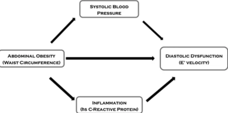
Path analysis is an extension of regression analysis which allows for the simultaneous estimation of the interrelations between vari-ables in a set[25]. This technique is being increasingly used to de-compose and compare the magnitudes of effects between variables with complex interrelations or to test the plausibility of mediation effects. We conducted path analysis assuming the possible causal model depicted inFig. 1. Models werefitted with Mplus software
(Muthén and Muthén, Los Angeles, CA); 95% confidence intervals
were calculated by bootstrapping; and goodness offit was evaluated
using the comparativefit index (CFI). Models were considered to
have goodfit if CFI was higher than 0.90. 3. Results
Thefinal analysis included 1063 participants with a mean age of 62.4 ± 10.6 years and 62.0% were women.Table 1shows the clinical,
anthropometric, analytical and echocardiographic characteristics of the study sample, by sex. There was a high prevalence of cardiovascular risk factors, especially hypertension and dyslipidemia.
The total prevalence of diastolic dysfunction was 23.1%: 14.2% had mild diastolic dysfunction and 8.9% had moderate or severe diastolic dysfunction, as shown in Table 2. No significant differences were
found in DD grades according to sex (p = 0.20). In 25 patients (2.4%) it was not possible to determine DD grade due to atrialfibrillation or fu-sion of the E/A mitralflow pattern. Patients with diastolic dysfunction were older, had increased systolic and diastolic blood pressure and higher levels of C-reactive protein. Left ventricle mass was higher in pa-tients with diastolic dysfunction, which also showed decreased E′ veloc-ities and increased E/E′ ratio.
3.1. Adiposity parameters and diastolic dysfunction grades
Overall, overweight and obese participants had higher prevalence of diastolic dysfunction: 27.0% in the overweight, 26.3% in the obese and 14.7% in individuals with BMIb 25 kg/m2. In addition, as shown in
Table 2, participants with diastolic dysfunction had a significant in-crease in BMI, fat mass percentage, waist circumference/height, hip-circumference/height and waist-to-hip ratio (pb 0.001). The association between diastolic function and adiposity parameters was different ac-cording to sex and age.
3.2. Adiposity and diastolic dysfunction in women
Overall, in women there was no significant relation between BMI and E′ velocity or E/E′ ratio, after adjustment for age and systolic blood pres-sure, either below or above 65 years of age. However, in women younger than 65 years we observed a significant association between total fat mass percentage and E′ velocity, even after adjustment for age and systolic blood pressure (Table 3).
In women the association between abdominal adiposity, assessed by waist-to-height ratio, and E′ velocity was different according to age (p for interaction = 0.01). In womenb 65 years, waist-to-height ratio was associated with decreased E′ velocity (r = −0.21; crude β: −8.0, 95% CI:−11.8 to −4.3; p b 0.01) but, after adjustment for SBP, we ob-served an attenuation of this association (adjustedβ: −2.3; 95% CI: −6.1 to 1.6; p = 0.25). In this subgroup, a similar association was ob-served between waist-to-height ratio and E/E′ ratio (crude β: 4.3, 95% CI: 1.7 to 6.8; p = 0.001), which was no longer significant after adjust-ment for SBP.
3.3. Adiposity and diastolic dysfunction parameters in men
In men, BMI was associated with decreased E′ velocity (p b 0.001), increased E/E′ ratio (p b 0.001) and a higher likelihood of diastolic dys-function (adjusted OR: 1.17; 95% CI: 1.09–1.27; p b 0.001), even after ad-justment for age and SBP. We also found an association between fat mass percentage with E′ velocity (p b 0.001) and E/E′ ratio (p b 0.001). The as-sociation between DD and BMI or fat mass % was significant in both age groups, but stronger in younger men (p for interaction of 0.03 and 0.04, respectively), as shown inTable 3.
In men younger than 65 years, E′ velocity was more strongly corre-lated with abdominal obesity, namely with waist-to-height ratio, than with BMI. After adjustment for age and SBP, waist-to-height ratio was associated with decreased E′ velocity and increased E/E′ ratio (adjusted β: 9.7, 95% CI: 5.4–10.0; p b 0.001). In men older than 65 years, we also observed an association between waist-to-height ratio and E′ velocity, but of lesser magnitude compared to younger patients (Table 3). 3.4. Pathophysiological path analysis for the association between waist-to-height ratio and diastolic function
Fig. 1shows schematically the tested model for the path analysis be-tween waist-to-height ratio and E′ velocity.Table 4presents the esti-mated total, direct and indirect effects of abdominal adiposity (waist-to-height ratio) on E′ velocity.
In men, the association between waist-to-height ratio and E′ velocity was significant in both age groups and mainly determined by a direct ef-fect. As shown inTable 4, in men younger than 65 years, 81.3% of the
Fig. 1. Hypothesized causal mechanisms linking abdominal adiposity and diastolic dysfunction.
Table 1
Characteristics of the study sample, by sex.
Women (n = 659) Men (n = 404) Clinical data
Age, years 62.0 ± 10.3 63.1 ± 11.1 Cardiovascular risk factors
Hypertension (%) 465 (70.6%) 293 (72.5%) Diabetes (%) 75 (11.4%) 51 (12.6%) Dyslipidemia (%) 379 (57.5%) 204 (50.5%) Obesity (%) 207 (31.4%) 55 (13.6%) Systolic blood pressure, mm Hg 134.9 ± 22.0 134.9 ± 18.0 Diastolic blood pressure, mm Hg 81.4 ± 11.0 82.0 ± 10.3 Adiposity parameters BMI, kg/m2 28.2 ± 5.1 26.5 ± 3.7 Waist circumference/height, cm/m 0.89 ± 0.08 0.57 ± 0.06 Hip circumference/height, cm/m 0.67 ± 0.07 0.59 ± 0.04 Waist-to-hip ratio 0.89 ± 0.07 0.97 ± 0.06 Fat mass, % 35.5 ± 6.9 24.3 ± 5.5 Analytical data Total cholesterol, mg/dL 220.1 ± 51.7 214.1 ± 56.7 HDL, mg/dL 61.5 ± 43.1 56.8 ± 48.6 LDL, mg/dL 134.0 ± 51.1 131.0 ± 57.1 Triglycerides, mg/dL 151.1 ± 43.8 167.9 ± 50.3 Glucose, mg/dL 102.0 ± 43.1 108.6 ± 53.0 Log hs-CRP, mg/dL −0.68 ± 0.46 −0.77 ± 0.50 Echocardiography Septum, mm 8.4 ± 1.4 9.1 ± 1.4 Posterior wall, mm 7.7 ± 1.2 8.4 ± 1.3 LV mass index, g/m2 86.7 ± 18.1 86.8 ± 24.2 Left atrium volume index, mL/m2
29.7 ± 10.9 29.7 ± 12.4 LV end-diastolic volume, mL/m2 63.6 ± 15.8 71.6 ± 18.7 LV end-systolic volume, mL/m2 25.6 ± 9.1 25.6 ± 9.1 Ejection fraction, % 61.1 ± 6.3 58.5 ± 7.7 E wave, cm/s 73.7 ± 15.8 67.9 ± 15.2 A wave, cm/s 81.2 ± 20.2 74.2 ± 18.6 E/A ratio 0.96 ± 0.32 0.96 ± 0.33 Deceleration time, ms 234.8 ± 52.7 242.9 ± 63.3 IVRT, ms 91.5 ± 16.1 92.6 ± 16.1 E′ velocity, cm/s 10.4 ± 3.2 10.5 ± 3.3 E/E′ ratio 7.7 ± 2.8 7.0 ± 2.6 Diastolic dysfunction (DD) grade
Normal diastolic function, n (%) 482 (73.1%) 310 (76.7%) Mild DD, n (%) 99 (15.0%) 52 (12.9%) Moderate/severe DD, n (%) 63 (9.6%) 32 (7.9%) Data are presented as mean and standard deviation for continuous variables and count and percentage for categorical variables.
BMI, body mass index; hs-CRP, high sensitivity C reactive protein; DD, diastolic dysfunction; IVRT, isovolumetric relaxation time; LV, left ventricle.
effect (−12.762, 95% CI: −19.520 to −6.005) was direct, 12.4% was mediated by inflammation (−1.954; −3.902 to −0.005) and 6.3% via SBP (−0.982; 95% CI: −3.301 to 1.336). In men older than 65 years, most of the effect was also direct (92.0%).
In women, we found an association between abdominal adiposity and E′ velocity only in the younger group. Contrary to men, most of the effect of waist-to-height ratio on E′ velocity was indirect, mainly through SBP (50.8%), with non-significant proportions of 15.1% via in-flammation and 34.2% by direct effect (Table 4).
4. Discussion
In this population-based study we found that diastolic function is in-dependently associated with several adiposity parameters, especially abdominal adiposity. This association is more important in men and in the younger population. Moreover, in men, abdominal adiposity was re-lated with worse diastolic function mainly by direct (adiposity mediat-ed) mechanisms, while in women the predominant effect was indirect, mainly through hypertension.
4.1. The association between diastolic dysfunction and adiposity distribution parameters
In this study we have shown that increased BMI is independently associated with worse diastolic function, which is in accordance with previous studies showing the same independent association[13,26]. We observed that this association is more important in men and in younger people. Considering that subclinical diastolic dysfunction is a predictor of future heart failure risk[27], and BMI is known as an inde-pendent risk factor for incident heart failure[9,28], it has been proposed that diastolic dysfunction can be one of the important pathophysiologi-cal links between obesity, heart failure and mortality[29]. Besides, some studies have shown that long-term weight loss, for example after bar-iatric surgery, can improve diastolic function[30,31], an observation that supports this relation. However, it is known that BMI is an imper-fect measure of fat mass, as it can be influenced by several other factors
[21]. In this study, we have shown the same independent association of diastolic dysfunction with fat mass percentage, evaluated by bioim-pedance analysis, which also strengthens the link between total adiposity and worse diastolic function.
Table 2
Clinical characteristics according to diastolic dysfunction grades. Normal diastolic function n = 792 (74.5%)
Mild diastolic dysfunction n = 151 (14.2%)
Moderate or severe diastolic dysfunction n = 95 (8.9%)
p value for trend
Age, years 59.9 ± 10.0 70.3 ± 8.7 68.0 ± 8.7 b0.001
Female, n (%) 482 (60.8%) 99 (65.5%) 63 (66.3%) 0.58
Hypertension, n (%) 380 (60.6%) 265 (85.2%) 69 (95.7%) b0.001
Systolic blood pressure, mm Hg 131.6 ± 19.4 147.0 ± 23.1 141.7 ± 18.3 b0.001 Diastolic blood pressure, mm Hg 81.1 ± 10.5 83.3 ± 11.4 63.3 ± 11.1 0.02
Log hsCRP, mg/dL 0.18 0.21 0.24 b0.001 Adiposity parameters BMI, kg/m2 27.1 ± 4.6 28.1 ± 4.2 28.3 ± 4.3 b0.001 Fat mass, % 30.6 ± 8.6 33.4 ± 7.3 33.9 ± 7.3 b0.001 Waist-to-height ratio, cm/cm 0.58 ± 0.07 0.61 ± 0.07 0.60 ± 0.06 b0.001 Hip-to-height ratio, cm/cm 0.63 ± 0.07 0.65 ± 0.06 0.64 ± 0.06 b0.001 Waist-to-hip ratio 0.91 ± 0.08 0.94 ± 0.07 0.94 ± 0.08 b0.001 Echocardiography LA volume index, mL/m2 28.7 ± 9.5 30.0 ± 11.7 30.4 ± 12.3 0.35 LV mass index, g/m2 76.7 ± 17.0 91.6 ± 21.8 91.7 ± 29.9 b0.001 E′ velocity, cm/s 11.7 ± 2.7 6.4 ± 1.0 6.6 ± 1.1 b0.001 E/E′ ratio 6.4 ± 1.7 9.6 ± 2.5 11.9 ± 3.4 b0.001 E/A ratio 1.0 ± 0.3 0.6 ± 0.1 1.0 ± 0.4 b0.001 Deceleration time, ms 230.0 ± 48.9 286.9 ± 75.6 234.0 ± 50.2 b0.001 IVRT, ms 90.8 ± 14.8 101.2 ± 15.9 90.9 ± 18.8 b0.001
Data are presented as mean and standard deviation for continuous variables and count and percentage for categorical variables.
BMI, body mass index; BP, blood pressure; hsCRP, high sensitivity C-reactive protein; IVRT, isovolumetric relaxation time; LA, left atrium; LV, left ventricle.
Table 3
Univariate and multivariate linear regression analyses for the association between E′ velocity and several adiposity parameters according to age and sex.
b65 years ≥65 years Crude Adjusteda Crude Adjusteda β (95% CI) r β (95% CI) p value β (95% CI) r β (95% CI) p value p value for interaction Women BMI, kg/m2 −0.10 (−0.15 to −0.04) −0.17 −0.03 (−0.08 to 0.03) 0.30 0.02 (−0.06 to 0.09) 0.03 0.03 (−0.04 to 0.1) 0.45 0.27 Fat mass, % −0.11 (−0.15 to −0.07) −0.24 −0.04 (−0.09 to −0.01) 0.04 −0.01 (−0.06 to 0.04) −0.01 −0.01 (−0.05 to 0.05) 0.94 0.24 Waist/height, cm/m −8.0 (−11.8 to −4.3) −0.21 −2.7 (−6.4 to 1.0) 0.15 −2.0 (−6.3 to 2.4) −0.06 0.4 (−4.0 to 4.9) 0.84 0.01 Men BMI, kg/m2 −0.30 (−0.41 to −0.18) −0.33 −0.23 (−0.34 to −0.11) b0.001 −0.11 (−0.22 to −0.01) −0.15 −0.13 (−0.24 to −0.03) 0.01 0.03 Fat mass, % −0.19 (−0.26 to −0.11) −0.32 −0.14 (−0.21 to −0.06) b0.001 −0.06 (−0.13 to 0.01) −0.12 −0.07 (−0.13 to −0.01) 0.04 0.04 Waist/height, cm/m −19.6 (−13.3 to −25.8) −0.38 −14.4 (−21.1 to −7.6) b0.001 −7.9 (−14.5 to −1.2) −0.17 −8.6 (−14.9 to −2.3) 0.01 0.06 β, regression coefficient; 95% CI, 95% confidence interval; BMI, body mass index; r, correlation coefficient.
a
Abdominal obesity parameters, especially waist circumference, are more closely related with excess visceral fat, which is the most metabol-ically active fat[21]. Several studies have shown that compared to BMI, abdominal obesity is a stronger determinant of myocardial infarction, cardiovascular disease and death[32,33]. In our population we observed that increased waist-to height ratio was a stronger determinant of dia-stolic dysfunction, especially in men. The association between waist-to height ratio and diastolic function was independent of BMI, which em-phasizes a specific role for visceral fat. This observation is consistent with recently published data, that have also shown that waist circum-ference[34], and increased abdominal visceral fat mass assessed by CT scan[35], are independent determinants of diastolic dysfunction, inde-pendently of BMI and subcutaneous fat, respectively. The confirmation of abdominal obesity as a more important determinant of subclinical left ventricle diastolic dysfunction might have important clinical impli-cations, particularly by the increasing prevalence of obesity worldwide. Interestingly, previous studies have observed an association between waist circumference and incident heart failure[36], which was indepen-dent of BMI[37]. Also, recent prospective studies have confirmed that subclinical diastolic dysfunction is associated with the development of heart failure, especially with heart failure with preserved ejection fraction (HFpEF)[27,38]. Therefore, the early identification and cor-rection of the main determinants of subclinical diastolic dys-function, such as abdominal obesity, can be particularly important in the prevention of HFpEF, a disease where no therapy has been shown to change the prognosis. This will need to be confirmed in fu-ture longitudinal studies.
4.2. The role of sex in the association between adiposity and diastolic dysfunction
The total amount of fat and its distribution is quite different in men and women. It is known that younger women have a more peripheral and subcutaneous distribution of fat but, later, menopause induces a more abdominal distribution of fat with increasing age. Therefore, we hypothesized that the effect on left ventricle diastolic function could be sex- and age-specific. In this study we observed a stronger relation between adiposity parameters and diastolic dysfunction in men than in women, and this difference is especially apparent in the younger pop-ulation. Most of the previous studies that have also evaluated the rela-tion between obesity and diastolic funcrela-tion did not assess the sex difference[29]or did notfind a significant difference in effect[13]. However, Canepa et al., found that the association between waist cir-cumference and diastolic function was more pronounced in women than in men[34]. Nevertheless, these results could be partially ex-plained by women being significantly younger than men, bearing in mind that in our study the association between adiposity and diastolic function is also age dependent. Also, body composition differs signi fi-cantly between racial and ethnical subgroups and our study was con-ducted in a European population, while the study from Canepa et al. included 29% of African origin participants.
4.3. Direct and indirect mechanisms linking adiposity and diastolic dysfunction
The pathophysiological pathways that link obesity with diastolic dysfunction are not entirely understood. This association can be mediat-ed by a combination of both direct and indirect effects that, according to our results, seem to be sex specific. The effect can be indirect, through established risk factors that can also induce diastolic dysfunction. Alter-natively, this effect can be caused by the secretion of adipokines that have been shown to modulate myocardial structure and function by inducing a systemic pro-inflammatory effect and/or by causing direct hemodynamic and mechanical changes in the heart and cardiovascular system[28].
In our study, we have shown that the association between the sever-al adiposity parameters and DD is partisever-ally attenuated when adjusting for age and mainly for blood pressure, which is consistent with previous studies[13]. As shown in the path analysis, this indirect effect through systolic blood pressure is especially important in younger women, being responsible for about 50% of the effect. Interestingly, another study including only patients with hypertension found that the adverse effect of abdominal obesity on left ventricle diastolic function was more pronounced among hypertensive females[39].
Adipose tissue is an important source of proinflammatory cytokines
[17]. Previous studies have linked inflammation with diastolic dysfunc-tion in patients with hypertension[40]and coronary artery disease[41]
and, in experimental studies, the inhibition of inflammatory pathways prevented diastolic dysfunction development[42]. Therefore, the role of adiposity on diastolic dysfunction can be at least partially mediated by inflammation. The path analysis that we have performed showed that in individuals younger than 65 years, the effect of waist-to-height ratio on diastolic dysfunction was mediated by hsCRP, which was responsible for 12.4% and 15.1% of this indirect effect in men and women, respectively. Interestingly, systemic inflammatory state has been shown to be predictive of incident HF, especially of HFpEF[43]
and, more importantly, previous studies have proposed that the as-sociation between obesity and HF may be related to pathophysio-logic pathways associated with inflammation[44]. Combining all these observations, Paulus and Tschope[45]have recently proposed a novel paradigm for HFpEF development which identifies a system-ic proinflammatory state, induced by obesity and other comorbidi-ties, as a cause of reduced nitric oxide availability and decreased protein kinase G activity inducing stiffer cardiomyocytes and inter-stitialfibrosis deposition.
Finally, obesity can directly influence myocardial structure and func-tion through other mechanisms, some not evaluated in this study. As ob-served in the path analysis performed, in men younger than 65 years, the direct effect can be responsible for up to 80% of the association between abdominal obesity and diastolic dysfunction. Several other pathophysio-logical mechanisms can be involved. Adipose tissue is an endocrine organ capable of secreting several adipokines (such as leptin, adiponectin, resistin and others) that have been shown to induce left
Table 4
Path analysis with estimated total, direct and indirect effects of waist-to-height ratio on E′ velocity.
Women Men
b65 years ≥65 years b65 years ≥65 years
Total −4.985 (−8.377 to −1.592) −0.253 (−4.536 to 4.030) −15.698 (−22.051 to −9.346) −9.208 (−15.584 to −2.833) Direct −1.703 (−5.818 to 2.412) 0.652 (−3.821 to 5.125) −12.762 (−19.520 to −6.005) −8.480 (−14.852 to −2.108) Indirect— role of SBP −2.531 (−4.189 to −0.873) −0.998 (−2.157 to 0.162) −0.982 (−3.301 to 1.336) −0.521 (−1.522 to 0.481) Indirect— role of hsCRP −0.751 (−2.723 to 1.221) 0.093 (−0.757 to 0.943) −1.954 (−3.902 to −0.005) −0.21 (−0.839 to 0.425)
CFI 0.990 1.000 0.946 1.000
Standardized regression coefficients and 95% confidence intervals (in parentheses) were calculated by bootstrapping using 1000 draws. CFI, comparativefit index; hsCRP, high sensitivity C reactive protein; SBP, systolic blood pressure.
ventricle remodeling and myocardial dysfunction[46]. Indeed, it has been suggested that the association between obesity and HF may be partially mediated by plasma leptin[28]. Also, obese patients have in-creased myocardial fatty infiltration that can lead to the worse diastolic dysfunction[47]. Furthermore, the excess in body fat determines an in-crease in both preload and afterload, due to a hyperdynamic circulation, chronic volume overload, and increase in peripheral resistance[48], which have also been shown to influence diastolic function[49]. 4.4. Strengths and limitations
To assess the pathophysiological mechanisms involved in the now well-established association between increased adiposity and diastolic dysfunction we used a path analysis, which is an extension of regression analysis, being increasingly used to decompose and compare the mag-nitudes of effects between variables with complex interrelations or to test the plausibility of mediation effects. Other strengths of this study in-clude the large sample of individuals free from other cardiac diseases, the contemporaneous assessment of cardiac diastolic function using the most recent consensus criteria for diastolic function evaluation and the use of several adiposity parameters including anthropometry and bioimpedance analysis to measure fat mass distribution.
The main limitation of this study is the cross-sectional design, which partially limits comments on causality, as this would be more robust in a prospective design. Although we have excluded patients with clinical signs of coronary artery disease, we did not perform any stress test to exclude myocardial ischemia, which is one determinant of diastolic dysfunction. We did not evaluate the intraobserver and interobserver variability in the evaluation of diastolic function. However, all the cardi-ologists had extensive experience in echocardiography, worked in the same institution and a detailed procedure protocol was discussed be-tween the team, prior to study beginning in order to harmonize the methodology and the measurements. Moreover, several previous stud-ies have shown a good reproducibility and a low inter-observer mean error for diastolic parameters[50].
5. Conclusion
Obesity, especially abdominal obesity, was associated with worse diastolic function. This association was stronger in men than in women and in the younger population. The causal mechanisms potentially in-volved were sex-specific, being mainly mediated by blood pressure in women, while in men there was mainly a direct effect.
Conflict of interest
None of the authors have conflicts of interest to declare. Acknowledgments
This work was supported by Portuguese Foundation for Science and Technology Grants POCI/SAU-ESP/61492/2004, PEST-C/SAU/UI0051/ 2011, and EXCL/BIM-MEC/0055/2012 (partially funded by FEDER through COMPETE) and European Commission Grant FP7-Health-2010; MEDIA-261409.
Alexandra Gonçalves receives funds from Portuguese Foundation for Science and Technology Grant HMSP-ICS/007/2012.
References
[1] J.N. Cohn, R. Ferrari, N. Sharpe, Cardiac remodeling—concepts and clinical implica-tions: a consensus paper from an international forum on cardiac remodeling. Behalf of an International Forum on Cardiac Remodeling, J. Am. Coll. Cardiol. 35 (2000) 569–582.
[2] C.W. Yancy, M. Jessup, B. Bozkurt, J. Butler, D.E. Casey Jr., M.H. Drazner, et al., ACCF/ AHA Guideline for the Management of Heart Failure: Executive Summary: a Report
of the American College of Cardiology Foundation/American Heart Association Task Force on Practice Guidelines, Circulation 2013 (2013).
[3] J.J. McMurray, S. Adamopoulos, S.D. Anker, A. Auricchio, M. Bohm, K. Dickstein, et al., ESC Guidelines for the diagnosis and treatment of acute and chronic heart failure 2012: the Task Force for the Diagnosis and Treatment of Acute and Chronic Heart Failure 2012 of the European Society of Cardiology. Developed in collaboration with the Heart Failure Association (HFA) of the ESC, Eur. Heart J. 33 (2012) 1787–1847.
[4] T.E. Owan, D.O. Hodge, R.M. Herges, S.J. Jacobsen, V.L. Roger, M.M. Redfield, Trends in prevalence and outcome of heart failure with preserved ejection fraction, N. Engl. J. Med. 355 (2006) 251–259.
[5] R.S. Bhatia, J.V. Tu, D.S. Lee, P.C. Austin, J. Fang, A. Haouzi, et al., Outcome of heart failure with preserved ejection fraction in a population-based study, N. Engl. J. Med. 355 (2006) 260–269.
[6] A.F. Leite-Moreira, Current perspectives in diastolic dysfunction and diastolic heart failure, Heart 92 (2006) 712–718.
[7] M.R. Zile, J.S. Gottdiener, S.J. Hetzel, J.J. McMurray, M. Komajda, R. McKelvie, et al., Prevalence and significance of alterations in cardiac structure and function in pa-tients with heart failure and a preserved ejection fraction, Circulation 124 (2011) 2491–2501.
[8] W.J. Paulus, C. Tschope, J.E. Sanderson, C. Rusconi, F.A. Flachskampf, F.E. Rademakers, et al., How to diagnose diastolic heart failure: a consensus statement on the diagno-sis of heart failure with normal left ventricular ejection fraction by the Heart Failure and Echocardiography Associations of the European Society of Cardiology, Eur. Heart J. 28 (2007) 2539–2550.
[9] S. Kenchaiah, J.C. Evans, D. Levy, P.W. Wilson, E.J. Benjamin, M.G. Larson, et al., Obesity and the risk of heart failure, N. Engl. J. Med. 347 (2002) 305–313.
[10] C.J. Lavie, M.A. Alpert, R. Arena, M.R. Mehra, R.V. Milani, H.O. Ventura, Impact of obe-sity and the obeobe-sity paradox on prevalence and prognosis in heart failure, JACC Heart Fail. 1 (2013) 93–102.
[11] M.A. Alpert, J. Omran, A. Mehra, S. Ardhanari, Impact of obesity and weight loss on cardiac performance and morphology in adults, Prog. Cardiovasc. Dis. 56 (2014) 391–400.
[12]M. Bastien, P. Poirier, I. Lemieux, J.P. Despres, Overview of epidemiology and contribution of obesity to cardiovascular disease, Prog. Cardiovasc. Dis. 56 (2014) 369–381.
[13] C. Russo, Z. Jin, S. Homma, T. Rundek, M.S. Elkind, R.L. Sacco, et al., Effect of obesity and overweight on left ventricular diastolic function: a community-based study in an elderly cohort, J. Am. Coll. Cardiol. 57 (2011) 1368–1374.
[14] T.B. Horwich, G.C. Fonarow, Glucose, obesity, metabolic syndrome, and diabetes rel-evance to incidence of heart failure, J. Am. Coll. Cardiol. 55 (2010) 283–293.
[15] S.S. Martin, A. Qasim, M.P. Reilly, Leptin resistance: a possible interface of inflamma-tion and metabolism in obesity-related cardiovascular disease, J. Am. Coll. Cardiol. 52 (2008) 1201–1210.
[16] S.B. Patel, G.P. Reams, R.M. Spear, R.H. Freeman, D. Villarreal, Leptin: linking obesity, the metabolic syndrome, and cardiovascular disease, Curr. Hypertens. Rep. 10 (2008) 131–137.
[17] A. Taube, R. Schlich, H. Sell, K. Eckardt, J. Eckel, Inflammation and metabolic dysfunc-tion: links to cardiovascular diseases, Am. J. Physiol. Heart Circ. Physiol. 302 (2012) H2148–H2165.
[18]J.P. Bastard, C. Jardel, J. Delattre, B. Hainque, E. Bruckert, F. Oberlin, Evidence for a link between adipose tissue interleukin-6 content and serum C-reactive protein concentrations in obese subjects, Circulation 99 (1999) 2221–2222.
[19] B.D. Powell, M.M. Redfield, K.A. Bybee, W.K. Freeman, C.S. Rihal, Association of obe-sity with left ventricular remodeling and diastolic dysfunction in patients without coronary artery disease, Am. J. Cardiol. 98 (2006) 116–120.
[20] C.Y. Wong, T. O'Moore-Sullivan, R. Leano, N. Byrne, E. Beller, T.H. Marwick, Alterations of left ventricular myocardial characteristics associated with obesity, Circulation 110 (2004) 3081–3087.
[21]M.A. Cornier, J.P. Despres, N. Davis, D.A. Grossniklaus, S. Klein, B. Lamarche, et al., Assessing adiposity: a scientific statement from the American Heart Association, Circulation 124 (2011) 1996–2019.
[22]E. Ramos, C. Lopes, H. Barros, Investigating the effect of nonparticipation using a population-based case-control study on myocardial infarction, Ann. Epidemiol. 14 (2004) 437–441.
[23]R.M. Lang, M. Bierig, R.B. Devereux, F.A. Flachskampf, E. Foster, P.A. Pellikka, et al., Recommendations for chamber quantification, Eur. J. Echocardiogr. 7 (2006) 79–108.
[24]S.F. Nagueh, C.P. Appleton, T.C. Gillebert, P.N. Marino, J.K. Oh, O.A. Smiseth, et al., Recommendations for the evaluation of left ventricular diastolic function by echo-cardiography, Eur. J. Echocardiogr. 10 (2009) 165–193.
[25] JC L, Latent Variable Models: An Introduction to Factor, Path, and Structural Equation Analysis, Psychology Press, Hove, United Kingdom, 2003.
[26]W. Aljaroudi, C. Halley, P. Houghtaling, S. Agarwal, V. Menon, L. Rodriguez, et al., Impact of body mass index on diastolic function in patients with normal left ventric-ular ejection fraction, Nutr. Diabetes 2 (2012) e39.
[27] G.C. Kane, B.L. Karon, D.W. Mahoney, M.M. Redfield, V.L. Roger, J.C. Burnett Jr., et al., Progression of left ventricular diastolic dysfunction and risk of heart failure, JAMA 306 (2011) 856–863.
[28] S.G. Wannamethee, A.G. Shaper, P.H. Whincup, L. Lennon, N. Sattar, Obesity and risk of incident heart failure in older men with and without pre-existing coro-nary heart disease: does leptin have a role? J. Am. Coll. Cardiol. 58 (2011) 1870–1877.
[29] K.A. Ammar, M.M. Redfield, D.W. Mahoney, M. Johnson, S.J. Jacobsen, R.J. Rodeheffer, Central obesity: association with left ventricular dysfunction and mortality in the community, Am. Heart J. 156 (2008) 975–981.
[30] H.J. Willens, S.C. Chakko, P. Byers, J.A. Chirinos, E. Labrador, J.C. Castrillon, et al., Effects of weight loss after gastric bypass on right and left ventricular function assessed by tissue Doppler imaging, Am. J. Cardiol. 95 (2005) 1521–1524.
[31] O.J. Rider, J.M. Francis, M.K. Ali, S.E. Petersen, M. Robinson, M.D. Robson, et al., Beneficial cardiovascular effects of bariatric surgical and dietary weight loss in obe-sity, J. Am. Coll. Cardiol. 54 (2009) 718–726.
[32]R. Huxley, S. Mendis, E. Zheleznyakov, S. Reddy, J. Chan, Body mass index, waist circumference and waist:hip ratio as predictors of cardiovascular risk—a review of the literature, Eur. J. Clin. Nutr. 64 (2010) 16–22.
[33]T. Pischon, H. Boeing, K. Hoffmann, M. Bergmann, M.B. Schulze, K. Overvad, et al., General and abdominal adiposity and risk of death in Europe, N. Engl. J. Med. 359 (2008) 2105–2120.
[34] M. Canepa, J.B. Strait, D. Abramov, Y. Milaneschi, M. AlGhatrif, M. Moni, et al., Contri-bution of central adiposity to left ventricular diastolic function (from the Baltimore Longitudinal Study of Aging), Am. J. Cardiol. 109 (2012) 1171–1178.
[35] M. Canepa, J.B. Strait, Y. Milaneschi, M. Alghatrif, R. Ramachandran, S. Makrogiannis, et al., The relationship between visceral adiposity and left ventricular diastolic function: results from the Baltimore Longitudinal Study of Aging, Nutr. Metab. Cardiovasc. Dis. 23 (2013) 1263–1270.
[36]G. Hu, P. Jousilahti, R. Antikainen, P.T. Katzmarzyk, J. Tuomilehto, Joint effects of physical activity, body mass index, waist circumference, and waist-to-hip ratio on the risk of heart failure, Circulation 121 (2010) 237–244.
[37]B.J. Nicklas, M. Cesari, B.W. Penninx, S.B. Kritchevsky, J. Ding, A. Newman, et al., Abdominal obesity is an independent risk factor for chronic heart failure in older people, J. Am. Geriatr. Soc. 54 (2006) 413–420.
[38]C.S. Lam, A. Lyass, E. Kraigher-Krainer, J.M. Massaro, D.S. Lee, J.E. Ho, et al., Cardiac dysfunction and noncardiac dysfunction as precursors of heart failure with reduced and preserved ejection fraction in the community, Circulation 124 (2011) 24–30.
[39]C.P. Tsioufis, D.L. Tsiachris, M.N. Selima, K.S. Dimitriadis, C.G. Thomopoulos, D.C. Tsiliggiris, et al., Impact of waist circumference on cardiac phenotype in hypertensives according to gender, Obesity 17 (2009) 177–182.
[40]S. Sciarretta, A. Ferrucci, G.M. Ciavarella, P. De Paolis, V. Venturelli, G. Tocci, et al., Markers of inflammation and fibrosis are related to cardiovascular damage in hy-pertensive patients with metabolic syndrome, Am. J. Hypertens. 20 (2007) 784–791.
[41] E.S. Williams, S.J. Shah, S. Ali, B.Y. Na, N.B. Schiller, M.A. Whooley, C-reactive protein, diastolic dysfunction, and risk of heart failure in patients with coronary disease: Heart and Soul Study, Eur. J. Heart Fail. 10 (2008) 63–69.
[42] D. Westermann, S. Van Linthout, S. Dhayat, N. Dhayat, A. Schmidt, M. Noutsias, et al., Tumor necrosis factor-alpha antagonism protects from myocardial inflammation andfibrosis in experimental diabetic cardiomyopathy, Basic Res. Cardiol. 102 (2007) 500–507.
[43] A. Kalogeropoulos, V. Georgiopoulou, B.M. Psaty, N. Rodondi, A.L. Smith, D.G. Harrison, et al., Inflammatory markers and incident heart failure risk in older adults: the Health ABC (Health, Aging, and Body Composition) study, J. Am. Coll. Cardiol. 55 (2010) 2129–2137.
[44]H. Bahrami, D.A. Bluemke, R. Kronmal, A.G. Bertoni, D.M. Lloyd-Jones, E. Shahar, et al., Novel metabolic risk factors for incident heart failure and their relationship with obesity: the MESA (Multi-Ethnic Study of Atherosclerosis) study, J. Am. Coll. Cardiol. 51 (2008) 1775–1783.
[45] W.J. Paulus, C. Tschope, A novel paradigm for heart failure with preserved ejection fraction: comorbidities drive myocardial dysfunction and remodeling through coro-nary microvascular endothelial inflammation, J. Am. Coll. Cardiol. 62 (2013) 263–271.
[46] I. Falcao-Pires, P. Castro-Chaves, D. Miranda-Silva, A.P. Lourenco, A.F. Leite-Moreira, Physiological, pathological and potential therapeutic roles of adipokines, Drug Discov. Today 17 (2012) 880–889.
[47]R.W. van der Meer, L.J. Rijzewijk, M. Diamant, S. Hammer, M. Schar, J.J. Bax, et al., The ageing male heart: myocardial triglyceride content as independent predictor of diastolic function, Eur. Heart J. 29 (2008) 1516–1522.
[48] V. Palmieri, G. de Simone, D.K. Arnett, J.N. Bella, D.W. Kitzman, A. Oberman, et al., Re-lation of various degrees of body mass index in patients with systemic hypertension to left ventricular mass, cardiac output, and peripheral resistance (The Hypertension Genetic Epidemiology Network Study), Am. J. Cardiol. 88 (2001) 1163–1168.
[49]A.F. Leite-Moreira, A.P. Lourenco, R. Roncon-Albuquerque Jr., T. Henriques-Coelho, M.J. Amorim, J. Almeida, et al., Diastolic tolerance to systolic pressures closely reflects systolic performance in patients with coronary heart disease, Basic Res. Cardiol. 107 (2012) 251.
[50]H. Dalen, A. Thorstensen, L.J. Vatten, S.A. Aase, A. Stoylen, Reference values and distribution of conventional echocardiographic Doppler measures and longitudinal tissue Doppler velocities in a population free from cardiovascular disease, Circ. Cardiovasc. Imaging 3 (2010) 614–622.