Ana Gabriela da Silva
Cavaleiro Henriques
Biologia do Aβ
β
β
β e a sua contribuição para a doença
de Alzheimer
Aβ
β
β
β biology and its contribution to Alzheimer´s
disease
Ana Gabriela da Silva
Cavaleiro Henriques
Biologia do Aβ
β
β
β e a sua contribuição para a doença
de Alzheimer
Aβ
β
β
β biology and its contribution to Alzheimer´s
disease
Tese apresentada à Universidade de Aveiro para cumprimento dos requisitos necessários à obtenção do grau de Doutora em Bioquímica, realizada sob a orientação científica da Doutora Odete Abreu Beirão da Cruz e Silva, Professora Auxiliar da Secção Autónoma de Ciências da Saúde da Universidade de Aveiro.
o júri
presidente Prof. Doutor Aníbal Manuel de Oliveira Duarte
Professor Catedrático do Departamento de Electrónica, Telecomunicações e Informática da Universidade de Aveiro
Prof. Doutora Cecília M. P. Rodrigues
Professora Associada com Agregação da Faculdade de Farmácia da Universidade de Lisboa
Prof. Doutor Tiago Fleming Outeiro
Professor Auxiliar da Faculdade de Medicina da Universidade de Lisboa
Prof. Doutor Amadeu Soares
Professor Catedrático com Agregação do Departamento de Biologia da Universidade de Aveiro
Prof. Doutor Edgar F. da Cruz e Silva
Professor Associado do Departamento de Biologia da Universidade de Aveiro
Prof. Doutor António Calado
Professor Auxiliar do Departamento de Biologia da Universidade de Aveiro
Prof. Doutora Odete A. B. da Cruz e Silva
agradecimentos Muito Obrigada,
Professora Odete, pela orientação e discussões científicas que permitiram o desenvolvimento e a defesa desta tese.
Professor Edgar, pelas suas observações e sugestões pertinentes que são sempre uma mais-valia.
A ambos, pela amizade e pelas oportunidades. Por terem acreditado em mim, e me terem apoiado ao longo destes anos, permitindo o meu enriquecimento científico e profissional.
Obrigada,
A todos os meus colegas e amigos dos laboratórios de Neurociências e Transdução de sinais (e laboratório vizinho!), que sempre se disponibilizaram para ajudar. Pelas discussões filosóficas e piadas que alegravam os dias de trabalho!
Um especial agradecimento à Sandra e à Elena. Queridas amigas, muito obrigada, pela contribuição para o trabalho, mas principalmente pela amizade e carinho!
Para ti também Ana Paula! Boa sorte! Obrigada,
Ao Centro de Biologia Celular da Universidade de Aveiro.
À FCT (BD/16071/2004), projectos nacionais (POCTI/NSE/40682, POCTI/NSE/33520, POCI/BIA-BCM/58469 e REEQ/1023/BIO/2005) e projectos internacionais (DIADEM, APOPIS e cNeupro), pelo financiamento para o desenvolvimento do trabalho experimental e para a participação em congressos internacionais.
Agradecimento especial,
À minha família que sempre me apoiou e incentivou em todos os momentos. Gosto muito de vocês!
Um beijinho especial para o André, por todo o carinho e amor, e para a minha querida filha Inês. O meu Mundinho!
Beijinhos Pai, Mãe e Mano.
Beijinho Avô e Avó (lembro-me sempre de ti!). Obrigada por toda a ajuda ao longo destes anos.
Beijinho Telma, que és para mim família, pela amizade, apoio e companhia desde sempre!
palavras-chave Doença Alzheimer, Aβ, metabolismo APP, AICD, Fe65, sinalização nuclear, secreção vesicular, citosqueleto, laminina, gelsolina, proteínas fosfatases.
resumo A doença de Alzheimer (DA) é uma desordem neurodegenerativa progressiva
patologicamente caracterizada pela presença de placas de amilóide (placas senis) insolúveis e também pela presença de tranças neurofibrilhares,formadas pela proteína Tau hiperfosforiladada. O principal constituinte das placas senis é o peptídeo beta-amilóide (Aβ), que deriva do processamento proteolítico da proteína precursora de amilóide de Alzheimer (APP). Embora Aβ exista como um agregado pouco solúvel nas placas senis, ele é secretado pelas células como uma molécula solúvel. O Aβ “per se” pode afectar o metabolismo da APP. Alguns autores sugerem que o Aβ exerce o seu efeito alterando o processamento ou catabolismo da APP, outros sugerem que ele também induz a transcrição da APP, onde aumentando os níveis da APP pode estar a contribuir para a sua própria produção (mecanismo de “feedback” positivo). Assim sendo, torna-se difícil consolidar todas estas observações e identificar as potenciais funções fisiológicas do Aβ “in vivo”, ou as consequências da sua produção. Neste trabalho caracterizaram-se os efeitos do Aβ no metabolismo da APP. Os nossos estudos revelaram que um dos mecanismos induzidos pelo Aβ é a acumulação intracelular do fragmento neuroprotector sAPP (isAPPα) em estruturas com características vesiculares associadas ao citosqueleto. Estudos adicionais em culturas primárias revelaram que o Aβ estava a exercer o seu efeito ao nível da secreção vesicular, provavelmente interferindo com o transporte de APP/sAPP ao longo da rede do citosqueleto. Esta hipótese é sustentada pelo facto do Aβ estar a afectar a estabilidade e a polimerização de proteínas envolvidas na dinâmica do citosqueleto.
Contrariamente a publicações anteriores o Aβ não induziu a transcrição da APP, na verdade em culturas primárias neuronais foi observado uma diminuição nos níveis de expressão da APP. Isto foi acompanhado por um aumento nos fragmentos C-terminais da APP (CTFs) e uma diminuição na localização nuclear do seu domínio intracelular (AICD), sugerindo alterações na sinalização nuclear da APP. O Aβ pode afectar outras vias de sinalização, particularmente alterando o balanço entre as actividades das proteínas cinases e fosfatases, o que pode ter consequências para o desenvolvimento da doença. Os dados obtidos indicam que o Aβ é capaz de inibir a actividade da proteína fosfatase1, a sua importância numa perspectiva de futuras terapias é discutida.
Devido à relevância da agregação do Aβ para a sua toxicidade, a formação de complexos com proteínas que promovem a sua desagregação/degradação e o seu efeito no processamento da APP foi avaliado. Na presença destes
complexos observou-se uma reversão da acumulação isAPP, demonstrando o potencial terapêutico destas proteínas como moduladores do metabolismo da APP. Este trabalho permitiu compreender melhor os mecanismos envolvidos nos efeitos do Aβ no processamento da APP e descobrir algumas moléculas que podem ser relevantes numa perspectiva de diagnóstico e terapia na DA.
keywords Alzheimer´s Disease, Aβ, APP metabolism, AICD, Fe65, nuclear signalling, vesicular secretion, cytoskeleton, laminin, gelsolin, protein phosphatases.
abstract Alzheimer´s disease (AD) is a progressive neurodegenerative disorder
pathologically characterized by the presence of extracellular deposition of insoluble amyloid plaques (senile plaques) and also by the appearance of neurofibrillary tangle-bearing neurons, mainly composed of
hyperphosphorylated Tau protein. The major component of senile plaques is the amyloid-beta (Aβ) peptide, derived from proteolytic processing of the Alzheimer´s amyloid precursor protein (APP). Although Aβ exists as an aggregated, poorly soluble form in brain deposits, it is secreted from cells during normal metabolism as a soluble molecule. Aβ “per se” has been
reported to affect APP metabolism, and while some authors suggest that it may exert its effects by altering APP processing/catabolism, others reported that it also induces APP transcription. This latter observation suggested a positive feedback mechanism resulting in increased APP levels, thus Aβ would stimulate its own production. Difficulties arise when trying to consolidate all these observations and to identify the potential “in vivo” physiological role of Aβ, or the consequences of its overproduction in AD. Hence, in this work we characterized the Aβ effects on APP metabolism. Our studies reveal that mechanistically Aβ leads to intracellular retention of the neuroprotective fragment (isAPPα) at vesicular-like densities associated with the cytoskeleton. Additional studies in primary neuronal cultures revealed that Aβ was exerting an inhibitory effect at the vesicular secretory level, probably by interfering with APP/sAPP transport along the cytoskeleton network. This hypothesis was supported by the fact that Aβ was affecting the stability and polymerization of proteins involved in cytoskeleton dynamics. Contrary to previous observations Aβ did not induce APP transcription. In fact for primary neuronal cultures we observed a decrease on APP expression levels. This was accompanied by an increase in APP C-terminal fragments (CTFs) and decreased APP intracellular domain (AICD) nuclear targeting, suggesting altered APP nuclear signaling. Aβ may affect other signaling cascades, for instance by altering the balance between kinase and phosphatase activities which may have consequences for the disease progression. Data obtained reveals that Aβ was able to inhibit protein phosphatase1 activity, and the importance of this for therapeutic approaches is discussed.
Further, it is also known that Aβ aggregation state is associated with its neurotoxicity. Hence, Aβ complex formation with proteins that promote its disaggregation/clearance and its effect on APP processing was evaluated. These complexes reverse isAPP retention, thus demonstrating the therapeutic potential of these proteins as modulators of APP metabolism. The work in this thesis allowed for a better understanding of the molecular mechanisms underlying Aβ effects on APP metabolism and identified molecules relevant for diagnosis and therapy in AD.
Chapter I – INTRODUCTION 15
OPENNING REMARKS 17
1.1 NEUROPATHOLOGICAL FEATURES OF AD 18
1.2 ADCLINICAL DIAGNOSIS 23
1.3 ADSPORADIC AND GENETIC RISK FACTORS 24
1.3.1 Genetics of AD 25
Genes implicated in familial disease 25
Genes implicated in sporadic disease 27
Genetic screening of AD 28
1.3.2 Non-genetic factors contributing to sporadic AD
29
1.4 ACENTRAL ROLE FOR Aβ IN ADNEURODEGENERATION 30
1.5ALZHEIMER´S AMYLOID PRECURSOR PROTEIN (APP) 33
1.5.1 APP gene family and alternatively spliced isoforms 33
1.5.2 APP functional domains 35
1.5.3 Putative functions for APP and APP cleaved fragments 37
APP as a receptor molecule 37
APP involvement in cell adhesion 38
APP and cell motility regulation 39
APP and sAPPα role in neurite outgrowth and synaptogenesis 39
APP, sAPP and Aβ functions in memory 41
APP and sAPP involvement in brain repair and neuroprotection 42
Aβ and CTF contribution to neurotoxicity and apoptosis 43
Signal transduction and AICD transcriptional regulation 44
Aβ physiological concentration and physiological function 47
1.5.4 APP proteolytic processing and Aβ genesis 49
APP Secretases 52
1.5.5 APP trafficking 57
APP subcellular trafficking and maturation 57
APP processing during its intracellular trafficking 60
Intracellular sites for non-amyloidogenic pathway 61
Intracellular sites for Aβ generating pathway 62
1.6INVOLVEMENT OF THE CYTOSKELETON NETWORK IN ALZHEIMER´S DISEASE 65
1.6.1 Microtubule network in APP axonal transport 66
Microtubule acetylation and neuronal transport 69
1.6.2 The role of actin remodelling in AD 70
Actin and the dendritic cytoskeleton 70
The actin cytoskeleton in exocytosis of secretory vesicles 72
1.7CURRENT AND FUTURE THERAPEUTIC APPROACHES IN ALZHEIMER´S DISEASE 74
1.7.1 Current strategies to ameliorate AD symptoms 74
1.7.2 Therapeutic strategies targeting Aβ peptide 76
Modulation of Aβ production by affecting secretase activity 77
Inhibition of Aβ aggregation 78
Promoting Aβ clearance via Aβ−binding substances 80
Promoting Aβ clearance via up-regulation of Aβ degrading enzymes 80
Promoting Aβ clearance via Aβ immunotherapy 81
1.8AIM OF THE THESIS 83
1.9REFERENCES 84
Chapter II – Aβ β β β AFFECTS sAPP SECRETION BY INTERFERING WITH
CYTOSKELETAL ORGANIZATION 117
Chapter Outline 119
Manuscript 1 – Intracellular sAPP retention in response to Aβ is
mapped to cytoskeletal associated structures 121
Manuscript 2 – Aβ affects cytoskeleton dynamics with consequences
for neuronal sAPP vesicular traffick 151
Chapter III – Aβ β β β ALTERS SIGNAL TRANSDUCTION MECHANISMS 175
Chapter Outline 177
Manuscript 3 – Aβ hinders APP and Fe65 nuclear targeting in primary
Chapter IV – THE BIOCHEMICAL BASIS OF Aββββ BASED THERAPEUTICS 199
Chapter Outline 201
Manuscript 4 – Aβ-disaggregating proteins as modulators of APP processing 203
Manuscript 5 – PP1 inibition by Aβ peptide as a potential pathological
mechanism in Alzheimer´s disease 223
Chapter V – DISCUSSION 241
5.1 Aβ-mediated Effects on APP processing and trafficking 243
Inhibition of sAPP secretion due to altered cytoskeletal dynamics 243
5.2 Aβ-mediated Effects on APP expression 247
Regulation of AICD and Fe65 nuclear targeting 247
5.3 Therapeutic approaches targeting Aβ-mediated effects 251
Prevention of isAPP retention by inhibiting Aβ- fibril formation 251
PP1 as a potential therapeutic target for Aβ-induced responses 253
CONCLUDING REMARKS 255
5.4REFERENCES 256
Appendix 263
Appendix I – Methods and Kits 265
Abbreviations aa Aβ AD ADAM ADDLS ADF AChE AICD/AID ANOVA APH-1 APLP1/2 APL-1 APP APP-BP1 APPL ApoE ATP BACE BBB BCA BChE BSA CAM cDNA CHO CSF CHX CNS (α/β) CTF CytD Dab1 DAPI DIC DMEM DNA DTT ECL Amino acid Abeta Alzheimer’s disease
A desintegrin and metalloproteinase Abeta-derived diffusible ligand Actin-depolymerizing factor Acetylcholinesterase APP intracellular domain One way analysis of variance Anterior pharynx defective 1 APP-Like Protein 1/2 C. elegans APP homolog
Alzheimer’s Amyloid Precursor Protein APP-binding protein 1
Drosophila APP homolog Apolipoprotein E
Adenosine triphosphate β-site APP cleaving enzyme Brain blood barrier Bicinchoninic acid Bytyrylcholinesterase Bovine serum albumin Cell adhesion molecule Complementary DNA
Chinese Hamster Ovary cell line Cerebral spinal fluid
Cicloheximide Central nervous system
APP Carboxy-terminal Fragment of α/β-secretase processing Cytochalasin D
Disabled 1
4',6-diamidino-2-phenylindole
Differential interference contrast microscopy Dulbecco’s Modified Eagle’s Medium
Deoxyribonucleic acid Dithiothreitol
EDTA EGTA ER FAD FBS FDG g G0 GDP GLUT3 GPI GSK3β GTP h/hr H4 HEPES HFIP IDE IgG IgA JIP JNK KAI1 KHC KLC KO KPI LRP LTD LTP MBP MDCK MENA min MINT mRNA MRI ND NFT NMDA Ethylenediaminetetraacetic acid
Ethylene glycol-bis(2-aminoethylether)-N,N,N´,N´-tetraacetic acid Endoplasmic reticulum
Familial Alzheimer’s Disease Fetal bovine serum
18F-2-Fluoro-2deoxy-D-Glucose Gravitational acceleration
Heterotrimeric (α, β, and γ subunits) G0 protein Guanosine
Glucose transporter3 Glycosylphosphatidylinositol Glycogen synthase kinase 3β Guanosine triphosphate Hour
Histone 4
4-(2-HydroxyEthyl)-1-PiperazineEthane sulfonic acid 1,1,1,3,3,3-hexafluoro-2-propanol Insulin-degrading enzyme Immunoglobulin G Immunoglobulin A JNK interacting protein Jun Kinase
“Kang ai” (Chinese for anticancer) protein 1 Kinesin heavy chain
Kinesin light chain Knockout
Kunitz-type serine proteinase inhibitor
Low density lipoprotein receptor-related protein Long-term depression
Long-term potentiation Myielin basic protein
Madine-Darby Canine Kidney cell line Mammalian enabled
Minute
Munc-18-interacting protein Messenger ribonucleic acid Magnetic resonance imaging Non-demented
Neurofibrillary tangle N-methyl-D-aspartate
NT2 PAGE PBS PC12 PCR PEN-2 PET PF PHF PIB PKC PM PS1/2 PP1/2 PTB PTM RAGE RIP ROS RP rpm RRP RT-PCR RT SNARE (α/β)sAPP SAM SDS SEM TACE TBS TBS-T TEMED TGN Tip60 TM Tris-HCl
Human neuron teratocarcinoma cell line PolyAcrylamide gel electrophoresis
Phosphate buffer saline (modified Dulbecco’s) Rat adrenal pheochromocytoma cell line Polymerase chain reaction
Presenilin enhancer
Positron emission tomography Protofibrils
Paired helical filament Pittsburg compound-B Protein kinase C Plasma membrane Presenilin 1 and 2
Protein phosphatase type 1/2 Phospho tyrosine binding Post-translational modification
Advanced glycation end product receptor Regulated intramembranar proteolysis Reactive oxygen species
Reserve pool Rotations per minute Readily releasable pool
Reverse transcriptase-polymerase chain reaction Room temperature
Soluble N-ethylmaleimide-sensitive fusion attachment protein receptor Secreted APP of α/β-secretase processing origin
Substrate adhesion molecule Sodium dodecyl sulfate Standard error of the mean
Tumor necrosis factor-α converting enzyme Tris buffered saline
TBS supplemented with Tween detergent N,N,N’,N’-tetramethylethylenediamine Trans-golgi network
Tat interactive protein, 60 kDa Transmembrane
Centro Biologia Celular 15 Universidade de Aveiro
Chapter I
16 Centro Biologia Celular Universidade de Aveiro
Centro Biologia Celular 17 Universidade de Aveiro
OPENING REMARKS
First described by the German pathologist Alois Alzheimer in 1906 (Alzheimer 1906), Alzheimer´s disease (AD) is a progressive neurodegenerative disorder that occurs predominantly in later life. The prevalence of the disease is below 1% in individuals aged 60-64 years old but it rises abruptly with age, so that in people aged 75 years or older, prevalence is about 19% and up to 30% above 85 (Blennow et al. 2006; Lambert and Amouyel 2007). As life span is increasing there is the possibility that this elderly population may develop AD, which will constitute a major public-health problem. Therefore, it is of extreme importance to understand the basic nature of the disease, so that effective preventive procedures can be developed. The latest worldwide estimate of AD prevalence shows that 26.6 million people were living with the disease in 2006. Researchers predict that global prevalence will quadruple by 2050 to more than 100
million, at which time 1 in 85 people worldwide will be living with the disease.
AD is a progressive neurodegenerative dementia that invariably leads to a complete loss of all cognitive abilities and ultimately to death. Initially AD patients exhibit subtle memory failure which becomes more severe. The short-term memory begins to decline when the cells in the hippocampus degenerate. As the cerebral cortex (the outer layer of the brain) becomes affected, judgment declines, emotional outbursts may occur, and language is impaired. Progression of the disease leads to death of nerve cells and subsequent behavioural changes, such as wandering and agitation. Thus, in the later stages perception and orientation are affected as well as loss of personality and intellect to a level that influences daily activities. The ability to recognize faces and to communicate is completely lost in the final stages of the disease. Patients will eventually need constant care, complete dependency may last for years before the patient dies. The average length of time from diagnosis to death is 4 to 8 years, although neurodegeneration is estimated to start 20-30 years before clinical symptoms become apparent (Bird 2007a). The overall lifetime risk of developing dementia is 10-12% (Goedert and Spillantini 2006).
18 Centro Biologia Celular Universidade de Aveiro
1.1 NEUROPATHOLOGICAL FEATURES OF AD
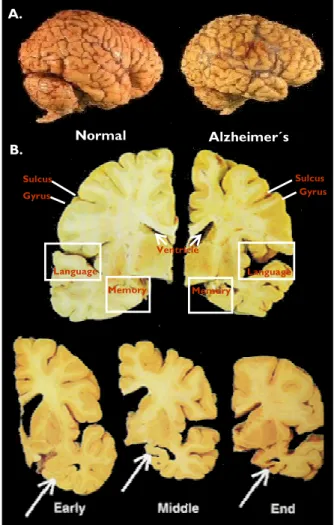
AD is characterized by a variety of pathological features, including extracellular senile plaques and intracellular neurofibrillary tangles, synaptic loss, and brain atrophy (Selkoe 2000; Hardy and Selkoe 2002; Forman et al. 2004). The characteristic brain atrophy and mass loss in AD patients is due to extensive neuronal damage and death. There is an overall shrinkage of brain tissue, with the most affected areas being the hippocampus, the cerebral cortex and amygdala, regions of the brain that play a major role in memory, cognition and behaviour. Widening of the sulcus and shrinkage of the gyrus, the well-developed fold of the brain's outer layer, is evident. The ventricles, which are cavities or spaces in the brain that contain cerebrospinal fluid, are enlarged (Figure 1).
Figure 1. Normal versus Alzheimer´s brain. A. Brain volume reduction and atrophy characteristic of AD patients (right). From www.cienciahoje.pt/index.php?oid=17362&op=all. B. Brain areas affected in AD patients. Images represent brain cross sections of a normal individual (left) and of an AD patient (right). The bottom three pictures represent brain shrinkage as AD continues to evolute. The arrows demonstrate shrinkage in the region of the brain associated with short term memories. Adapted from www.alzheimer.sk.ca/english/Just4Kids/alz_dis...
Early Middle End Gyrus Sulcus Gyrus Sulcus Memory Language Language Memory Ventricle A. B. Normal Alzheimer´s
Centro Biologia Celular 19 Universidade de Aveiro
The senile or neuritic plaques are mainly composed of Aβ (Allsop et al. 1983; Glenner and Wong 1984), while the neurofibrillary tangles (NFT) are intraneuronal bundles of paired helical filaments (PHF) consisting predominantly of hyperphosphorylated Tau protein (Goedert et al. 1992; Goedert et al. 1996) (Figure 2). While Aβ deposition is specific for AD, the NFTs are also seen in other degenerative disorders, but the coexistence of both lesions, along with cerebral atrophy and neuronal degeneration, are
the conclusive hallmarks of the AD.
Senile plaques NFT
Figure 2. Signature lesions of AD. Plaques are extracellular deposits of Aβ surrounded by dystrophic neuritis, reactive astrocytes, and microglia, whereas neurofibrillary tangles (NFT) are intracellular aggregates composed of a hyperphosphorylated form of the microtubule-associated protein Tau. Bielschowsky silver stains (From www.neuropathologyweb.org/.../chapter9bAD.html).
NFTs - Tangles occur at dystrophic neuritis (small dendrites and axons with degenerative changes), and are mainly found in the pyramidal regions of the amygdala, the hippocampus and the neocortex (Haroutunian et al. 1999; Oddo et al. 2003). NFTs consist of pyramidal cells filled with paired helical and straight filaments of aggregated hyperphosphorylated Tau. Tau is usually quite soluble and a key normal function of this protein is to bind to axonal microtubules to stabilize the axonal cytoskeleton framework. Tau is known to aid in cell microtubule assembly and stabilization, by promoting tubulin polymerization and reducing dynamic instability of the microtubule (Smith et al. 1996). This binding of Tau protein increases the rate of association at the end of the microtubule and decreases the rate of dissociation at the growing end (Goedert et al. 1997). It was also discovered that this protein can acts as a regulator of intracellular vesicles and organelle traffic, by
20 Centro Biologia Celular Universidade de Aveiro
interacting with cytoskeletal proteins, such as actin which also aid in cytoskeletal maintenance and trafficking (Drewes et al. 1998; Drouet et al. 2000). The extent to which Tau promotes its activity depends on its phosphorylation state (Lindwall and Cole 1984), with abnormal hyperphosphorylation interfering with its normal biological function (Gustke et al. 1992; Alonso et al. 1994). Problems arise when Tau becomes hyperphosphorylated at serine and threonine residues of the protein by a still unclear mechanism (Tanaka et al. 2000). Hyperphosphorylated Tau protein loses its ability to bind tubulin and stabilize microtubule assembly (Drouet et al. 2000), leading to microtubule breakdown into PHFs and NFTs. Hence, abnormal Tau phosphorylation may contribute to the formation of NFTs resulting in neuronal degeneration (Higuchi et al. 2002b; Sorrentino and Bonavita 2007).
Senile plaques - As already mentioned, another feature of AD brains are the neuritic plaques found distributed throughout the brain, but notably in the cerebral cortex and hippocampus of AD patients (Dickson 1997; Haroutunian et al. 1998). These plaques exhibit a central core of extracellular amyloid, surrounded of dystrophic neuritis, containing Tau aggregates (mostly in the straight filament form), and also reactive astrocytes and microglia, among other protein/peptides constituents. This central core is
composed of aggregates of Aβ peptide of 40-43 amino acids (called Aβ1-40, Aβ1-42 and
Aβ1-43). The Aβ1-40 peptides are most soluble and apparently less neurotoxic (majority of
Aβ peptides), whereas the Aβ1-42 peptides are more hydrophobic (less soluble), and
exhibit a higher potential for aggregation and neurotoxicity than does Aβ1-40. Although
Aβ1-42 peptides are less prevalent, overall they predominate in the central core of the
plaques (Jarrett and Lansbury 1992; Jarrett et al. 1993). Aβ deposition occurs as oligomeric, protofibrilar, amylospheroid and fibrillar forms (Kuo et al. 1996; Lambert et al. 1998; Hartley et al. 1999; Walsh et al. 1999; Hoshi et al. 2003). The term “soluble Aβ” is generally applied either to newly generated, cell secreted Aβ, or to the fraction of tissue Aβ that is taken into the aqueous phase of a non-detergent-containing extraction buffer. “Misfolded” and “aggregated” Aβ are the terms used to describe very early, non-specific changes in Aβ folding states or solubility states, respectively. ”Oligomeric” Aβ refers to
Centro Biologia Celular 21 Universidade de Aveiro
peptide assemblies with limited stoichiometry (e.g. dimmers, trimers, etc.), while protofibrils (PFs) are structures of intermediates preceding biologically inert amyloid fibrils that are found in plaques. The term “Aβ-derived diffusible ligand” (ADDLs) is also applied to pre-protofibrillar intermediates (Figure 3). Indeed, oligomers, PFs and ADDLs are believed to be the Aβ assembly states with the most potent toxicity, being therefore the mediators of Aβ induced neurotoxicity (Klein et al. 2001; Kayed et al. 2004). The final assemblies, named fibrils, are the basic insoluble building blocks of the amyloid plaques.
Figure 3. Different assembly (biophysical) states of Aββββ. The assembled forms obtained from incubation with synthetic Aβ are highly sensitive to preparation and incubation. Widely different proportions of insoluble fibrils (A), soluble PFs (B), and oligomers (C), also known as ADDLs are revealed by atomic force microscopy. Scale bars: 200 nm. (Adapted from Gandy, 2005).
It has been hypothesized that the most dangerous Aβ form may be smaller groups of a “few pieces”, rather than the large plaques themselves. The small clumps are suggested to synapse signal and possibly trigger immune system inflammation.
In addition to neuritic plaques, Aβ is also found in diffuse, non-fibrillar deposits, known as diffuse plaques, without accompanying dystrophic neuritis. Although these plaques may be found sometimes in large numbers in old, non-demented persons, and therefore not associated with dementia, they may also represent an early stage of AD plaques.
Consistently, these plaques contain predominantly Aβ1-42 and small levels of Aβ1-43 rather
than Aβ1-40 (Gowing et al. 1994; Iwatsubo et al. 1994; Iizuka et al. 1995; Iwatsubo et al.
22 Centro Biologia Celular Universidade de Aveiro
Amyloid and NFTs - The unequivocal fact that both senile plaques and NFT are consistently found in the early-stages of the disease and increase as a function of disease severity have led a number of researchers to postulate a role for Aβ and Tau abnormalities in the pathogenesis of AD. Although it remains inconclusive whether Aβ or Tau initiates AD pathology, several evidences demonstrated that altered Aβ metabolism plays an essential role. Consistently, evidence points to amyloid deposition preceding and precipitating the formation of NFTs in some patients, with Aβ preceding Tau aggregation. In agreement with this, Tau deposition in transgenic mice is influenced by Aβ (Lewis et al. 2001; Oddo et al. 2003). Further, in young Down’s syndrome patients, Aβ deposits exist in the absence of NFTs, notably in areas of the brain most affected by AD (Iwatsubo et al. 1995; Leverenz and Raskind 1998; Gouras et al. 2000). On the other hand, a number of Tau mutations result in familial forms of non-AD neurodegenerative dementia (Higuchi et al. 2002a), and neuropathological investigations of AD brains have indicated that filamentous Tau aggregates are more closely related to neuronal loss than Aβ plaques (Arriagada et al. 1992; Cummings and Cotman 1995; Gomez-Isla et al. 1996). By inducing Tau abnormalities, which promote disruption of neuronal structure and function leading to neuronal death, Aβ peptide may be placed in the centre of a molecular cascade of events that contributes to AD pathogenesis.
Centro Biologia Celular 23 Universidade de Aveiro
1.2 ADCLINICAL DIAGNOSIS
AD is usually diagnosed only after clinical symptoms, such as memory loss and confusion, become apparent, symptoms that in most cases develop after Aβ begins to accumulate in the brain, and even then a diagnosis cannot be conclusive. Indeed, AD can only be diagnosed with entire certainty by examining post-mortem brain. Nowadays physicians can use brain imaging such as MRI (magnetic ressonance imaging) techniques, alongside with developed cognitive tests in order to identify and document specific changes in the brain as early as possible. More recently, neuroimaging techniques, such as PET (positron emission tomography) have been developed (Figure 4). Using PET, an Alzheimer's-predicting 18F-2-Fluoro-2deoxy-D-glucose (FDG)-PET scan, scientists can visualize and measure brain metabolic activity and try to predict future AD development (Mosconi 2005). Further, it is also possible to visualize Aβ inside the brain before the disease becomes debilitating, by using a chemical, named Pittsburgh Compound-B, or PIB for short, a novel PET biomarker. PIB can enter the brain in living humans, bind to the beta-amyloid plaques, and be detected by PET (Klunk et al. 2004). This compound can help determine the efficacy of anti-amyloid drug therapies in clinical trials, and in the future, it may also be used as a diagnostic agent for AD. The development of molecular imaging agents for AD is critically important in early diagnosis, neuropathogenesis studies and
treatment of AD.
FDG
Figure 4. PET an Alzheimer´s predicting technique. FDG-PET (left) and PIB-PET (right) images show the regional distribution of the rate of glucose metabolism and of the amount of Aβ petide in AD brains, respectively. FDG-PET scans show the decline in metabolic activity in an Alzheimer’s brain (AD) compared to a normal brain (Control). Because active neurons have a very high metabolic rate, FDG uptake is high in brains of healthy subjects, especially in the cortex. In contrast, FDG uptake in AD is greatly diminished, especially in the temporal and parietal regions of the brain. From www.researchmagazine.uga.edu/.../ra_slime2.htm and http://www.sciencedaily.com/releases/2004/01/040122084019.htm.
Control AD FDG-PET MR PIB-PET PIB-PET MR Control AD
24 Centro Biologia Celular Universidade de Aveiro
1.3 AD SPORADIC AND GENETIC RISK FACTORS
Several pathogenic mechanisms that underlie the changes observed in AD have been extensively studied, including Aβ aggregation and deposition with plaque development, tau hyperphosphorylation with tangle formation, neurovascular dysfunction, and other mechanisms such as cell-cycle abnormalities, inflammatory processes, mitochondrial dysfunction, and oxidative stress. However to date, none of the known mechanisms alone are sufficient to explain all the biochemical and pathological alterations observed in AD. Nonetheless, the cause-and-effect relationship between Aβ deposition and AD pathology is once again strongly supported by the discovery of genetic mutations that are causative of familiar AD. Indeed, all identified mutations greatly alter the metabolic processing of Alzheimer´s amyloid precursor protein (APP) and Aβ production, resulting in decreased clearance and increased accumulation of fibrillary Aβ in the brain (Hardy and Selkoe 2002; Tsubuki et al. 2003).
From an etiological perspective AD forms have been characterized as sporadic or familial AD (FAD). FAD is considered when more that one person in a family has been affected, while sporadic refers to AD cases when no other cases in close family have been seen. Approximately 5% of all AD cases are associated to the familial form, with the remainder being sporadic (Bird 2007a). AD is further divided into early-onset (denotes onset of disease before age 65) and late-onset (denotes age onset after age 65). Most of the sporadic AD cases, which represent the vast majority, are late-onset. In most cases, AD is a complex multifactorial disease resulting from the interaction of several factors, principal genetic but also environmental.
Centro Biologia Celular 25 Universidade de Aveiro
1.3.1 Genetics of AD
From a genetic standpoint, AD is a heterogeneous disorder, with a number of genes that may increase the risk of developing the disease. The most well-established genetics link is in familial early-onset AD cases. Although these only represent a small percentage of all AD cases, their molecular and genetic study allows for improved knowledge about the etiology of the more abundant sporadic forms, since increased Aβ production and accumulation is a common feature in all cases.
Genes implicated in familial disease
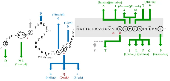
Three genetic loci have been linked to early onset inherited forms of the disease namely genes encoding for APP located on chromosome 21, presenilin-1 (PS1) on chromosome 14 and presenilin-2 (PS2) on chromosome 1 (reviewed in Marambaud and Robakis, 2005). The APP gene encodes the APP which is normally cleaved to form Aβ. Within the APP gene twenty-three locations carrying mutations have been identified, however these only explain around 5-10% of familial early-onset cases (Bird 2007b). Although some of them are directly linked to AD, others are associated with cerebral haemorrhages (Figure 5 and
see www.alzforum.org/res/com/mut/app). The first APP mutation discovered was Glu693
to Gln (“Dutch” mutation) within the Aβ sequence (Hardy and Allsop 1991). Synthetic peptides containing this mutation were shown to have high propensity to aggregate (Wisniewski et al. 1991; Clements et al. 1993). Subsequently, some families with early
onset AD were found to have pathogenic mutations at APP Val717, resulting in a change
from Val717 to Ile, Gly or Phe (Chartier-Harlin et al. 1991; Goate et al. 1991; Murrell et al.
1991). This mutation was called the “London” mutation. The “Swedish” double mutation
(Lys/Met670 to Asn/Leu, on the immediate Aβ N-terminus) results in secretion of larger
amounts of total Aβ (Citron et al. 1992; Mullan et al. 1992; Cai et al. 1993). A more
recently discovered pathogenic mutation, named the APP “Arctic” mutation (Glu693 to
Gly), leads to decreased Aβ1-40/1-42 levels in plasma and in cells conditioned media, but as a
consequence a higher tendency of Aβ to aggregate. In fact, Aβ in the Arctic mutation, forms protofibrils at a much higher rate and in larger quantities than wild-type Aβ (Nilsberth et al. 2001). These mutations, all lye near or within the Aβ domain, result in
26 Centro Biologia Celular Universidade de Aveiro
APP being more efficiently processed by secretases, thus generating increased amounts of Aβ that is more likely to form plaques (Citron et al. 1992; Cai et al. 1993; Haass et al. 1995; Goedert and Spillantini 2006), thereby promoting amyloidogenesis.
Further, the level of APP being expressed also appears to be an important aspect. For instance, in Down’s Syndrome, caused by trissomy of chromosome 21, there is an extra copy of the APP gene. These individuals show increased levels of Aβ and invariably develop plaques and tangles in their brains, with clinical dementia in many cases before the age of 50 (Tanzi et al. 1987; Selkoe 1997; Esler and Wolfe 2001).
Figure 5. APP mutations lying near or in the Aββββ domain. Location of the mutations within the fragment 665-723 of APP is highlighted with a circle in the corresponding amino acid sequence (APP770 isoform numbering).
The most frequent Aβ peptides (Aβ1-40 and Aβ1-42) extend from position 672 to amino acid 711 (for Aβ1-40) or 713
(for Aβ1-42). Mutations indicated in green produce Alzheimer disease phenotypes; mutations notated in blue are
primarily associated with cerebral amyloid angiopathy phenotypes. Mutations depicted in gray produce neither phenotype. Dutch mutation, the first APP mutation discovered, at position 22 of Aβ is highlighted in red. The gray box, spanning from positions 700 to 723, represents the location of the single transmembrane domain of APP. Adapted from www.nature.com/.../n9/fig_tab/nn0904-902_F1.html.
Centro Biologia Celular 27 Universidade de Aveiro
Mutations in the highly homologous PS1 and PS2 genes account for most cases of familial AD (Levy-Lahad et al. 1995; Sherrington et al. 1995). A total of 142 mutations have been found for PS1 in 281 families (www.alzforum.org/res/com/mut/pre/table1.asp), which represents the gene with highest number of pathogenic mutations for AD. For PS2 10 mutations have been found in 16 families (www.alzforum.org/res/com/mut/pre/table2.asp). These genes encode for proteins that are involved in the normal cleavage of the APP
protein, and mutations on these genes will result in increased Aβ1-42 production (Citron
et al. 1997; Xia et al. 1997; De Strooper et al. 1998). In particular, PS1 participates in the
catalytic core of γ-secretase complex and its mutations induce relative amounts of AβX-42
peptides (Wolfe et al. 1999). Some PS2 mutations, like those of PS1, were functionally
associated with increased production of AβX-42 peptides, while others did not modify
either AβX-40 or AβX-42 peptide production (reviewed in Lambert and Amouyel, 2007).
Genes implicated in sporadic disease
Inheritance of ε4 allele of apolipoprotein E (APOE ε4) represents the greatest genetic risk factor in sporadic AD (Corder et al. 1993; Poirier 1994; Raber et al. 2004; Goedert and Spillantini 2006), although its mode of action in AD progression is unknown. The APOE ε4 allele appears to operate mainly by modifying age onset (Meyer et al. 1998; Xiong et al. 2005), with each allele copy lowering the age of onset by almost 10 years, suggesting that APOE ε4 association with AD may be related to longer disease duration in these cases (Basun et al. 1995). Further, there is no well described molecular mechanism underlying APOE ε4 as an increased risk factor for AD. It is known that APOE acts as a cholesterol transporter in the brain, with APOE ε4 being less efficient than the other variants in recycling membrane lipids and neuronal repair (Poirier 1994). On the other hand, APOE is essential for Aβ deposition, promoting Aβ aggregation and plaque formation (Holtzman et al. 2000; Holtzman 2001), possibly by acting as a pathological chaperone that binds to Aβ. Further, other genetic susceptibility factors have been proposed. Polymorphisms on genes encoding several proteins, including α2-macroglobulin, angiotensin I converting enzyme, Fe65 (Chapman et al. 1998; Alvarez et al. 1999; Kovacs 2000; Lambert et al. 2000), as well as some mitochondrial genetic polymorphisms (reviewed in Zhu et al. 2004) have been
28 Centro Biologia Celular Universidade de Aveiro
associated with the disease. More recently, new susceptibility loci for late AD have been identified on chromosome 1, 9, 10, 12, and 13 (Bertram and Tanzi 2004). These risk factor genes are likely to affect one or more of the known pathogenic mechanisms (i.e. altered Aβ production, increased Aβ aggregation and inflammatory responses) which will result in decreased Aβ degradation/clearance and ultimately in neurodegeneration.
Genetic screening of AD
Genetic testing for the mutations associated with both familial and sporadic AD are available, although the circumstances under which testing is recommended differ. If there is suspicion of familial early-onset AD, genetic testing to detect gene mutations can and should be performed to identify the disease causing mutation and the molecular lesion (Bird 2007b). For APOE, and although a large number of patients with sporadic late-onset AD have at least one allele APOE ε4, the association of the mutation with the development of the disease is not strong enough to recommend that APOE genotyping be used as a predictive test in asymptomatic individuals. Instead APOE genotyping is most useful as an adjunct diagnostic test in individuals exhibiting symptoms of progressive dementia (Xiong et al. 2005; Bird 2007a).
Centro Biologia Celular 29 Universidade de Aveiro
1.3.2 Non-genetic factors contributing to sporadic AD
For AD sporadic forms, besides aging, which is the most obvious risk factor, epidemiological studies have proposed several other putative contributing factors. Some can be linked to decreased reserve brain capacity (including reduced brain size and number of neurons and their synaptic and dendritic arborisation) due to for instance brain injury, low educational and occupational attainment, low mental ability in early life, and reduced mental and physical activity during late life (Mayeux 2003; Mortimer et al. 2003; Jellinger 2004). Other risk factors are associated with vascular disease, including hypertension, hypercholesterolemia, atherosclerosis, coronary heart disease, smoking, obesity and diabetes (Mayeux 2003). Nonetheless is not clear if these are true causal risk factors that lead to the pathological features of the disease, or whether they induce cerebrovascular pathology that will add to the clinically silent disease pathology beyond the threshold for dementia. Evidence suggests that dietary intake of antioxidants, such as vitamin C and E; homocysteine-related vitamins (vitamin B12 and folate); unsaturated fatty acids; and also moderate alcohol intake, especially wine, could reduce the risk for AD (Luchsinger et al. 2007), but data so far does not support the recommendation of any specific diet for the prevention of AD. Although many environmental factors may increase the risk of developing sporadic AD, this form of the disease has been shown to have a significant genetic background.
30 Centro Biologia Celular Universidade de Aveiro
1.4 ACENTRAL ROLE FOR Aββββ IN ADNEURODEGENERATION
The association of the pathogenic mutations with alterations in APP processing pathways
that relatively increase Aβ1-42 production, together with the in vitro and in vivo observations
of Aβ−induced neurotoxicity (for review see Canevari at al. 2004 and Smith et al. 2006) sustain that this peptide is at the heart of the disease process. These findings have lead to the proposal of the amyloid cascade hypothesis, which has been the basis of several research activities that have significantly contributed to the understanding of the molecular basis of AD (reviewed in Hardy 2006). In the cascade theory, Aβ is the central trigger for the pathological changes observed in AD brains, such as synapse loss, activation of inflammatory processes, the induction of NFTs and, ultimately, neuronal death. Therefore, all factors that can contribute to altered APP processing/metabolism resulting in increased Aβ production and/or aggregation, like APP and presinilin mutations, APOE ε4 allele, Trissomia 21, oxidative stress, environmental factors, and even normal aging will contribute to AD progression.
At the molecular cascade level diffuse amyloid deposits progress over time and eventually
become neuritic plaques. One hypothesis is that deposition of Aβ1-42 may form a
“precipitation core” to which soluble Aβ1-40 could aggregate, in an AD-specific process.
The in vivo evolution of Aβ deposition and aggregation may trigger an oxidative inflammatory response that can be initiated due to the release of reactive oxygen intermediates, nitric oxide and inflammatory cytokines by activated microglia (Lukiw and Bazan 2000; Butterfield et al. 2001; Eikelenboom et al. 2008). Some of these pro-inflammatory molecules may be locally toxic to neuronal processes in the vicinity of amyloid plaques. Further, the relative overproduction of this peptide may lead, first to neurofibrillary degeneration and then to neuronal death (Hardy 1997).
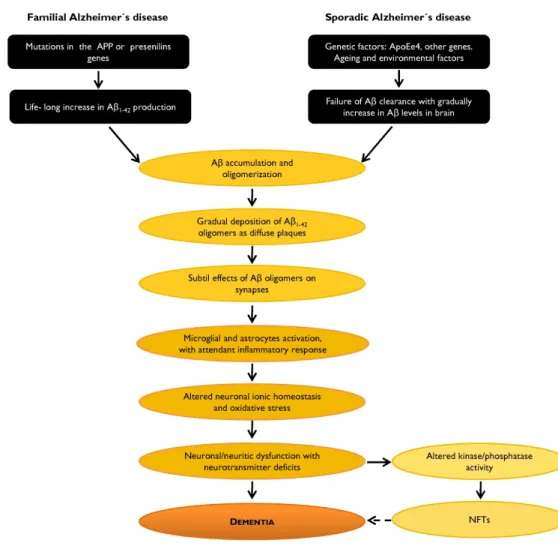
The unequivocal involvement of Aβ in AD neurodegenerative process is summarized in the following figure.
Centro Biologia Celular 31 Universidade de Aveiro
Figure 6. Aββββ involvement in the AD neurodegenerative process: the amyloid cascade hypothesis. It is likely that during the disease process the balance between Aβ production and Aβ catabolism is altered, which results in increased Aβ production/accumulation. Adapted from http://www.alzforum.org/res/adh/cur/knowntheamyloidcascade.asp.
32 Centro Biologia Celular Universidade de Aveiro
Nonetheless, this hypothesis, often considered a dogma, has some limitations and other pathophysiological mechanisms, not necessarily exclusive, are still under study. These include, for example alterations in cell trafficking (Naruse et al. 1998) or neuronal calcium homeostasis induced by mutant presenilins (Schneider et al. 2001), and also oxidative stress (reviewed in Chauhan and Chauhan 2006 and Hamel et al. 2008). Corroborating the latter hypothesis is the observation that genetic mutations in AD have lead to increased cellular vulnerability to oxidative stress and apoptotic insults. It is still unclear whether mutations result in Aβ deposition that then cause oxidative stress, or whether mutations cause oxidative stress that result in Aβ deposition. Indeed, while some studies demonstrated that Aβ can directly cause oxidative stress others shows that the reverse is also true. Obviously, whether Aβ is the culprit, as argued by the amyloid cascade, or just a promoter factor that may culminate in neuronal death needs to be further investigated to advance our understanding and contribute to the design of efficacious therapeutics for this disease.
Centro Biologia Celular 33 Universidade de Aveiro
1.5 ALZHEIMER´S AMYLOID PRECURSOR PROTEIN (APP)
1.5.1 APP gene family and alternatively spliced isoforms
Aβ is constitutively secreted by both neuronal and non-neuronal mammalian cells into the extracellular fluid (Selkoe 1994b) and arises from the proteolytic processing of APP, an integral transmembrane glycoprotein containing a membrane-spanning domain towards its carboxyl-terminus (Kang et al. 1987). The mammalian APP superfamily comprises APP and APP-like proteins, known as APLP1 and APLP2 (Sprecher et al. 1993; Wasco et al. 1993), of which APLP2 is the nearest relative (50% of homology). These three related proteins are well-conserved in evolution, functionally and structurally related, and share similar functions (Bayer et al. 1999; Coulson et al. 2000). The mammalian APP family members are type I integral membrane proteins that have relatively large extracellular domains and short intracellular domains. Of note is that APLP1 and APLP2 share homology at the amino acid sequence, domain structure and protein organization with APP, but lack the Aβ domain. Other known members of the APP superfamily are non-mammalian and include APPL in Drosophila (Rosen et al. 1989; Luo et al. 1992), APL-1 in C. elegans (Daigle and Li 1993) and an APP homologue protein in Xenopus (Okado and Okamoto 1992).
APP is encoded by a gene on chromosome 21 (21q21.3) and contains 18 exons (GenBank accession number D87675), with the Aβ sequence occurring between exons 16 and 17. Alternative post-transcriptional splicing of exons 7, 8 and 15 of the APP mRNA (Neve et al. 1988; Palmert et al. 1988; Koo et al. 1990b; Ohgami et al. 1993a; Sandbrink et al. 1994b) produces different isoforms of this protein ranging from 365-770 amino acid residues that differ in size extracellularly, but share the same cytoplasmic, transmembranar and Aβ peptide sequences. At least eight isoforms of APP have been described (Figure 7), numbered according to their length in amino acids: 677, 695, L-696, 714, L-733, 751, L-752, 770 (Kitaguchi et al. 1988; Ponte et al. 1988).
34 Centro Biologia Celular Universidade de Aveiro
Figure 7. APP isoforms resulting from alternative splicing of the APP gene. Alternatively spliced exons are indicated in colour: dark blue, exon 7; light blue, exon 8; orange, exon 15. The Aβ sequence lying in exon 16 and 17 is indicated in red. Adapted from Sandbrink et al. 1994b.
APP is ubiquitously expressed in mammalian cells with a broad tissue distribution (Tanzi et al. 1987; Neve et al. 1988; Tanzi et al. 1988; Weidemann et al. 1989; Golde et al. 1990; Sisodia and Price 1995). Analysis of APP mRNA expression levels revealed that APP can be detected in almost all tissues examined, as well as in cultured cells. The tissue-specific pattern of APP mRNA splicing was studied by RT-PCR analysis (Sandbrink et al. 1994a). The less abundant L-APP isoforms, lacking exon 15, are mainly expressed in leukocyte cells, such as T-lymphocytes, macrophages and microglial cells. They are also ubiquitously expressed in rat tissues, including brain, but not in neurons (Ohgami et al. 1993b; Sandbrink et al. 1994b).
The three major isoforms expressed were found to be 695, 751 and 770 amino acids (APP695, APP751 and APP770, respectively). While the 751- and 770- amino acid spliced isoforms are predominantly expressed in peripheral tissues, the 695 is the APP isoform predominantly produced in the mammalian brain with the 695:751:770 mRNA ratios
APP 770 APP 751 APP L-752 APP L-733 APP 714 APP 695 APP L-696 1 2 3 4 5 6 7 8 9 10 11 12 13 14 15 16 17 18 APP L-677
Centro Biologia Celular 35 Universidade de Aveiro
being approximately 20:10:1 (Neve et al. 1988; Konig et al. 1989; Tanaka et al. 1989; Kang and Muller-Hill 1990). The exon 7-containing isoforms also predominate in cultured astrocytes (Gray and Patel 1993a; Gray and Patel 1993b; Rohan de Silva et al. 1997), with
the 695:751:770 ratio being 1:4:2 (Gray and Patel 1993a; Gray and Patel 1993b). APP695,
lacking exon 7 and 8, is most highly expressed in neurons, representing 95% of total
neuronal APP (Tanzi et al. 1987; Weidemann et al. 1989; LeBlanc et al. 1991), and is
therefore often referred to as the “cerebral” or “neuronal” isoform. Due to APP695
predominance in the brain and CNS (Neve et al. 1988; Tanzi et al. 1993), this isoform has received considerable attention in AD research. Additional studies have indicated that alternative splicing of exons 7 and 8 changes in brain during aging and with AD, but results obtained are still inconsistent and controversial to consider altered alternative splicing as an AD risk factor (Sandbrink et al. 1994a; Rockenstein et al. 1995; Moir et al. 1998; Panegyres et al. 2000).
1.5.2 APP functional domains
The APP exon 7 encodes a 56 amino acid (aa) Kunitz-type serine protease inhibitor (KPI) domain, which inhibits proteases, such as trypsin or plasmin, and blood coagulation factors (Van Nostrand and Cunningham 1987; Kitaguchi et al. 1988; Ponte et al. 1988; Tanzi et al. 1988; Wagner et al. 1992) and thus may regulate the degradation of APP (Edelberg and Wei 1996). Exon 8 encodes a 19 aa domain with homologies to the MRC OX-2 antigen found on the surface of neurons and certain cells involved in the immune response such as thymocytes (Clark et al. 1985). Besides KPI and OX-2, several other structural domains have been identified within APP (Figure 8 and Reinhard et al. 2005). Several heparin-binding domains (Small et al. 1994), a collagen-binding site (Beher et al. 1996), an integrin-binding motif (amino acid sequence RHDS, Ghiso et al. 1992) and N-linked carbohydrate attachment sites (Weidemann et al. 1989) were also found in this region. Consistently, APP has been shown to bind heparin (Mok et al. 1997), collagen (Beher et al. 1996), and laminin (Kibbey et al. 1993). Additionally, two subdomains (328-332 and 444-612) were presumed to have a neuroprotective function, including the “RERMS” sequence with putative growth-promoting properties (Ninomiya et al. 1993).
36 Centro Biologia Celular Universidade de Aveiro
The APP ectodomain also contains binding-sites for metals such as zinc (Bush et al. 1993; Bush et al. 1994) and copper (Hesse et al. 1994; Multhaup et al. 1996; Barnham et al. 2003) in the APP N-terminal but also within the Aβ domain. The C-terminus can be cleaved to release the APP intracellular domain (AICD). Almost all known APP binding proteins bind at its C-terminus, and specifically at one of two APP domains: YTSI and YENPTY. The latter is highly conserved from nematodes to humans, and has been shown to be responsible for several protein-protein interactions. Several intracellular proteins have been shown to bind to this domain including Fe65 (a phosphotyrosine binding domain-containing protein), X11 also known as LIN-10 or MINT (Munc-18-interacting protein), Dab1 (disabled homolog 1) and JIP-1b. Interacting proteins for YTSI domain included APP-BP1 (APP-binding protein 1), the microtubule associated protein PAT1 (protein interacting with APP tail 1), and kinesin-I (an axonal transport protein) (De Strooper and Annaert 2000; Van Gassen et al. 2000; King and Scott Turner 2004). Another domain, VTPEER, has not been implicated so far in APP binding to other
proteins, but it includes a G0 binding sequence. These intracellular domains can be
classified according to their attributed functions and are thought to be involved in
regulating APP rate of secretion, endocytosis,and Aβ production (Ando et al. 1999; Iijima
et al. 2000; Mueller et al. 2000; Ando et al. 2001; Sabo et al. 2001; Roncarati et al. 2002).
Figure 8. APP functional domains. Yellow, signal peptide (1-17); light blue, heparin-binding domains (28-123; 174-185; 391-412); brown, copper-binding domain (135-155); violet, zinc-binding domain (181-188); pink, KPI domain; grey, OX-2 domain; “RERMS”, putative growth-promoting motif (403-407); orange, gelateinase A (matrix metalloproteinase) inhibitor (407-417); green, collagen-binding site (523-540); red, Aβ; “RHDS”, integrin-binding motif (aa 5-8 of Aβ); heparin-binding motif “VHHQK” (aa 12-16 of Aβ); dark Blue, APP intracellular domain (AICD) which include YTSI, YENPTY, VTPEER. APP770 isoform numbering. TM, transmembrane domain.
Centro Biologia Celular 37 Universidade de Aveiro
1.5.3 Putative functions for APP and APP cleaved fragments
The overall physiological function of APP has not yet been definitively determined but due to the structures and the specific characteristics of its domains several APP putative functions have been attributed and considered valid. These include cell surface receptor, cell adhesion molecule, precursor to growth factor and regulator of neuronal copper homeostasis. Besides that, other functions arise from many in vitro and in vivo studies which evaluated the involvement of APP and APP fragments resulting from APP processing (see section 1.5.4). These include neurotoxicity, neuritic outgrowth, synaptogenesis, involvement in learning and memory processes, and in cell signaling (for review see Zheng and Koo 2006; Senechal et al. 2006; Reinhard et al. 2005).
APP as a receptor molecule
Due to the type I integral membrane structure and due to it binding site to G0 protein, via
the intracellular tail, it has been suggested that APP might function as a cell surface
G-protein coupled receptor (Kang et al. 1987; Okamoto et al. 1995). G0 is a major GTP
binding protein in brain involved in signal transduction cascades, such as adenylyl cyclase (Carter and Medzihradsky 1993), phospholipase C (Moriarty et al. 1990), voltage-dependent calcium channels (Hescheler et al. 1987) and pathways for apoptosis (Giambarella et al. 1997). Therefore, the activation of APP may contribute to one or
more of these cascades, although the exact downstream mechanisms involving G0
activation or inhibition by APP are unknown. Additionally, the analogy of the secondary structures and proteolytic processing profile between APP and Notch is also consistent with APP functioning as a cell surface receptor similar to Notch (Selkoe and Kopan 2003). Fibrillar forms of Aβ were reported to bind cell surface APP (Lorenzo et al. 2000), and Nogo-66 receptor was also shown to interact with the APP ectodomain, interaction which affects Aβ production (Park et al. 2006). Further evidence came from the Ho and Sudhof (2004) study, which demonstrated that the APP extracellular domain binds to F-spondin, a neuronally secreted glycoprotein, and that this interaction regulates APP cleavage and subsequent Aβ production and downstream signaling. More recently, Ma et al. (2008) identified TAG1 (a GPI-linked recognition molecule of CNS) as a functional
38 Centro Biologia Celular Universidade de Aveiro
ligand for APP, and suggested that this TAG1–APP signaling pathway was involved in the modulation of neurogenesis.
APP involvement in cell adhesion
Convincing data places APP as a CAM (cell adhesion molecule) and SAM (subtrate adhesion molecule). Neuronal CAMs play an important role in neuronal plasticity and thus learning and memory processes may be closely linked to CAM function and any disruption in CAM interactions may have potential neuropathological consequences (Cotman et al. 1998). Cell surface APP has been described to enhance neuronal cell adhesion and neurite outgrowth (Breen et al. 1991). APP possesses several ectodomains (Figure 8) that promote binding to specific substrates such as heparin, collagen and laminin (extracellular matrix components), supporting its role in cell-substratum adhesion. The same sequences have been shown to be involved in cell-cell interactions. “RHDS” motif (Figure 8) within the Aβ sequence appears to also promote cell adhesion. In particular, cell surface APP has been recently reported to trans-interact with other APP (or APLP) molecules by homo- or hetero-dimer formation at the surface of adjacent cells, and that these trans-dimerizations promote trans-cellular adhesion in vivo (Soba et al. 2005). Furthermore, down-regulation of APP using antisense oligonucleotides also reduced neuronal adhesion to specific substrata, and APP overexpression in the neuronal-like B103 cells led to more rapid cellular adhesion (Schubert et al. 1993). Moreover, fibroblasts from FAD patients that were observed to have down-regulated APP mRNA levels, presented decreased cellular adhesiveness (Ueda et al. 1989), and Hep-1 cells expressing an APP FAD mutant cDNA also exhibited decreased cell adhesion properties (Kusiak et al. 2001).
Centro Biologia Celular 39 Universidade de Aveiro
APP and cell motility regulation
Via its intracellular domain APP can bind several proteins, including the adapter protein Fe65. The latter binds a second protein, MENA, which is a cytoskeletal protein expressed in active actin remodelling areas such as axonal growth cones. In H4 neuroglioma and MDCK cells, Fe65 has been found associated with MENA in active actin areas. The functional role of this ternary complex was documented in non-neuronal cells, in which co-expression of APP and Fe65 drastically increases cell motility, and this process appears to be partially dependent on the MENA/actin complex (Sabo et al. 2001). Subsequent analysis from the same group showed that in primary neurons, the APP and Fe65 complex is localized to the dynamic adhesion sites (actin-rich sites) in the growth cone (Sabo et al. 2003). Taken together these data supports a role for APP/Fe65 complex in cell motility and growth cone dynamics.
APP and sAPPαααα have a role in neurite outgrowth and synaptogenesis
This function is probably the most consistent and well documented since several overexpression and downregulation studies have addressed this issue. Neurotrophic and synaptogenic roles have been attributed to both APP and sAPPα (APP secreted fragment resulting from α-secretase cleavage, see section 1.5.4). As such APP may exert these activities in both an autocrine and a paracrine mode. APP is expressed at neuronal synapses and exhibits widespread expression in vesicular structures of cell bodies, axons and dendrites. It undergoes rapid anterograde transport and is targeted to the synaptic sites (Koo et al. 1990a; Sisodia et al. 1993; Yamazaki et al. 1995). During neuronal maturation and development APP expression has been described to be upregulated (Hung et al. 1992; Bibel et al. 2004), and correlated with periods of intense neuritic outgrowth and synaptogenesis (Loffler and Huber 1992; Moya et al. 1994). Moreover, cultured hippocampal neurons derived from APP knockout (KO) mice exhibit both reduced viability and neuritic outgrowth (Perez et al. 1997). Other studies directly demonstrated the importance of sAPPα (lacking APP C-terminal domain) in these functions. In vivo application of sAPPα causes neurite outgrowth in cultured fibroblasts (Saitoh et al. 1989;
40 Centro Biologia Celular Universidade de Aveiro
Bhasin et al. 1991), PC12 cells (Milward et al. 1992), cortical and hippocampal neuronal cells (Araki et al. 1991; Qiu et al. 1995; Ohsawa et al. 1997) and human neuroblastoma cell lines (Wang et al. 2004). More recently, studies in mice overexpressing ADAM-10 (a secretase involved in APP cleavage to sAPPα) and perfused with exogenous sAPPα, were shown to present neurotrophic effects on cortical synaptogenesis (Bell et al. 2008). Of interest, the pentapeptide sequence “RERMS” located C-terminal to the KPI and OX-2 domains (Figure 8) was identified as the site responsible for the growth-promoting trophic role of sAPP (Ninomiya et al. 1993). Infusion of this peptide or sAPP into brain animals resulted in improved memory and increased synaptic density (Roch et al. 1994; Meziane et al. 1998). Further, neurotrophic and neuroprotective effects are induced by sAPPα approximately 100 times more strongly than by sAPPβ (resulting upon APP cleavage by β-secretase, section 1.5.4). Indeed, Li and colleagues (1997) showed that sAPPβ lowered neurite outgrowth below control levels. Although the conserved regions of both proteins contain domains that have been associated with these functional effects (such as “RERMS” region), the difference between these sAPP effects seems to subsist in their C-terminal region, where sAPPα contain 17 more aminoacids. This region of sAPPα contains a heparin-binding domain (“VHHQK” residues 12-16 of Aβ), that is lacking in sAPPβ, and appears to play a key role in mediating the neurotrophic effects (Furukawa et al. 1996).
Centro Biologia Celular 41 Universidade de Aveiro
APP, sAPP and Aββββ functions in memory
APP appears to have an important role in the regulation of synaptic structure and neuronal function. Sequence-specific antibodies were used to block protein function and antisense oligonucleotides to prevent APP translation. APP association with improving memory was supported by studies were infusion of “RERMS” sequence rescued amnesia that was induced by anti-APP antibodies (Mileusnic et al. 2000). Additionally, intraventricular injection of anti-N-terminal APP antibodies (for example delivery of the 22C11 antibody targeting the N-terminal part of APP) in rat, close to the training period, has been shown to result in an impairment of rat memory in a passive avoidance task (Doyle et al. 1990; Huber et al. 1993; Gschwind et al. 1996; Turner et al. 2003). In the case of APP-null mutations, mice show a variety of alterations in neuronal structure and function, including gliosis, decreased neocortical and hippocampal levels of synaptophysin, reduced dendritic length in hippocampal neurons, reduced survival of cultured neurons and impaired long-term potentiation (LTP) (Perez et al., 1997; Dawson et al., 1999; Chapman et al., 1999; Seabrook et al., 1999; but see Phinney et al., 1999). However these effects could also be due to the loss of the neurotrophic sAPPα fragment. Consistently, exposure of hippocampal slices to sAPPα results in raising the threshold for long term depression (LTD) but facilitating LTP (Ishida et al. 1997) thus changing synaptic efficacy. Since memories are believed to be stored within synapses, LTP and its opposing process, LTD, are widely considered the major cellular mechanisms underlie learning and memory (Cooke and Bliss 2006).
Contrary to APP or sAPPα effects, Aβ peptide has been associated with synaptic dysfunction and inhibition of LTP (Cullen et al. 1997; Lambert et al. 1998; Hsia et al. 1999; Walsh et al. 2002a; Raymond et al. 2003; Turner et al. 2003), although downstream mechanisms are not well understood. Further, studies in transgenic mice models of AD have shown that Aβ immunization reduces plaque deposition and improves cognitive function (Bard et al. 2000; Lombardo et al. 2003; Oddo et al. 2004; Brendza et al. 2005; Buttini et al. 2005). All these data strengthen a role for APP and APP proteolytic fragments in learning and memory processes.
42 Centro Biologia Celular Universidade de Aveiro
APP and sAPP involvement in brain repair and neuroprotection
Several studies have provided evidence that APP plays a role in the repair of adult brain following trauma. In fact, APP has been reported to be upregulated in response to neuronal injury and damage (Siman et al. 1989; Gentleman et al. 1993; Struble et al. 1998). Up-regulation of APP in response to neuronal injury appears to promote axonal arborization (Leyssen et al. 2005). Perez et al. (1997) showed that APP-deficient neurons exhibited shorter axons and branching processes. These effects were rescued when hippocampal neurons from APP-deficient mice were co-cultured with wild-type astrocytes, suggesting that APP contribute to axon and dendritic outgrowth and arborization. Consistent with a possible role for APP in axonal repair mechanisms, following traumatic brain injury in vivo administration of sAPPα has been shown to improve functional outcome and to reduce neuronal apoptosis and axonal injury (Thornton et al. 2006).
Metal ions such as copper are essential in brain, since it is an essential cofactor of oxidoreductive enzymes, including superoxide dismutase that is involved in cellular protection induced by oxidative stress. APP structure harbours several metal-binding domains for copper (Figure 8), in agreement with an APP involvement in the transport of this metal into the brain. Studies using APP KO mice have shown increased copper levels in the cerebral cortex, supporting a possible physiological role for APP in modulating copper neuronal homeostasis in vivo (White et al. 1999; Bayer and Multhaup 2005; Inestrosa et al. 2005; Maynard et al. 2005).
sAPP secreted fragment has also been implicated in neuroprotection (reviewed in Mattson 1997), including against Aβ effects and induced apoptosis. Stimulation of sAPPα secretion results in protective effects against Aβ-induced neurotoxicity (Levites et al. 2003). Also, in human keratinocytes, Wehner and colleagues (2004) have shown that at a nanomolar range sAPPα was able to protect these cells against induced apoptosis. Further, sAPPα was shown to increase expression levels of several neuroprotective genes and protect against Aβ induced-tau phosphorylation and neuronal death (Stein et al. 2004). Further, the APP N-terminal heparin binding domain was also reported to display a neuroprotective function (Small et al. 1999). Hence, it is clearly evident that