Inte ractio ns o f lam inin with the
am ylo id ß pe ptide . Im plicatio ns fo r
Alzhe im e r’s dise ase
Centro de Regulación Celular y Patología, Departamento de Biología Celular y Molecular, Facultad de Ciencias Biológicas, Pontificia Universidad Católica de Chile, Santiago, Chile C. Morgan and
N.C. Inestrosa
Abstract
Extensive neuronal cell loss is observed in Alzheimers disease.
Laminin immunoreactivity colocalizes with senile plaques, the
char-acteristic extracellular histopathological lesions of Alzheimer brain,
which consist of the amyloid ß (Aß) peptide polymerized into amyloid
fibrils. These lesions have neurotoxic effects and have been proposed
to be a main cause of neurodegeneration. In order to understand the
pathological significance of the interaction between laminin and
amyloid, we investigated the effect of laminin on amyloid structure
and toxicity. We found that laminin interacts with the Aß1-40 peptide,
blocking fibril formation and even inducing depolymerization of
preformed fibrils. Protofilaments known to be intermediate species of
Aß fibril formation were also detected as intermediate species of
laminin-induced Aß fibril depolymerization. Moreover,
laminin-amy-loid interactions inhibited the toxic effects on rat primary
hippocam-pal neurons. As a whole, our results indicate a putative
anti-amyloido-genic role of laminin which may be of biological and therapeutic
interest for controlling amyloidosis, such as those observed in cerebral
angiopathy and Alzheimers disease.
Co rre spo nde nce
N.C. Inestrosa
Centro de Regulación Celular y Patología
Departamento de Biología Celular y Molecular, FCB, PUC Alameda, 340 Casilla 114-D Santiago Chile
Fax: + 56-2-686-2717 E-mail: ninestr@ genes.bio.puc.cl
Presented at
SIMEC 2000 - International Symposium on Extracellular Matrix, Angra dos Reis, RJ, Brazil, September 24-27, 2000.
Research supported by a Presidential Chair in Science (1999-2001) and a FO NDAP grant in Biomedicine (No. 13980001) to N.C. Inestrosa and FO NDECYT (Nos. 2970070 and 4000011) to C. Morgan.
Received O ctober 19, 2000 Accepted February 5, 2001
Ke y wo rds
·Extracellular matrix ·Laminin
·Amyloid ß peptide ·Neuronal injury
·Neurodegenerative disease
Intro ductio n
Laminin is a major basement membrane
glycoprotein (M
r~850 kDa) widely
distrib-uted in the extracellular matrix (ECM). At
least 11 isoforms of laminin have been
de-scribed due to the presence of five different
a
chains (
a
1-
a
5) of 200-400 kDa which
combine with ß (220 kDa) and
g
(210 kDa)
chains into disulfide-bonded heterotrimers
arranged in a cross-shaped structure (1,2).
These three chains are coded by different
genes (3-5). The C-terminal globular
do-mains of the
a
chain are critical for
appropri-ate assembly of the whole glycoprotein (6).
This region is also important for driving
laminin polymerization and heparin binding
(7,8). Several other ECM proteins may bind
laminin through these globular domains, such
as perlecan,
a
-dystroglycan, fibulin-1,
fibu-lin-2, and nidogen-2 (9,10). Because of its
large size, complexity and multidomain
na-ture, laminin exhibits a variety of functions
still not completely understood, namely the
ability of laminin to keep the ECM assembled,
to promote neurite outgrowth, growth cone
guidance, pathfinding, and synapse
forma-tion (11,12).
The impo rtance o f amylo id ß pe ptide
in Alzhe im e r’s dise ase
by a loss of memory which finally
deter-mines the clinical onset of dementia.
Alzhei-mer brains are histopathologically
charac-terized by numerous senile plaques and
neu-rofibrillary tangles. Both lesions are
consid-ered to be of pivotal importance to
under-stand AD. In addition, genetic evidence points
at the regulation of amyloid precursor
pro-tein (APP) metabolism as a key factor in the
pathogenesis of AD (13). Amyloid-derived
neurotoxicity is widely considered as a main
factor explaining the neurodegenerative
changes observed in AD. The neurotoxic
effect of amyloid ß (Aß) peptide, a
pro-teolytic fragment of APP, is associated with
its polymerization state as amyloid fibrils
(14,15). Studies of amyloid-derived
neuro-toxicity
in vivo
have shown an increased
vulnerability to neurotoxicity with aging
(16), suggesting the participation of some
age-dependent factors.
ECM co m po ne nts and APP
Because of the extracellular deposition
of Aß peptide in senile plaques, particular
attention has been paid to interactions
be-tween amyloid and ECM components (17).
Indeed, several basement membrane
com-ponents such as heparan sulfate
proteogly-cans, type IV collagen and laminin have
been found in senile plaques (18).
More-over, proteoglycans and laminin interact with
APP (19-21) and these interactions seem to
disrupt the normal associations among ECM
components (22), suggesting that an
over-production of APP may change the normal
functions of ECM. On the other hand, APP
metabolism is affected by ECM components
both in microglial (23) and neuronal (24)
cell types.
The anti-amylo ido ge nic ro le o f
laminin
Laminin immunoreactivity is observed
mainly associated with blood vessels and
reactive astrocytes in normal brains but also
in neuronal cell bodies in most parts of the
developing and adult rat central nervous
sys-tem (25). Its expression seems to be
in-creased after brain injury (26) and in the
brain of AD and Down syndrome patients
(27). In order to examine the pathological
significance of laminin induction in
Alzhei-mer brains, we studied the effect of laminin
on Aß peptide polymerization, and we
showed for the first time that laminin was a
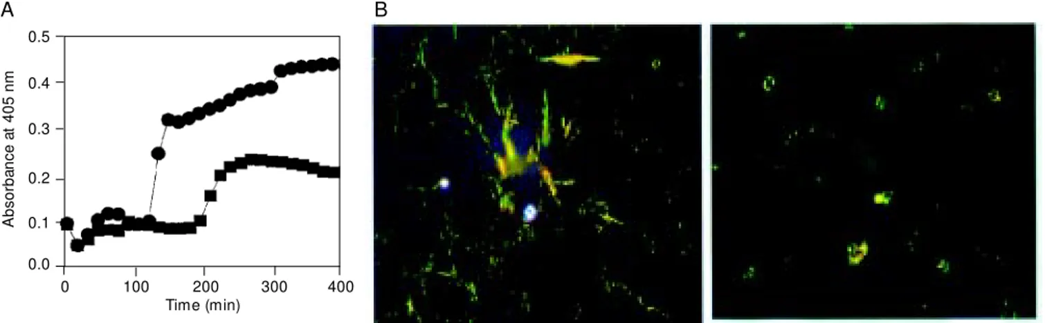
Figure 1. Inhibition of amyloid ß (Aß) peptide polymerization by laminin. A, Aß1-40 (100 µM ) peptide polymerization assay carried out in the presence (squares) or absence (circles) of 100 nM mouse laminin-1 (w ith stirring at room temperature in PBS, pH 7.4). Turbidity measurements w ere undertaken at the indicated time points. B, The final products of the assay w ere examined by polarized light microscopy after Congo red staining. Abundant green birefringent material w as observed in the absence of laminin (left), indicating the presence of amyloid; conversely, a scarce positive reaction w as observed in the presence of laminin (right).
B
0.5
0.4
0.3
0.2
0.1
0.0
0 100 200 300 400 Time (min)
A
b
s
o
rb
a
n
c
e
a
t
4
0
5
n
m
potent inhibitor of Aß fibril formation
(28,29). Figure 1A shows a turbidity assay
indicating that Aß aggregation in the
pres-ence of laminin was clearly decreased, and
that the formation of amyloid was also
di-minished in the presence of laminin, as
evi-denced by Congo red staining (Figure 1B).
These observations confirm those obtained
by other authors (30,31). Since an inhibition
of fibril formation is expected to be
accom-panied by an attenuation of amyloid
neuro-toxicity, we have proposed that laminin may
have a therapeutic potential in the treatment
of AD. In fact, a diminished neurotoxicity
of amyloid has been observed in the
pres-ence of laminin in rat primary hippocampal
neurons (Figure 2). Similar results have
been observed in primary cortical cells (32).
Since a kinetic inhibition of
amyloido-genesis may retard but not reverse Aß fibril
formation, we decided to investigate the
abil-ity of laminin to depolymerize preformed Aß
fibrils (33). The studies were carried out
using molar ratios of Aß to laminin-1 of
2,500 or less. Amyloid depolymerization
in-duced by laminin is a concentration- and
time-dependent reaction (Figure 3A)
char-acterized by the appearance of protofilaments
at the expenses of preformed fibrils, which
finally leads to their clearance, forming
amor-phous non-amyloid material, as observed by
electron microscopy examination (Figure
3B). These observations suggest that
lami-nin behaves as a thermodynamic inhibitor of
amyloidogenesis. But if so, why does
amy-loidosis proceed in Alzheimer brains?
Sev-eral answers are possible, including one that
may involve laminin degradation. In fact, it
has been shown that plasmin-catalyzed
deg-radation of laminin mediates neuronal death
in the hippocampus after excitotoxic injury
with kainate (34). In that study, the authors
demonstrated that disruption of
laminin-neu-ron interaction makes neulaminin-neu-rons sensitive to
excitotoxic death. Therefore, it is reasonable
to assume that the interaction of laminin
with the Aß peptide may disrupt normal
interactions with cells. Moreover, we have
recently found that several laminin-derived
peptides, including the IKVAV sequence
(neurite promotor activity; 21), inhibit
amyloidogenesis. The efficacy of laminin in
depolymerizing Aß suggests that this
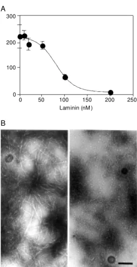
glyco-Figure 3. Depolymerization of amyloid ß (Aß) fibrils by lami-nin. Aß1-40 (250 µM ) fibrils w ere incubated w ith mouse la-minin-1 at the indicated con-centrations for 4 h at room temperature (20 mM sodium phosphate buffer, 150 m M NaCl, pH 7.2). A, The presence of amyloid fibrils w as assessed by thioflavine-T fluorescence as expressed in arbitrary units.
B, Transmission electron mi-crographs of Aß fibrils used as starting material (left); amyloid fibrils w ere depolymerized to protofilaments in the presence of 100 nM lam inin (right ). These prot of ilam ent s w ere completely cleared in the pres-ence of 200 nM laminin (data not show n). Bar = 0.25 µm.
B
Figure 2. Inhibition of amyloid ß (Aß) fibril neurotoxicity by la-minin. Rat hippocampal neu-rons in culture w ere incubated for 24 h w ith 10 µM Aß fibrils formed in the absence (Aß1-40) or presence of lam inin (Aß1-40 + laminin) as indicated in the legend of Figure 1. Con-trol cells (ConCon-trol) w ere incu-bated w ith an equivalent vol-ume of PBS, pH 7.4. Cell sur-vival w as determined by moni-toring M TT reduction (32) and is reported as percentage of the control assay.
C
e
ll
s
u
rv
iv
a
l
(%
)
100
80
60
40
20
0
Control Aß1-40 Aß1-40 + Laminin
300
200
100
0
0 50 100 150 200 250 Laminin (nM )
Re fe re nce s
1. Timpl R (1996). M acromolecular organiza-tion of basement membranes. Current Opinion in Cell Biology, 8: 618-624. 2. Beck K, Hunter I & Engel J (1990).
Struc-ture and function of laminin: anatomy of a multidomain glycoprotein. FASEB Jour-nal, 4: 148-160.
3. Barlow D, Green M , Kurkinen M & Hogan B (1984). Sequencing of laminin B chain cDNAs reveals C-term inal regions of coiled-coil a-helix. EM BO Journal, 3: 2355-2362.
4. Sasaki M & Yamada Y (1987). The laminin B2 chain has a multidomain structure ho-mologous to the B1 chain. Journal of Bio-logical Chemistry, 262: 17111-17117. 5. Sasaki M , Kleinman HK, Huber H,
Deutz-mann R & Yamada Y (1988). Laminin, a multidomain protein. The A chain has a unique globular domain and homology w ith the basement membrane proteogly-can and the laminin B chains. Journal of Biological Chemistry, 263: 16536-16544. 6. Utani A, Nomizu M , Timpl R, Roller PD &
Yamada Y (1994). Laminin chain assem-bly. Specific sequences at the C terminus of the long arm are required for the for-mation of specific double- and triple-stranded coiled-coil structures. Journal of Biological Chemistry, 269: 19167-19175. 7. Yurchenco PD, Tsilibary EC, Charonis AS
& Furthmayr H (1985). Laminin polymer-ization in vitro. Evidence for a tw o-step assembly w ith domain specificity. Journal of Biological Chemistry, 260: 7636-7644. 8. Yurchenco PD, Cheng Y-S & Schittny JC
(1990). Heparin modulation of laminin po-lymerization. Journal of Biological
Chem-istry, 265: 3981-3991.
9. Talts JF, Andac Z, Göhring W, Brancaccio A & Timpl R (1999). Binding of the G domains of laminin a1 and a2 chains and perlecan to heparin, sulfatides, a -dystro-glycan and several extracellular matrix pro-teins. EM BO Journal, 18: 863-870. 10. Andac Z, Sasaki T, M ann K, Brancaccio A,
Deutzmann R & Timpl R (1999). Analysis of heparin, a-dystroglycan and sulfatide binding to the G domain of the laminin a1 chain by site-directed mutagenesis. Jour-nal of M olecular Biology, 287: 253-264. 11. Venstrom KA & Reichardt LF (1993).
Ex-tracellular matrix. 2: Role of exEx-tracellular matrix molecules and their receptors in the nervous system. FASEB Journal, 7: 996-1003.
12. Luckenbill-Edds L (1997). Laminin and the mechanism of neuronal outgrow th. Brain Research Review s, 23: 1-27.
13. Selkoe DJ (1999). Translating cell biology into therapeutic advances in Alzheimer’s disease. Nature, 399: A23-A31.
14. Pike CJ, Burdick D, Walencew icz AJ, Glabe CG & Cotman CW (1993). Neurode-generation induced by ß-amyloid peptides
in vitro: the role of peptide assembly state. Journal of Neuroscience, 13: 1676-1687.
15. Lorenzo A & Yankner B (1994). ß-Amyloid neurotoxicity requires fibril formation and is inhibited by congo red. Proceedings of the National Academy of Sciences, USA, 91: 12243-12247.
16. Geula C, Wu CK, Saroff D, Lorenzo A, Yuan M & Yankner BA (1998). Aging ren-ders the brain vulnerable to amyloid
ß-protein neurotoxicity. Nature M edicine, 4: 827-831.
17. Brandan E & Inestrosa NC (1993). Extra-cellular matrix components and amyloid in neuritic plaques of Alzheimer’s disease.
General Pharmacology, 24: 1063-1068. 18. Perlmutter LS, Barrón E, Saperia D & Chui
HC (1991). Association betw een vascular basement membrane components and the lesions of Alzheimer’s disease. Jour-nal of Neuroscience Research, 30: 673-681.
19. Cáceres J & Brandan E (1997). Interaction betw een Alzheimer’s disease ßA4 pre-cursor protein (APP) and the extracellular matrix: evidence for the participation of heparan sulfate proteoglycans. Journal of Cellular Biochemistry, 65: 145-158. 20. Narindrasorasak S, Low ery DE, Altman
RA, Gonzalez-DeWhitt PA, Greenberg BD & Kisilevsky R (1992). Characterization of high affinity binding betw een laminin and Alzheimer’s disease amyloid precursor proteins. Laboratory Investigation, 67: 643-652.
21. Kibbey M C, Jucker M , Weeks BS, Neve RL, Van Nostrand WE & Kleinman HK (1993). ß-Amyloid precursor protein binds to the neurite-promoting IKVAV site of laminin. Proceedings of the National Acad-emy of Sciences, USA, 90: 10150-10153. 22. Narindrasorasak S, Altman RA, Gonzalez-DeWhitt P, Greenberg BD & Kisilevsky R (1995). An interaction betw een basement membrane and Alzheimer amyloid pre-cursor proteins suggests a role in the pathogenesis of Alzheim er’s disease.
Laboratory Investigation, 72: 272-282.
protein may exhibit a number of active
se-quences interacting with amyloid. This may
involve the loss of several normal functions
of laminin including its role in the survival of
neurons.
The rape utic po te ntial o f lam inin in
Alzhe ime r’s dise ase
The anti-amyloidogenic properties of
lam-inin make it suitable for therapeutic use;
however, several points have to be
consid-ered. First, the interactions between laminin
23. M öning U, Sandbrink R, Weidemann A, Banati RB, M asters CL & Beyreuther K (1995). Extracellular matrix influences the biogenesis of amyloid precursor protein in microglial cells. Journal of Biological Chemistry, 270: 7104-7110.
24. Bronfman FC, Soto C, Tapia L, Tapia V & Inestrosa NC (1996). Extracellular matrix regulates the amount of the ß-amyloid precursor protein and its amyloidogenic fragments. Journal of Cellular Physiology, 166: 360-369.
25. Hagg T, M uir D, Engvall E, Varon S & M anthorpe M (1989). Laminin-like antigen in rat CNS neurons: distribution and changes upon brain injury and nerve grow th factor treatment. Neuron, 3: 721-732.
26. Liesi P, Kaakkola S, Dahl D & Vaheri A (1984). Laminin is induced in astrocytes of adult brain by injury. EM BO Journal, 3: 683-686.
27. M urtomäki S, Risteli J, Risteli L, Koivisto U-M , Johansson S & Liesi P (1992). Lami-nin and its neurite outgrow th-promoting domain in the brain in Alzheimer’s dis-ease and Dow n syndrome patients. Jour-nal of Neuroscience Research, 32: 261-273.
28. Bronfman FC, Garrido J, Alvarez A, M or-gan C & Inestrosa NC (1996). Laminin inhibits amyloid-ß-peptide fibrillation. Neu-roscience Letters, 218: 201-203. 29. Bronfman FC, Alvarez A, M organ C &
Inestrosa NC (1998). Laminin blocks the assembly of w ild-type Aß and the Dutch variant peptide into Alzheimer’s fibrils.
Amyloid, 5: 16-23.
30. M onji A, Tashiro K, Yoshida I, Hayashi Y & Tashiro N (1998). Laminin inhibits Aß40 fibril formation promoted by apolipopro-tein E4 in vitro. Brain Research, 796: 171-175.
31. M onji A, Tashiro K, Yoshida I, Hayashi Y &
Tashiro N (1998). Laminin inhibits Aß42 fibril formation in vitro. Brain Research, 788: 187-190.
32. Drouet B, Pinçon-Raymond M , Chambaz J & Pillot T (1999). Laminin 1 attenuates ß-amyloid peptide Aß(1-40) neurotoxicity of cultured fetal rat cortical neurons. Jour-nal of Neurochemistry, 73: 742-749. 33. M organ C & Garrido J (1998). Laminin
disaggregates amyloid ß-fibrils. Neurobi-ology of Aging, 19: S45 (Abstract 185). 34. Chen Z-L & Strickland S (1997). Neuronal
death in the hippocampus is promoted by plasmin-catalyzed degradation of laminin.
Cell, 91: 917-925.