Submitted24 April 2016
Accepted 9 July 2016
Published18 August 2016
Corresponding author
Shengming Yin, [email protected]
Academic editor
Jie Liu
Additional Information and Declarations can be found on page 13
DOI10.7717/peerj.2306
Copyright
2016 Chen et al.
Distributed under
Creative Commons CC-BY 4.0
OPEN ACCESS
Effects of social isolation and
re-socialization on cognition and
ADAR1 (p110) expression in mice
Wei Chen1,*, Dong An1,*, Hong Xu2,*, Xiaoxin Cheng1, Shiwei Wang3,
Weizhi Yu1, Deqin Yu1, Dan Zhao1, Yiping Sun1, Wuguo Deng4,
Yiyuan Tang5and Shengming Yin1
1College of Basic Medical Sciences, Dalian Medical University, Dalian, China 2Department of Physiology Laboratory, Dalian Medical University, Dalian, China 3Menzies Research Institute, University of Tasmania, Tasmania, Australia 4Institute of Cancer Stem Cell, Dalian Medical University, Dalian, China
5Department of Psychological Sciences, Texas Tech University, Lubbock, United States
*These authors contributed equally to this work.
ABSTRACT
It has been reported that social isolation stress could be a key factor that leads to cognitive deficit for both humans and rodent models. However, detailed mechanisms are not yet clear. ADAR1 (Adenosine deaminase acting on RNA) is an enzyme involved in RNA editing that has a close relation to cognitive function. We have hypothesized that social isolation stress may impact the expression of ADAR1 in the brain of mice with cognitive deficit. To test our hypothesis, we evaluated the cognition ability of mice isolated for different durations (2, 4, and 8 weeks) using object recognition and object location tests; we also measured ADAR1 expression in hippocampus and cortex using immunohistochemistry and western blot. Our study showed that social isolation stress induced spatial and non-spatial cognition deficits of the tested mice. In addition, social isolation significantly increased both the immunoreactivity and protein expression of ADAR1 (p110) in the hippocampus and frontal cortex. Furthermore, re-socialization could not only recover the cognition deficits, but also bring ADAR1 (p110) immunoreactivity of hippocampus and frontal cortex, as well as ADAR1 (p110) protein expression of hippocampus back to the normal level for the isolated mice in adolescence. In conclusion, social isolation stress significantly increases ADAR1 (p110) expression in the hippocampus and frontal cortex of the mice with cognitive deficit. This finding may open a window to better understand the reasons (e.g., epigenetic change) that are responsible for social isolation-induced cognitive deficit and help the development of novel therapies for the resulted diseases.
SubjectsAnimal Behavior, Biochemistry, Neuroscience, Toxicology, Pharmacology
Keywords Cognitive ability, ADAR1, Social isolation
INTRODUCTION
both human beings and animals (Valtorta et al., 2016). It has been reported that early adverse social events can significantly damage the development and maturation of brains, leading to morphological and functional abnormalities of the central nervous system (Murai et al., 2007). The syndrome resulted from social isolation has been found in rats (Hatch et al., 1965), mice (Valzelli, 1973), as well as monkeys (Bowden & McKinney, 1972). In addition, social isolation leads to behavior changes of adult rodents, with similar symptoms for patients experiencing from neuropsychiatric diseases, such as attention deficit hyperactivity disorder, obsessive-compulsive disorder, autism, schizophrenia, and depression (Koike et al., 2009). Investigations on both humans (Grant, Hamer & Steptoe, 2009) and rodent models (Fone & Porkess, 2008) indicate that social isolation can lead to cognitive dysfunction (Yusufishaq & Rosenkranz, 2013). Many studies have explored the possible reasons that are responsible for social isolation stress-induced cognitive deficit, including the alterations of glutamate receptors (Araki et al., 2014;Meffre et al., 2012), neurotransmitters (Baarendse et al., 2013;Hellemans, Nobrega & Olmstead, 2005), ion channels (Quan et al., 2010), Neural cell adhesion molecule (Pereda-Pérez et al., 2013; Djordjevic et al., 2012), and the hypothalamo-pituitary-adrenal (HPA) axis (Sandstrom & Hart, 2005;Pisu et al., 2016;Haj-Mirzaian et al., 2016). However, detailed mechanism is still not clear. So far, little is known whether there is any direct link between social isolation and ADAR1 expression.
ADAR1 (Gene ID: ADAR) is widely distributed in the central nervous system (Yang et al., 2003) and belongs to ADAR family. ADAR1 proteins possess two types of nucleic acid binding domains. Three copies of a double-stranded RNA binding motif are present in the central region of the p110 and p150 proteins (Patterson & Samuel, 1995;Liu & Samuel, 1996;Fierro-Monti & Mathews, 2000). The dsRNA binding motifs found in ADAR1 proteins are similar to the prototypical dsRNA binding motif discovered initially in protein kinase R (Toth et al., 2006). Both p150 and p110 are active enzymes that catalyze the C6 deamination of adenosine in duplex RNA structures. Genetic disruption of the mouse ADAR1 gene by knocking out the expression of both p150 and p110 ADAR1 proteins results in embryonic lethality (Hartner et al., 2004; Wang et al., 2004; George, John & Samuel, 2014).
ADAR1-mediated RNA editing has been shown to affect a number of biological responses including virus growth and persistence, cell proliferation, neurotransmitters, and innate immune responses (Bombail et al., 2014;Cattenoz et al., 2013;Mattick & Mehler, 2008). Moreover, pre-mRNA of 5-HT2C receptor (Schirle et al., 2010;Schmauss et al., 2010), AMPA receptor, GABA receptor, and KV1.1 potassium channel catalyzed by ADAR family have been reported to have closed relation to cognition (Bombail et al., 2014;Cattenoz et al., 2013;Mattick & Mehler, 2008). In addition, some study indicates that social isolation can disturb the normal neurotransmitter and innate immune responses (Okada et al., 2015) and induce cognitive deficit (Yusufishaq & Rosenkranz, 2013). So we have hypothesized that there may be a direct link between social isolation and ADAR1 expression.
cortex and hippocampus was measured using immunohistochemistry and western blot. Furthermore, the changes of ADAR1 (p110) expression in hippocampus and frontal cortex for behavioral deficit recovery were evaluated for the mice with re-socialization. Our results have demonstrated that cognitive deficit resulted from social isolation is related to ADAR1 (p110) over expression in the brain. This finding may open a window to better understand the reasons that are responsible for social isolation-induced cognitive deficit and help the development of novel therapies (e.g., epigenetics) for the resulted disorders from social isolation.
MATERIALS AND METHODS
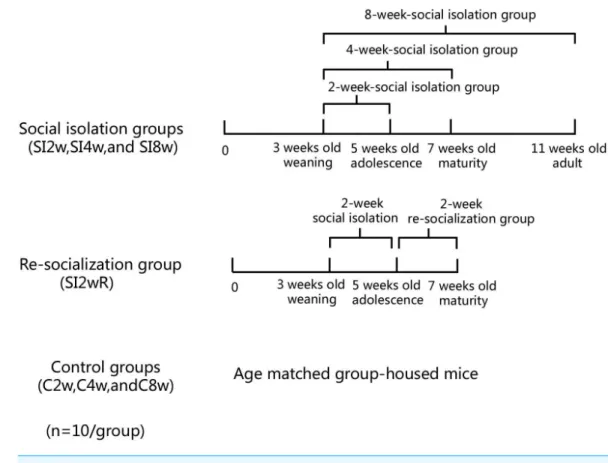
AnimalsMale KM mice (15±5 g) at the age of 21 days old were purchased from Laboratory Center of Dalian Medical University (ID: 0003746). They were housed in the plastic cage (Beijing Heli Technology Development Co. Ltd. China, 290×178×160 mm) with 5 mice for each cage reared in conditions of 21±1◦C, 55±5% humidity, and 12-h rhythm of day/night cycle. The mice were fed with food and water ad libitum. They were divided into seven groups randomly with 10 mice for each group, as shown inFig. 1. The mice were housed individually for 2, 4, and 8 weeks respectively. They were labeled as SI 2W group (isolation for 2 weeks), SI 4W group (isolation for 4 weeks), and SI 8W group (isolation for 8 weeks). In addition, recovery of behavioral deficit by re-socialization (rearing with their littermates at adolescence period) was also examined. The re-socialization mice were labeled as SI 2WR group (re-socialization for 2 weeks after isolation for 2 weeks). Control groups were age matched group-housed mice (C2W, C4W, and C8W). All experimental procedures were approved by the Tab of Animal Experimental Ethical Inspection, Number: L20140021.
Methods
Object Recognition Test (ORT)
The minor modified protocol (Fig. 2) as published in the literature (Võikar et al., 2005;Van Hagen et al., 2015;Cechella et al., 2014) was used in this study to measure the non-spatial cognitive ability. The test was performed in the behavior procedure room using the behavior observation apparatus (XR-XX117, Shanghai Xinruan Technology Co. Ltd. China). The observed box was 40×40×35 cm. Woody block A and B (Black, 5×5×5 cm) and block C (Black and white pattern, 5×5×5 cm) were used as recognition objects, with each object heavy enough to be stable. The process for ORT included both sample trial and test trial. Firstly, the mouse was trained to be in the empty box for 5 min to acclimate to the new environment. Then, during the sample trial, object A and object B were placed oppositely with a distance of 14 cm between them. The exploring time for each mouse was 5 min. The mouse was placed in the middle of two objects in the test. After the sample trial, the mouse returned back to its home cage for 4 h. During the test trial, object B was replaced with object C that was a novel one for the mouse. After each trial, the objects and the box were cleaned in order to avoid olfactory cues using 75% ethanol. The behavior of the mouse was recorded by videotapes.
Figure 1 Treatment of divided mice groups with isolation and re-socialization.The mice were housed individually for 2, 4, and 8 weeks respectively. They were labeled as SI2W group (isolation for 2 weeks), SI 4W group (isolation for 4 weeks), and SI 8W group (isolation for 8 weeks). In addition, recovery of behav-ioral deficit by re- socialization (rearing with their littermates at adolescence period) was also examined. The re-socialization mice were labeled as SI 2WR group (re-socialization for 2 weeks after isolation for 2 weeks). Control groups were the age matched group-housed mice (C 2W, C 4W, and C 8W).
Object Location Test (OLT)
The performance of OLT was similar with that of ORT (Võikar et al., 2005;Van Hagen et al., 2015;Cechella et al., 2014). This test is used to measure the spatial cognitive ability. Object A and B used in this study were the same as those used in the ORT. The acclimation was carried out in the same way as that in the ORT. In the sample trial, object A and B were put in the same location as that in the ORT. The mouse explored each object for 5 min and then was returned to the home cage for 4 h. After that, in the test trial, object B was moved to the opposite direction toward the object A, then the mouse was left to explore object A and object B with a novel location for 5 min. The behavior of the mouse was recorded in the same way as that used in ORT.
Immunohistochemistry staining
The mice were injected with 4% chloral hydrate for anesthesia (400 mg/kg, i.p.), followed by perfused transcardially with 1% and 4% paraformaldehyde (PFA) respectively. After that, the mice brains were fixed with PFA for 24 h at 4◦
C and then cryoprotected in 20% sucrose at 4◦
Figure 2 Diagram of Object Recognition Test and Object Location Test.(A) Object Recognition Test. The diagram shows the apparatus and recognition objects used in this test. Woody block A and B (Black, 5×5×5 cm) and block C (Black and white pattern, 5×5×5 cm) were used as recognition objects. Objects A and B were identical. The process for ORT included both sample trial and test trial. During the sam-ple trial, object A and object B were placed oppositely with a distance of 14 cm between them. In the test trial, object B was replaced by novel object C. (B) Object Location Test. The apparatus and objects A and B were the same as that used in the ORT. In the sample trial, objects A and B were put in the same location as that in the ORT. In the test trial, object B was moved to be located at the opposite direction toward the object A.
10 min for each time and then were incubated in 1% bovine serum albumin. After that, the slices were incubated with primary antibody of ADAR1-Ab (p110) (1:100, Proteintech, USA) overnight at 4◦
C. Then the sections were washed with PBS and were cultivated with biotinylated second antibody (ZSJQ-BIO Company, China) at room temperature for 1.5 h. After washing with PBS, the slices were treated with avidin-biotin complex at room temperature for 2 h. The positive expression of ADAR1 (p110) was visualized with diaminobenzidine (DAB) for the detection. Negative control slices were incubated with PBS without antibody, followed by stained with 1% thionine (sigma), a specific staining of nissl body of the neuron. Image analysis for quantification of the results was performed.
Western blot
transferred to polyvinyl diflouride (PVDF) membranes blocked for 1 h with 5% bovine serum albumin and then immunoblotted with the primary antibody (ADAR1-Ab-p110, 1:1,000; Proteintech, Chicago, IL, USA). Subsequently, membranes were washed with tris-buffered saline with Tween 20 (TBST) and incubated with horseradish peroxidase-labeled secondary antibody (anti-goat 1:5,000; ZSJQ-BIO Company, Beijing, China) for 2 h at room temperature in the dark. The Infrared band signals were detected and quantified using BIO-RAD (Hercules, CA, USA) gel analysis software. Membranes were then stripped using stripping buffer, washed in TBST, and probed with GADPH-Ab (1:1,000, Beyotime Company, China). After washing with TBST, membranes were incubated with horseradish peroxidase-labeled secondary antibody (anti-mouse, 1:5,000; ZSJQ-BIO Company, Beijing, China) and then were detected. ADAR1 (p110) protein expression was normalized by internal control-GADPH.
Statistical analysis
Graph-Pad Prism (GraphPad Software Inc.) and SPSS 21.0 were used to analyze statistically. All data were expressed as the mean±SEM and were analyzed by two-way ANOVA followed by Tukey’s post hoc testing.T test was used to analyze the variance between matched social isolation group and control group; two-way ANOVA was used to understand whether there is an interaction between social isolation and age level (two independent variables) on cognitive function (dependent variable) among mice. Post-hoc tests (or post-hoc comparison tests) are used at the second stage of the analysis of variance (ANOVA) or multiple analyses of variance (MANOVA) if the null hypothesis is rejected. In our study, the values of isolation 2 weeks, 4 weeks, and 8 weeks were analyzed as multiple analyses of variance, as well as the values of control 2 weeks, 4 weeks, and 8 weeks. ANOVA was used to analyze the differences among groups.P<0.05 was considered statistically significance.
RESULTS
Decreased DI of cognition by social isolation and its recovery by re-socialization
Figure 3 Decreased DI of spatial and non-spatial cognition in social isolation mice and its recovery by re-socialization.Compared to the control group (C2W), 2 weeks social isolation (SI2W) resulted in decreased discrimination index (DI) of spatial and non-spatial cognition. Similarly, 4 and 8 weeks social isolation also decreased values of DI (SI4W vs. C4W and SI8W vs. C8W). Re-socialization (SI2WR) recov-ered the decreased DI of isolated mice (SI 4W). (A) results of ORT, (B) results of OLT. Data is presented as mean±SEM (n=10/group).∗P<0.05 (C2W vs. SI2W, C4W vs. SI4W, and C8W vs. SI 8W).
Increased ADAR1 (p110) immunoreactivity by social isolation and its recovery by re-socialization
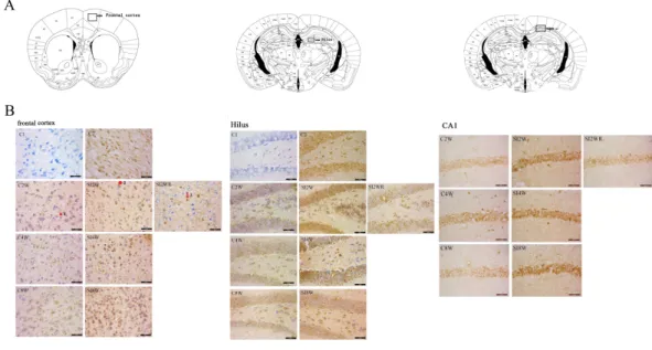
Figure 4 Increased ADAR1 (p110) immunoreactivity in frontal cortex and hippocampus of social iso-lated mice and its recovery by re-socialization. ADAR1 (p110) immunoreactivity was predominantly de-tected in frontal cortex and hippocampus of control and social isolation mice, in the meanwhile, the num-ber of detectable ADAR1 (p110) immunoreactivity positive signals significantly increased in the social iso-lation stress groups as shown inFig. 4, compared to age matched group-housed control mice. In frontal cortex, the ADAR1 (p110) immunoreactivity positive signals displayed almost all layers from molecular layer to multiform layer. In hippocampus, ADAR1 (p110) positive signals widely distributed in CA1, den-tate gyrus, and hilus. Re-socialization at adolescence recovered the increased ADAR1 (p110) immunoreac-tivity in frontal cortex and hippocampus of the isolated mice (Fig. 4). (A) The brain regions were analyzed on the basis of mice brain atlas ofPaxinos & Franklin (1997). Black boxes represented the brain regions magnified and presented as followingFig. 4B. Scale bar=50µm. (B) Double staining was performed
us-ing DAB stainus-ing and 1% thionine—a specific stainus-ing for nissl body of cell. ADAR1-Ab (p110) was used for marking ADAR1 (p110) immunoreactivity-positive signals. Arrow2 represented ADAR1 (p110) im-munoreactivity positive signals in nissl staining cell, arrow 1 represented nissl staining cell without ADAR1 (p110) immunoreactivity positive signals, and arrow 3 represented ADAR1 (p110) immunoreactivity posi-tive signals expressed in negaposi-tive nissl staining region.
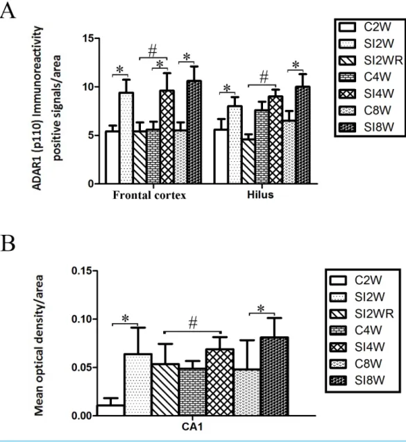
Figure 5 Increased ADAR1 (p110) immunoreactivity-positive signals in frontal cortex and hippocam-pus of the isolated mice by statistical analysis.(A) The number of ADAR1 (p110) immunoreactivity pos-itive signals/area in frontal cortex significantly increased in the mice isolated for 2, 4, and 8 weeks, com-pared to age matched group-housed control mice (C2W vs. SI2W, C4W vs. SI4W, and C8W vs. SI8W). In the meanwhile, the number of ADAR1 (p110) immunoreactivity positive signals/area in hilus significantly increased in the mice isolated for 2 and 8 weeks, compared to age matched group-housed control mice (C2W vs. SI2W and C8W vs. SI8W). (B) The mean optical density/area of ADAR1 (p110) immunoreactiv-ity positive signals in CA1 significantly increased in the mice isolated for 2 and 8 weeks, compared to age matched group-housed control mice (C2W vs. SI2W, C8W vs. SI8W). Re-socialization recovered the in-creased ADAR1 (p110) immunoreactivity in frontal cortex and hippocampus of the isolated mice (SI4W vs. SI2WR). The number of ADAR1 (p110) immunoreactivity positive signals was counted in the sequen-tial cutting sections. The sections analyzed in frontal cortex were from Bregma 1.18 mm for 4 sections, and the sections analyzed in hippocampus were from Bregma−2.18 mm for 4 sections (16µm per section). The brain regions analyzed were focused on the internal pyramidal cell layer 5 of frontal cortex, as well as hilus and CA1 of hippocampus. The square analyzed was 10,000 µm2. Data were expressed as the mean ±SEM and were analyzed by two-way ANOVA followed by Tukey’s post hoc testing (n=5/group).∗P<
number of ADAR1 (p110) immunoreactivity-positive signals in frontal cortex of the mice isolated for 2, 4, and 8 weeks were significantly increased, compared to age matched group-housed mice. The data was shown as follows: (Number: (2 weeks control group: 5.40 ±0.60; 2 weeks isolation group: 9.40±1.33; p=0.02); (4 weeks control group: 5.60 ±0.81; 4 weeks isolation group: 9.60±1.81; p=0.04); (8 weeks control group: 5.50±0.85; 8 weeks isolation group: 10.6±1.50;p=0.01)). However, the optical density of ADAR1 (p110) immunoreactivity-positive signals of CA1 and the number of ADAR1 (p110) immunoreactivity-positive signals of hilus were only increased obviously in the mice isolated for 2 and 8 weeks, compared to age matched group-housed mice. The data was shown as follows: (Number of hilus : (2 weeks control group: 5.60±1.08; 2 weeks isolation group: 8.00±0.94;p=0.04); (8 weeks control group: 7.60±0.88; 8 weeks isolation group: 10.00±1.31;p=0.04)) and (Optical density of CA1: (2 weeks control group: 0.01±0.02; 2 weeks isolation group: 0.06±0.03;p=0.04); (8 weeks control group: 0.05±0.03; 8 weeks isolation group: 0.08±0.02;p=0.03)). Furthermore, no obvious difference of number and optical density of ADAR1 (p110) positive immunoreactivity signals was found between the re-socialization group (SI2WR) and the control group (C4W), which suggested that re-socialization mice recovered the increased ADAR1 immunoreactivity in both frontal cortex and hippocampus of the isolated mice.
Increased ADAR1 (p110) protein expression by social isolation and its recovery by re-socialization
Western blot results of ADAR1 (p110) were consistent with those of immunohistochemistry staining results mostly. The protein expressions of ADAR1 (p110) significantly increased in not only the frontal cortex but also the hippocampus of the mice isolated for 2, 4, and 8 weeks, compared to age matched group-housed mice, as shown in Figs. 6 and7. The data was shown as follows: (the optical density of the ratio between ADAR1 (p110) and GADPH: frontal cortex: (2 weeks control group: 0.73 ±0.04; 2 weeks isolation group: 1.02±0.12;p=0.03); (4 weeks control group: 0.25±0.08; 4 weeks isolation group: 0.84±0.14;p=0.04); (8 weeks control group: 0.25±0.09; 8 weeks isolation group: 0.72 ±0.12;p=0.04); Hippocampus: (2 weeks control group: 0.91 ±0.09; 2 weeks isolation
group: 1.41±0.28;p=0.04); (4 weeks control group: 0.65±0.19; 4 weeks isolation group: 1.46±0.40;p=0.04); (8 weeks control group: 0.41±0.14; 8 weeks isolation group: 0.71 ±0.13; p=0.03)). The above results suggested that social isolation increased ADAR1
(p110) protein expression. In addition, the protein expression of ADAR1 (p110) of SI2WR in hippocampus was no difference with that of age matched group-housed mice. The results suggested that re-socialization bring ADAR1 (p110) protein expression of hippocampus back to the normal level for the isolated mice in adolescence.
DISCUSSION
Figure 6 Increased ADAR1 (p110) protein expression in frontal cortex and hippocampus of isolated mice and its recovery by re-socialization.The protein expression of ADAR1 (p110) significantly increased in the social isolation stress groups, as shown inFig. 6, compared to age matched group-housed control mice. The detailed results were that ADAR1 (p110) protein expression increased in both frontal cortex and hippocampus of the mice isolated for 2, 4, and 8 weeks, in the meanwhile, re-socialization mice recovered the increased ADAR1 (p110) protein expression in the hippocampus of the isolated mice. The analyzation was shown in the followingFig. 7.
not only recover the cognition deficit, but also bring ADAR1 (p110) immunoreactivity of hippocampus and frontal cortex, as well as ADAR1 (p110) protein expression of hippocampus back to the normal level for the isolated mice in adolescence. Theses novel findings suggest that ADAR1 (p110) is related to isolation-induced cognitive deficit.
Cognitive deficit induced by social isolation
Figure 7 Increased ADAR1 (p110) protein expression in frontal cortex and hippocampus of isolated mice and its recovery by re-socialization in statistical analysis.The ADAR1 (p110) protein expression increased in both frontal cortex and hippocampus of the mice isolated for 2, 4, and 8 weeks , compared to age matched group-housed control mice (SI2W vs. C2W, SI4W vs. C4W, and SI8W vs. C8W). In the meanwhile, re-socialization mice recovered the increased ADAR1 (p110) protein expression of the isolated mice in hippocampus to normal level (no different between C4W and SI2WR). Interestingly, the ADAR1 (p110) protein expression in both frontal cortex and hippocampus showed an age-dependent manner, which can be seen that ADAR1 (p110) protein expression of 11 weeks (C8W) and 7 weeks old mice (C4W) were less than that of 5 weeks old mice (C2W). ADAR1 (p110) protein expression was normalized by internal control—GADPH. The data was expressed as the mean±SEM and analyzed by
two-way ANOVA followed by Tukey’s post hoc testing.∗P
<0.05 (C2W vs. SI2W, C4W vs. SI4W, and C8W vs. SI8W),∧
P<0.05 (C2W vs. C4W and C2W vs. C8W).
Increased ADAR1 (p110) by social isolation in the brain of mice with cognitive deficit
that of age matched group-housed control mice, as shown inFig. 4. It is important to note that both frontal cortex and hippocampus are vulnerable to social isolation stress (Buechel et al., 2014). According to the literature, ADAR-deficient mice exhibit defects in nervous system and decrease tolerance to stress (Tseng et al., 2013). It appears that ADAR1 (p110) expression is related to cognitive deficit induced by isolation stress.
Recovered level of ADAR1 (p110) expression by re-socialization In the past years, a number of methods have been reported to be able to alleviate the symptoms of cognitive deficit induced by social isolation, including re-socialization (Maisonnette, Morato & Brandão, 1993), electro-acupuncture (Manni, Aloe & Fiore, 2009), and treatment with drugs such as citalopram, cariprazine, aripiprazole, acetylcysteine (NAC), selective blockade of dopamine D3 receptors, and selective mGluR2/3 agonist (Jones et al., 2011). In this study, the effects of re-socialization on ADAR1 (p110) expression and the recovery of cognitive deficit were evaluated. It was interestingly found that re-socialization was effective to recover cognitive dysfunction and bring ADAR1 (p110) immunoreactivity of hippocampus & frontal cortex and ADAR1 (p110) protein expression of hippocampus back to the normal level for the isolated mice in adolescence. The reason for the recovery of cognitive deficit is possible that the mice in adolescence have strong neuro-plasticity and could be highly sensitive to re-socialization (Forbes & Dahl, 2005).
In conclusion, social isolation led to spatial and non-spatial cognition deficits in mice and impacted ADAR1 (p110) immunoreactivity and protein expression in hippocampus and frontal cortex. Moreover, re-socialization was effective to recover cognitive dysfunction and bring ADAR1 (p110) expression to the normal level for the isolated mice in adolescence. However, it is too early to make a conclusion that there is direct link between ADAR1 (p110) expression and cognitive deficit induced by social isolation, based on the evidence provided by this study so far. In the future, ADARs gene knock-out mice could be used to investigate how ADARs regulate RNA editing in social isolation-induced cognitive dysfunction. The related studies should be beneficial to the studies of social environment and body-mind healthy in human beings.
ACKNOWLEDGEMENTS
We thank Dr Song Li for his technical support.
ADDITIONAL INFORMATION AND DECLARATIONS
Funding
This work was supported by grants from the National Natural Science Foundation of China (31201724). The funders had no role in study design, data collection and analysis, decision to publish, or preparation of the manuscript.
Grant Disclosures
Competing Interests
The authors declare there are no competing interests.
Author Contributions
• Wei Chen conceived and designed the experiments, wrote the paper, prepared figures
and/or tables.
• Dong An conceived and designed the experiments, prepared figures and/or tables.
• Hong Xu, Xiaoxin Cheng, Weizhi Yu and Deqin Yu performed the experiments.
• Shiwei Wang analyzed the data.
• Dan Zhao performed the experiments, prepared figures and/or tables.
• Yiping Sun and Wuguo Deng contributed reagents/materials/analysis tools.
• Yiyuan Tang contributed reagents/materials/analysis tools, advice the manuscript.
• Shengming Yin conceived and designed the experiments, wrote the paper, reviewed
drafts of the paper.
Animal Ethics
The following information was supplied relating to ethical approvals (i.e., approving body and any reference numbers):
The Tab of Animal Experimental Ethical Inspection—Number: L20140021.
Data Availability
The following information was supplied regarding data availability: The raw data has been supplied asData S1.
Supplemental Information
Supplemental information for this article can be found online athttp://dx.doi.org/10.7717/
peerj.2306#supplemental-information.
REFERENCES
Araki R, Ago Y, Hasebe S, Nishiyama S, Tanaka T, Oka S, Takuma K, Matsuda T, Int J. 2014.Involvement of prefrontal AMPA receptors in encounter stimulation-induced hyperactivity in isolation-reared mice.Neuropsychopharmacology 17:883–893
DOI 10.1017/S1461145713001582.
Baarendse PJ, Counotte DS, O’Donnell P, Vanderschuren LJ. 2013.Early social experi-ence is critical for the development of cognitive control and dopamine modulation of prefrontal cortex function.Neuropsychopharmacology38:1485–1494
DOI 10.1038/npp.2013.47.
Barratt RL, Shaban R, Moyle W. 2011.Patient experience of source isolation: lessons for clinical practice.Contemporary Nurse39:180–190DOI 10.5172/conu.2011.39.2.180.
Bombail V, Qing W, Chapman KE, Holmes MC. 2014.Prevention of
Bowden DM, McKinney WT. 1972.Behavioral effects of peer separation, isolation, and reunion on adolescent male rhesus monkeys.Developmental Psychobiology 5(4):353–362.
Buechel HM, Popovic J, Staggs K, Anderson KL, Thibault O, Blalock EM. 2014.Aged rats are hypo-responsive to acute restraint: implications for psychosocial stress in aging.Frontiers in Aging Neuroscience6:1–16.
Cattenoz PB, Taft RJ, Westhof E, Mattick JS. 2013.Transcriptome-wide identification of A > I RNA editing sites by inosine specific cleavage.RNA19:257–270
DOI 10.1261/rna.036202.112.
Cechella JL, Leite MR, Rosario AR, Sampaio TB, Zeni G. 2014.Diphenyl diselenide-supplemented diet and swimming exercise enhance novel object recognition memory in old rats.Age 36:1–10DOI 10.1007/s11357-014-9666-8.
Dang YH, Liu P, Ma R, Chu Z, Liu YP, Wang JB, Ma XC, Gao CG. 2015.HINT1 is involved in the behavioral abnormalities induced by social isolation rearing. Neuroscience Letters21, 607:40–45DOI 10.1016/j.neulet.2015.08.026.
Djordjevic J, Djordjevic A, Adzic M, Radojcic MB. 2012.Effects of chronic social isolation on Wistar rat behavior and brain plasticity markers.Neuropsychobiology 66:112–119DOI 10.1159/000338605.
Elston GN, Benavides-Piccione R, DeFelipe J. 2001.The pyramidal cell in cognition: a comparative study in human and monkey.Journal of Neuroscience21:163.
Fierro-Monti II, Mathews MB. 2000.Proteins binding to duplexed RNA: one motif, multiple functions.Trends in Biochemical Sciences25:241–246
DOI 10.1016/S0968-0004(00)01580-2.
Fone KC, Porkess MV. 2008.Behavioural and neurochemical effects of post-weaning social isolation in rodents-relevance to developmental neuropsychiatric disorders. Neuroscience and Biobehavioral Reviews32:1087–1090
DOI 10.1016/j.neubiorev.2008.03.003.
Forbes EE, Dahl RE. 2005.Neural systems of positive affect: relevance to understanding child and adolescent depression?Development and Psychopathology 17:827–850
DOI 10.1017/S095457940505039X.
George CX, John L, Samuel CE. 2014.An RNA editor, adenosine deaminase acting on double-stranded RNA (ADAR1).Journal of Interferon and Cytokine Research 34:437–446. ReviewDOI 10.1089/jir.2014.0001.
Gong WG, Wang YJ, Zhou H, Li XL, Bai F, Ren QG, Zhang ZJ. 2016.Citalopram ameliorates synaptic plasticity deficit in different cognition-associated brain regions induced by social isolation in middle-aged rats.Molecular NeurobiologyEpub ahead of print Feb 22 2016.
Grant N, Hamer M, Steptoe A. 2009.Social isolation and stress-related cardiovas-cular, lipid, and cortisol responses.Annals of Behavioral Medicine37:29–37
DOI 10.1007/s12160-009-9081-z.
stress through mitigating the negative impact of interlukin-1βand nitric oxide on hypothalamic-pituitary-adrenal axis function.Neuroscience315:271–285
DOI 10.1016/j.neuroscience.2015.12.024.
Hartner JC, Schmittwolf C, Kispert A, Müller AM, Higuchi M, Seeburg PH. 2004.Liver disintegration in the mouse embryo caused by deficiency in the RNA-editing enzyme ADAR1.Journal of Biological Chemistry279:4894–4902.
Hatch AM, Wiberg GS, Zawidzka Z, Cann M, Airth JM, Grice HC. 1965.Isolation syndrome in the rat.Toxicology and Applied Pharmacology7:737–745
DOI 10.1016/0041-008X(65)90132-8.
Hellemans K, Nobrega JN, Olmstead MC. 2005.Early environmental experience alters baseline and ethanol-induced cognitive impulsivity: relationship to forebrain 5-HT1A receptor binding.Behavioural Brain Research159:207–220
DOI 10.1016/j.bbr.2004.10.018.
Jones CA, Brown AM, Auer DP, Fone KC. 2011.The mGluR2/3 agonist LY379268 reverses post-weaning social isolation-induced recognition memory deficit in the rat.Psychopharmacology214:269–283DOI 10.1007/s00213-010-1931-7.
Khodaie B, Lotfinia AA, Ahmadi M, Lotfinia M, Jafarian M, Karimzadeh F, Coulon P, Gorji A. 2015.Structural and functional effects of social isolation on the hippocam-pus of rats with traumatic brain injury.Behavioural Brain Research278:55–65
DOI 10.1016/j.bbr.2014.09.034.
Koike H, Ibi D, Mizoguchi H, Nagai T, Nitta A, Takuma K, Nabeshima T, Yoneda Y, Yamada K. 2009.Behavioral abnormality and pharmacologic response in social isolation-reared mice.Behavioural Brain Research202:114–121
DOI 10.1016/j.bbr.2009.03.028.
Liscovitch N, Bazak L, Levanon EY, Chechik G. 2014.Positive correlation between ADAR expression and its targets suggests a complex regulation mediated by RNA editing in the human brain.RNA Biology11:1447–1456
DOI 10.4161/15476286.2014.992286.
Liu Y, Samuel CE. 1996.Mechanism of interferon action: functionally distinct binding and catalytic domains in the interferon-inducible, double-stranded RNA-specific adenosine deaminase.Journal of Virology 70:1961–1968.
Maisonnette S, Morato S, Brandão ML. 1993.Role of resocialization and of 5-HT1A re-ceptor activation on the anxiogenic effects induced by isolation in the elevated plus-maze test.Physiology & Behavior54:753–758DOI 10.1016/0031-9384(93)90087-V.
Manni L, Aloe L, Fiore M. 2009.Changes in cognition induced by social isolation in the mouse are restored by electro-acupuncture.Physiology & Behavior98:537–542
DOI 10.1016/j.physbeh.2009.08.011.
Mattick JS, Mehler MF. 2008.RNA editing, DNA recoding and the evolution of human cognition.Trends in Neurosciences5:227–233. ReviewDOI 10.1016/j.tins.2008.02.003.
perturbed cognition in schizophrenia.EMBO Molecular Medicine4:1043–1056
DOI 10.1002/emmm.201201410.
Murai T, Okuda S, Tanaka T, Ohta H. 2007.Characteristics of object location memory in mice: behavioral and pharmacological studies.Physiology & Behavior90:116–124
DOI 10.1016/j.physbeh.2006.09.013.
Okada R, Fujiwara H, Mizuki D, Araki R, Yabe T, Matsumoto K. 2015.Involvement of dopaminergic and cholinergic systems in social isolation-induced deficit in social affiliation and conditional fear memory in mice.Neuroscience299:134–145
DOI 10.1016/j.neuroscience.2015.04.064.
O’Keefe LM, Doran SJ, Mwilambwe-Tshilobo L, Conti LH, Venna VR. 2014.Social isolation after stroke leads to depressive-like behavior and decreased BDNF levels in mice.Behavioural Brain Research260:162–170DOI 10.1016/j.bbr.2013.10.047.
Patterson JB, Samuel CE. 1995.Expression and regulation by interferon of a double-stranded-RNA-specific adenosine deaminase from human cells: evidence for two forms of the deaminase.Molecular and Cellular Biology 15:5376–5588
DOI 10.1128/MCB.15.10.5376.
Paxinos G, Franklin KBJ. 1997.The mouse brain in stereotaxic coordinates. Second edition.
Pereda-Pérez I, Popović N, Otalora BB, Popović M, Madrid JA, Rol MA, Venero C. 2013.Long-term social isolation in the adulthood results in CA1 shrinkage and cognitive impairment.Neurobiology Learning Memory106:31–39
DOI 10.1016/j.nlm.2013.07.004.
Pisu MG, Garau A, Boero G, Biggio F, Pibiri V, Dore R, Locci V, Paci E, Porcu P, Serra M. 2016.Sex differences in the outcome of juvenile social isolation on HPA axis function in rats.Neuroscience320:172–182DOI 10.1016/j.neuroscience.2016.02.009.
Quan MN, Tian YT, Xu KH, Zhang T, Yang Z. 2010.Post weaning social isolation influences spatial cognition, prefrontal cortical synaptic plasticity and hippocampal potassium ion channels in Wistar rats.Neuroscience169:214–222
DOI 10.1016/j.neuroscience.2010.04.048.
Rybak-Wolf A, Stottmeister C, Glažar P, Jens M, Pino N, Giusti S, Hanan M, Behm M, Bartok O, Ashwal-Fluss R, Herzog M, Schreyer L, Papavasileiou P, Ivanov A, Öhman M, Refojo D, Kadener S, Rajewsky N. 2015.Circular RNAs in the mammalian brain are highly abundant, conserved, and dynamically expressed. Molecular Cell58:870–885DOI 10.1016/j.molcel.2015.03.027.
Sandstrom NJ, Hart SR. 2005.Isolation stress during the third postnatal week alters radial arm maze performance and corticosterone levels in adulthood.Behavioural Brain Research156:289–296DOI 10.1016/j.bbr.2004.05.033.
Schirle NT, Goodman RA, Krishnamurthy M, Beal PA. 2010.Selective inhibition of ADAR2-catalyzed editing of the serotonin 2c receptor pre-mRNA by a helix-threading peptide.Organic and Biomolecular Chemistry8:4898–4904
Schmauss C, Zimnisky R, Mehta M, Shapiro LP. 2010.The roles of phospholipase C activation and alternative ADAR1 and ADAR2 pre-mRNA splicing in modulating serotonin 2C-receptor editingin vivo.RNA16:1779–1785DOI 10.1261/rna.2188110.
Toth AM, Zhang P, Das S, George CX, Samuel CE. 2006.Interferon action and the double-stranded RNA-dependent enzymes ADAR1 adenosine deaminase and PKR protein kinase.Progress in Nucleic Acid Research and Molecular Biology81:369–434
DOI 10.1016/S0079-6603(06)81010-X.
Tseng CN, Chang HW, Stocker J, Wang HC, Lu CC, Wu CH. 2013.A method to identify RNA A-to-I editing targets using I-specific cleavage and exon array analysis. Molecular and Cellular Probes27:38–45DOI 10.1016/j.mcp.2012.08.008.
Valtorta NK, Kanaan M, Gilbody S, Ronzi S, Hanratty B. 2016.Loneliness and social isolation as risk factors for coronary heart disease and stroke: systematic review and meta-analysis of longitudinal observational studies.Heart 102:1009–1016
DOI 10.1136/heartjnl-2015-308790.
Valzelli L. 1973.The ‘‘isolation syndrome’’ in mice.Psychopharmacologia31:305–320
DOI 10.1007/BF00421275.
Van Hagen BT, Van Goethem NP, Lagatta DC, Prickaerts J. 2015.The object pattern separation (OPS) task: a behavioral paradigm derived from the object recognition task.Behavioural Brain Research285:44–52. ReviewDOI 10.1016/j.bbr.2014.10.041.
Võikar V, Polus A, Vasar E, Rauvala H. 2005.Long-term individual housing in C57BL/6J and DBA/2 mice: assessment of behavioral consequences.Genes Brain and Behavior 4:240–252.
Wang Q, Miyakoda M, Yang W, Khillan J, Stachura DL, Weiss MJ, Nishikura K. 2004.
Stress-induced apoptosis associated with null mutation of ADAR1 RNA editing deaminase gene.Journal of Biological Chemistry279:4952–4961.
Watson DJ, King MV, Gyertyán I, Kiss B, Adham N, Fone KC. 2016.The dopamine D3-preferring D2/D3 dopamine receptor partial agonist, cariprazine, reverses behavioural changes in a rat neurodevelopmental model for schizophrenia.European Neuropsychopharmacology 26:208–224DOI 10.1016/j.euroneuro.2015.12.020.
Watson DJ, Marsden CA, Millan MJ, Fone KC. 2012.Blockade of dopamine D3but
not D2receptors reverses the novel object discrimination impairment produced by
post-weaning social isolation: implications for schizophrenia and its treatment.The International Journal of Neuropsychopharmacology15:471–484
DOI 10.1017/S1461145711000435.
Yang JH, Luo X, Nie Y, Su Y, Zhao Q, Kabir K, Zhang D, Rabinovici R. 2003.
Widespread inosine-containing mRNA in lymphocytes regulated by ADAR1 in response to inflammation.Immunology109:15–23
DOI 10.1046/j.1365-2567.2003.01598.x.
Yusufishaq S, Rosenkranz JA. 2013.Post-weaning social isolation impairs observational fear conditioning.Behavioual Brain Research242:142–149